|
|

|

|
| e-Pub |
Section: New Results
Modelling and identification of the sensory-motor system
Whole Body Center of Mass Estimation with Portable Sensors: Using the Statically Equivalent Serial Chain and a Kinect
Participants : Alejandro Gonzalez de Alba, Mitsuhiro Hayashibe, Vincent Bonnet, Philippe Fraisse.
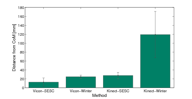
The trajectory of the whole body center of mass (CoM) is useful as a reliable metric of postural stability. If the evaluation of a subject-specific CoM were available outside of the laboratory environment, it would improve the assessment of the effects of physical rehabilitation. A method is developed tot enable tracking CoM position using low-cost sensors such that it can be moved around by a therapist or easily installed inside a patientâs home. We compare the accuracy of a personalized CoM estimation using the statically equivalent serial chain (SESC) method and measurements obtained with the Kinect to the case of a SESC obtained with high-end equipment (Vicon). We also compare these estimates to literature-based ones for both sensors. The method was validated with seven able-bodied volunteers for whom the SESC was identified using 40 static postures. The literature-based estimation with Vicon measurements had a average error 24.9 3.7 mm; this error was reduced to 12.8 9.1 mm with the SESC identification. When using Kinect measurements, the literature-based estimate had an error of 118.4 50.0 mm, while the SESC error was 26.6 6.0 mm. The subject-specific SESC estimate using low-cost sensors has an equivalent performance as the literature-based one with high-end sensors. The SESC method can improve CoM estimation of elderly and neurologically impaired subjects by considering variations in their mass distribution.
|
A Personalized Balance Measurement for Home-based Rehabilitation
Participants : Alejandro Gonzalez de Alba, Philippe Fraisse, Mitsuhiro Hayashibe.
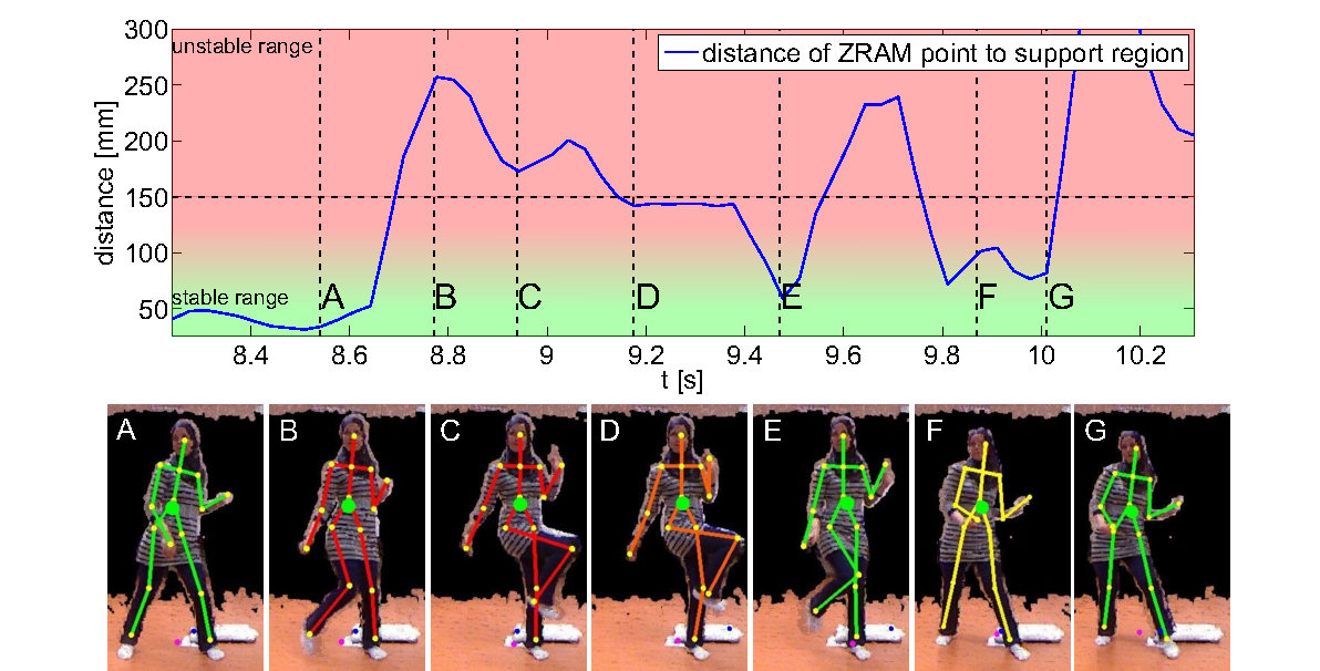
We use the subject-specific center of mass (CoM) estimate offered by the statically equivalent serial chain (SESC) method and the zero rate of change of angular momentum (ZRAM) concept to evaluate balance for a series of dynamic motions. Two healthy subjects were asked to stand on a Wii balance board and their SESC parameters were identified. A set of dynamic motions was to evaluate the rate of change of centroidal angular momentum and the distance of the ZRAM point to the center line of the support polygon. We found a good match between both balance metrics. As an application example, the subjects performed a dynamic motion (such as walking and abruptly stopping) and the stability was determined in real-time using the ZRAM point from the personalized CoM trajectory. This was implemented with a real-time balance visualization tool based on Kinect measurements for home-based rehabilitation.
|
Methodology for automatic movement cycle extraction using Switching Linear Dynamic System
Participants : Roberto de Souza Baptista, Mitsuhiro Hayashibe, Antônio P. L. Bó [Univ. Brasilia] .
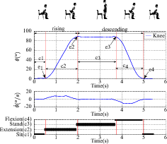
Human motion assessment is key for motor-control rehabilitation. Using standardized definifions and spatiotemporal features - usually presented as a movement cycle diagram- specialists can associate kinematic measures to progress in rehabilitation therapy or motor impairment due to trauma or disease. Although devices for capturing human motion today are cheap and widespread, the automatic interpretation of kinematic data for rehabilitation is still poor in terms of quantitative performance evaluation. In this paper we present an automatic approach to extract spatiotemporal features from kinematic data and present it as a cycle diagram. This is done by translating standard definitions from human movement analisys into mathematical elements of a Switching Linear Dynamic System model. The result is a straight-forward procedure to learn a tracking model from a sample execution. This model is robust when used to automatically extract the movement cycle diagram of the same motion (the Sit-Stand-Sit, as an example) executed in different subject-specific manner such as his own motion speed.
|
Real-time Muscle Deformation via Decoupled Modeling of Solid and Muscle Fiber Mechanics
Participants : Yacine Berranen, Mitsuhiro Hayashibe, David Guiraud, Benjamin Gilles.
This work presents a novel approach for simulating 3D muscle deformations with complex architectures. The approach consists in choosing the best model formulation in terms of computation cost and accuracy, that mixes a volumetric tissue model based on finite element method (3D FEM), a muscle fiber model (Hill contractile 1D element) and a membrane model accounting for aponeurosis tissue (2D FEM). The separate models are mechanically binded using barycentric embeddings. Our approach allows the computation of several fiber directions in one coarse finite element, and thus, strongly decreases the required finite element resolution to predict muscle deformation during contraction. Using surface registration, fibers tracks of specific architecture can be transferred from a template to subject morphology, and then simulated. As a case study, three different architectures are simulated and compared to their equivalent one dimensional Hill wire model simulations.
|
|
Adaptive model for viscoelastic solids
Participants : Benjamin Gilles, Maxime Tournier, Matthieu Nesme, Francois Faure.
A new adaptive model for viscoelastic solids is presented in [50] , [34] . Unlike previous approaches, it allows seamless transitions, and simplifications in deformed states. The deformation field is generated by a set of physically animated frames. Starting from a fine set of frames and mechanical energy integration points, the model can be coarsened by attaching frames to others, and merging integration points. Since frames can be attached in arbitrary relative positions, simplifications can occur seamlessly in deformed states, without returning to the original shape, which can be recovered later after refinement. We propose a new class of velocity-based simplification criterion based on relative velocities. Integration points can be merged to reduce the computation time even more, and we show how to maintain constant elastic forces through the levels of detail. This meshless adaptivity allows significant improvements of computation time.
Functional Brain Stimulation: Filling the gap between micro- and direct electrical stimulation of the brain in order to better understand and innovate
Participants : Marion Vincent, Olivier Rossel, Mitsuhiro Hayashibe, Guillaume Herbet, Hugues Duffau [Neurosurgery Department, CHU, Montpellier] , David Guiraud, François Bonnetblanc.
Micro-stimulation (MES) and Direct electrical stimulation (DES) of the brain are both used to perform in vivo functional mapping of the brain in fundamental neuroscience and neurosurgery respectively. The former is performed in animal experiments while the latter is performed on humans in the operative room. Very recently, a strong debate occurred to determine whether DES used during “wide-awake surgery” with success is a gold standard to study brain functions (Mandonnet et al., 2010; Borchers et al. 2012; Desmurget et al., 2013). In this debate, confusion is very often made between DES and MES, as these are considered to induce similar effects on the nervous tissues, with comparable behavioural consequences. However, electrical stimulation (ES) parameters used in both techniques are clearly different. More surprisingly, a strong biophysical rational of their choices is lacking. It may be due to historical, methodological and technical constraints that have guided empirically them. These constraints may have strongly shaped and limited experimental protocols in a standard way. By contrast, the gap between MES and DES may reveal a great potential for new experimental paradigms in ES of the brain in vivo. By considering this gap and new technical developments in the design of stimulators, it may be time to move on to alternative and innovative stimulations protocols, especially regarding and inspired from what is performed in functional electrical stimulation (FES) of peripheral nerves, for which more theoretical supports exist.
Modelling of structural and functional brain connectivity networks in Diffuse Low-Grade Glioma (DLGG)
Participants : Jija Syamala James, Anirban Dutta, François Bonnetblanc, David Guiraud, Nicolas Menjot de Champfleur [Department of Neuroradiology,CHU, Montpellier] , Emmanuelle Le Bars [Institute of Human Functional Imaging 12 FH, CHU, Montpellier] , Hugues Duffau [Neurosurgery Department, CHU, Montpellier] .
The impairment of functional brain connectivity networks in Diffuse Low Grade Glioma (DLGG) subjects can lead to distinct functional deficits where the challenge remains in greater understanding of distribution of dynamic brain connectivity. Our multimodality (resting state functional MRI, Diffusion Tensor Imaging and tractography) neuroimaging study aims to evaluate the differences in spatial and temporal patterns of brain connectivity networks in DLGG patients, pre and postoperatively.
In order to identify the brain connectivity networks, we analysed resting state fMRI data of 22 mesio-frontal DLGG patients by using MELODIC (Multivariate Exploratory Linear Optimised Decomposition into Independent Components)-ICA module, implemented in Freesurfer Software Library [FSL] (www.fmrib.ox.ac.uk/fsl). First we evaluated the clinical efficacy of rsfMRI technique to non-invasively map the dynamic functional reorganizations of brain connectivity networks in glioma subjects. Further we observed the existence of altered inter-hemispheric functional connectivity in DLGG subjects postoperatively.
We are currently performing this analysis using larger sample size in order to find out
a) the invivo structure-function relationship of motor, language and visual brain networks by fusing the information from DTI and fMRI.
b) the correlation between brain connectivity networks and neurobehavioural performance.
c) the neuroplastic alterations in topographic organization and strength of connections before and after DLGG surgery.
SPINSTIM: Direct spinal stimulation for rehabilitation of bladder, bowel and sexual functions in spinal cord injury
Participants : Christine Azevedo Coste, Luc Bauchet [Neurosurgery Department, CHU, Montpellier] , Claire Delleci [CHU Bordeaux] , Charles Fattal, Thomas Guiho, David Guiraud, Jean-Rodolphe Vignes [CHU Bordeaux] .
For the general public, spinal cord injury (SCI) is often restricted to limb paralysis. In reality, by interrupting communication between encephalon and peripheral organs, medullary wounds lead to physiological deficiencies such as urinary (retention and/or incontinence), gastrointestinal (constipation) or sexual impairments; disorders which are the center of patient’s expectations.
Spinal cord stimulation (SCS) is a general term which includes both epidural and intradural stimulation. Originally associated with the treatment of chronic neurogical pain (in the 1970ies), SCS led also to immediate and profound improvements of sensory and motor functions in recent studies both on SCI patients (few subjects involved) and rodents.
Despite these promising results some limitations have still to be overcome. Among them, the use of small animal models, the empirical aspect of the stimulation procedure and the impact of these protocols on intestinal and urinary functions are critical.
To counteract these limits, we want to explore intradural and epidural stimulations in an intermediate model- the house pig- and assess their impact on bladder, guts and genitals. In order to evaluate our approach, we will record EMG signals of lower limbs and sphincters (both urethral and anal), and simultaneously, we will monitor bladder and rectal pressure.
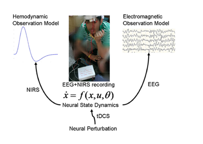
Development of point of care testing (POCT) device for neurovascular coupling from simultaneous recording of EEG and NIRS during non-invasive brain stimulation (NIBS) for closed-loop control of NIBS.
Participants : Mehak Sood, Utkarsh Jindal, Mitsuhiro Hayashibe, Stephane Perrey, Michael A. Nitsche, Abhijit Das, Shubhajit Roy Chowdhury, Anirban Dutta.
Our preliminary work showed that transcranial direct current stimulation (tDCS) can perturb local neuronal activity which can be used for assessing regional neurovascular coupling (NVC) functionality. It was postulated that tDCS leads to rapid dynamic variations of the brain cell microenvironment that perturbs the hemodynamic and electromagnetic responses. Based on these preliminary studies, we developed a POCT device for EEG-NIRS based screening and monitoring of neurovascular coupling functionality under perturbation with tDCS. The stroke case study showed detectable changes in the degree of NVC to a 0.526A/m2 square-pulse (0-30sec) of anodal tDCS where these alterations in the vascular system may result in secondary changes in the cortical excitability. The objective of this case study was to evaluate an empirical method to assess NVC using cross-correlation function (CCF) between mean (cortical) tissue oxy-haemoglobin concentration time-series and averaged PSD time-course from the EEG spectrogram. The CCF based assessment of the patient-specific status of NVC are currently being studied in a larger cohort with small vessel diseases. The overarching goal is closed-loop control of tDCS based on simultaneous recording of EEG and NIRS during non-invasive brain stimulation.