Section:
New Results
Cell-to-cell ascidian embryo registration
Participants :
Gaël Michelin, Grégoire Malandain.
This work is made in collaboration with Léo Guignard and
Christophe Godin (Virtual Plants) and Patrick Lemaire (CRBM), within
the Morphogenetics Inria Project Lab.
Recent microscopy techniques allow imaging temporal 3D stacks of developing organs or embryos with a cellular level of resolution and with a sufficient acquisition frequency to accurately track cell lineages.
Imaging multiple organs or embryos in different experimental
conditions may help to decipher the impact of genetic backgrounds and environmental inputs on the developmental program. For this, we need to precisely compare distinct individuals and to compute population statistics. The first step of this procedure is to develop methods to register individuals.
From a previous work of cell segmentation from microscopy
images [6] , we propose an approach to extract
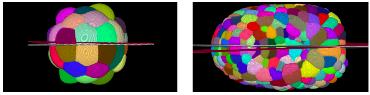
the Left-Right symmetry plane of embryos at early stages (Figure
7 ). Then we use the symmetry information to
both register these embryos at a similar developmental stage and
obtain a cell-to-cell mapping. We assessed the symmetry plane
extraction on more than 100 images from 10 individuals between
32-cells and late-neurula development stage. The cell-to-cell
registration was performed on 5 distinct individuals at 64-cells and
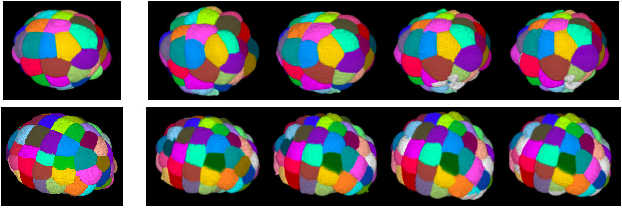
112-cells stage (Figure 8 ).
Figure
7. Left-Right symmetry plane initialization (red) and
final estimation (white) on (left) a 32-cells stage embryo
and (right) a neurula stage embryo.
|
|
Figure
8. Cell-to-cell mapping between reference image (left)
and test images (right) at 64-cells stage (first line) and
at 112-cells stage (last line). The reference images are
taken from the same individual, the test images are taken at
different time-points of a second individual. On the test
images, white cells are those that have not been matched to
a reference cell.
|
|