|
|

|

|
| e-Pub |
Section: New Results
Other approaches
Acne detection on polarized or non-polarized images
Participants : Zhao Liu, Josiane Zerubia [contact] .
This work is in collaboration with Dr. Queille-Roussel and Prof. Bahadoran in CHU Nice, France. Now Dr. Zhao Liu is a post-doc at Manchester University [www.manchester.ac.uk/ ], Manchester, UK.
This work is in collaboration with Dr. Queille-Roussel and Prof. Bahadoran in CHU Nice, France.
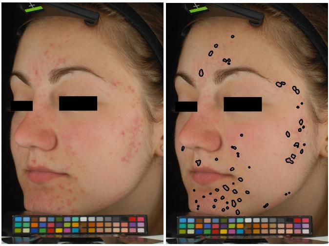
Acne vulgaris, a highly prevalent skin disease, has a significant life quality impact on patients. It is generally believed that this type of skin disorder results from proliferation of propionibacterium acnes in pilosebaceous units, which can lead to inflammatory lesions due to increase of oxyhemoglobin level. So far there is no golden standard for acne diagnosis in clinics. It entirely depends on dermatologists' experience for acne assessment. But significant variability among individual diagnosis may lead to less trustworthy results, and less reproducibility of human evaluation makes the comparison of acne follow-up difficult. This work, incorporating the knowledge of optical characteristics of human skin, identifies cutaneous chromophore distribution using bilateral decomposition. Then the inflammatory acne lesions are detected by a Markov random field (MRF) model associating the chromophore descriptors. Experimental results (see Figure 4 ) show that the proposed method is robust to large dynamic range intensity, and the derived automatic segmentation of inflammatory acne appears to be highly consistent to human visual assessment. This research work was started in 2013. This year, more tests have been conducted on polarized and non-polarized images.
|
Finer registration of facial wrinkles in time series images
Participants : Nazre Batool, Josiane Zerubia [contact] .
Dr. Batool was funded by the Inria-DPEI fellowship for the period Feb. 2014 – May 2015. Currently she is a postdoc researcher at CMIV, Linköping University [ www.liu.se/cmiv ], Linköping, Sweden.
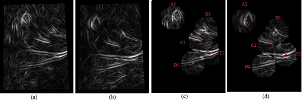
The goal of this work is to evaluate quantitatively the subtle variations in facial wrinkles for the same subject in response to treatment using image-based analysis. Any image analysis technique for the analysis of such subtle image variations would require high accuracy and precision for good performance. As in other imaging problems geared towards detection of temporal changes, accurate registration of key image features (wrinkles) is mandatory as a first step. We propose to compare image features in key wrinkle sites only while excluding the noise introduced by changes in surrounding skin texture. Therefore, previously we proposed a 2-step registration algorithm where the initial registration was based on the alignment of facial landmarks such as corners of eyes, nose, and mouth. Then a method based on Large Deformation Diffeomorphic Metric Mapping (LDDMM) was used to achieve finer local registration for wrinkles. However, the LDDMM algorithm had the shortcoming of the unavailability of time invariant finer facial landmarks and that the deformations were guided by image intensities which were varying among images as well due to subtle changes in skin texture. The deformation of skin due to underlying movement can be categorized loosely as locally rigid because the local skin texture remains constant but globally non-rigid because of the movement of skin areas due to slight expression and misalignment. Due to this dual nature of deformation, registration schemes such as thin plate spline or affine transformations are not applicable. Our improved approach is to guide the LDDMM registration on skin features with higher intensity gradients only (such as due to moles, wrinkles, rough surface) which have the higher probability of being constant and detected across temporal changes. First we detect key landmarks and landmark correspondences using the Gabor feature images where the phase correlation is used to find estimates of landmark correspondences. The phase correlation is based on the well-known Fourier shift property i.e. a shift in the spatial domain of two images results in a linear phase difference in the frequency domain of their respective Fourier Transforms. Figure 5 shows Gabor features of two images captured 4 weeks apart in (a) and (b). Figure 5 (c) shows key landmarks placed at high Gabor amplitude sites and (d) shows their corresponding landmarks detected using Fourier phase correlation.
|
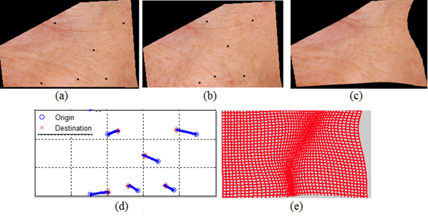
Then, as a next step, the detected key landmarks and their corresponding positions are used in the landmark based LDDMM algorithm to find locally non-rigid deformations between two images. Figure 6 shows an example where the corresponding landmarks are shown as black dots in (a) and (b). Fig. 5 (c) shows the image in (a) wrapped to (b) using LDDMM based on landmark correspondences. In (d) the drifts of landmarks are shown during the LDDMM algorithm and (e) shows the non-rigid deformation of underlying image grid. In the future, the proposed wrinkle registration algorithm will be used to compare wrinkle intensities in time series of images to quantify very minute changes in wrinkles in response to dermatological treatments.
|
Hyperspectral Image Processing for Detection and Grading of Skin Erythema
Participants : Ali Madooei, Josiane Zerubia [contact] .
Ali Madooei worked at Inria Sophia Antipolis on an internship funded by the Canadian Mitacs Globalink Research Award & Inria. He is currently in his last year of PhD at Simon Fraser [www.sfu.ca/ ] University, Canada. This work has been conducted in collaboration with Ramy M. Abdlaty, Lilian Doerwald-Munoz, Dr. Joseph Hayward and Prof. Qiyin Fang from Mc Master university [http://future.mcmaster.ca/ ]/Juravinsky cancer center [www.jcc.hhsc.ca/ ], Canada, and Prof. Joseph Hayward from Simon Fraser University, Canada.
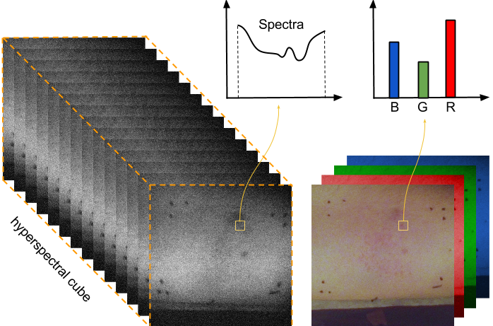
Acute skin erythema is a common side effect in patients undergoing radiotherapy treatment. It displays itself as an increase in skin redness and irritation. Erythema has been reported to correlate to individual patient response to radiation and therefore may be useful to guide and modify courses of treatment in a timely manner. Currently, upon visual examination, a qualitative score can be assigned to characterize the severity of erythema, which then may be used for assessing radiation response. Due to the subjective nature of this method, additional non-invasive techniques are needed for more accurate evaluation. Previous studies have mainly focused on tissue reflectance spectroscopy or imaging photography. The former retrieves spectral information from point measurements while the latter is obtained with conventional Red, Green, Blue(RGB) colour cameras. Photography has the advantage of offering spatial information but this comes at the cost of losing much of spectral information. We use hyperspectral imaging (HSI) which provides both spatial and spectral representation of the affected area. A hyperspectral camera effectively divides the spectrum into very many thin image slices (the actual number depending on the camera and application see Fig.7 ). This fine-grained slicing reveals spectral structure that may not be evident to the eye or to an RGB camera but can provide a rich set of information for image processing. As an emerging imaging modality for medical applications, the combination of HSI devices with adequate image processing techniques offers the perfect landscape for developing new methods for noninvasive disease monitoring and diagnosis.
The purpose of our study was to investigate the possibility of monitoring the degree of erythema using HSI data. To this aim, we proposed an image processing pipeline and conducted controlled experiments to demonstrate the efficacy of the proposed approach for (1) reproducing clinical assessments, and (2) outperforming RGB imaging data. We combined the problem of erythema detection and grading into a multi-class classification problem where each pixel is classified as one of the four erythema classes or a non-erythema class. We used a weighted LDA (linear discriminant analysis) classifier to deal with noisy labels. Moreover, we devised pre-processing steps to deal with noisy measurements. We evaluated the system against the clinical assessment of an experienced clinician. We also compared the performance to that of using digital photography (instead of HSI). The results from this preliminary study are encouraging and indicate that hyperspectral image data do contain relevant information, and indeed outperform imaging photography. In the future, we want to extend the technique to further detect other skin responses to radiation (such as dry/moist desquamation, skin necrosis, etc.) and also to experiment with real patients undergoing radiotherapy. Our ultimate objective is to build a system for monitoring radiation response in individuals using HSI technology and image processing.
SAR data classification using generalized Gamma mixture model
Participants : Vladimir Krylov, Josiane Zerubia [contact] .
Vladimir Krylov is a former AYIN post-doc, now post-doc at DITEN department, University of Genoa [www.diten.unige.it/ ], Italy. This work has been performed in collaboration with Prof. Heng-Chao Li, Prof. Ping-Zhi Fan (Southwest Jiaotong University, Chengdu [english.swjtu.edu.cn/ ], China) and Prof. William Emery (University of Colorado [www.colorado.edu/ ], Boulder, USA).
The accurate statistical modeling of synthetic aperture radar (SAR) images is a crucial problem in the context of effective SAR image processing, interpretation and application. In this work a semi-parametric approach is designed within the framework of finite mixture models based on the generalized Gamma distribution (GD) in view of its flexibility and compact analytical form. Specifically, we have developed a generalized Gamma mixture model (GMM) to implement an effective statistical analysis of high-resolution SAR images and proved the identifiability of such mixtures. A low-complexity unsupervised estimation method has been derived by combining the proposed histogram-based expectation-conditional maximization algorithm and the Figueiredo-Jain mixture estimation algorithm. This resulted in a numerical maximum likelihood (ML) estimator that can simultaneously determine the ML estimates of component parameters and the optimal number of mixture components. The state-of-the-art performance of the proposed method has been validated experimentally on a wide range of high-resolution SAR amplitude and intensity images.
|
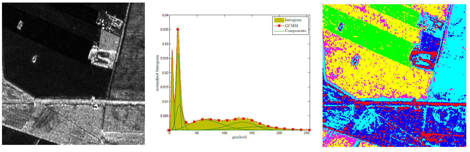
In Fig. 8 we demonstrate a typical result of the developed statistical modeling technique on a portion of a 2 meter resolution L-band image acquired by an airborne EMISAR system. The unsupervised GMM estimate contains five components and reports a very accurate result that outperforms the considered benchmark statistical modeling methods. In order to visualize the estimated five statistical components we also report a maximum likelihood classification map.
Multitemporal image change detection with a False Discovery Rate approach
Participants : Vladimir Krylov, Josiane Zerubia [contact] .
This work has been performed in collaboration with Prof. Sebastiano Serpico and Prof. Gabriele Moser, DITEN department, University of Genoa [www.diten.unige.it/ ], Italy.
Multitemporal change detection is one of the fundamental image processing problems and multiple detection, monitoring and tracking applications rely on its accurate and timely performance. In this work we address the problem of unsupervised change detection on two or more coregistered images of the same object or scene at several time instants. The designed method is appropriate for short image sequences with a relatively small amount of changes. Such analysis is instrumental in various applications where acquisitions are relatively sparse and report limited meaningful changes, in particular, in remote sensing and medical image processing. We develop a novel patch-based hypothesis testing approach which is based on a false discovery rate formulation for statistical significance testing. This alternative error metric allows to adjust the family-wise error rate by imposing control over the proportion of the false positives in the detection. The designed change detector allows the use of various statistical features. The appropriate choice of the latter enables the detector to address application-specific detection problems with a particular set of disturbance factors, like noise, illumination variation, etc. In particular, we demonstrate the use of two rank-based statistics for change detection on image pairs and one multisample statistic for the analysis of image sequences. The experiments with remotely sensed radar, dermatological, and still camera surveillance imagery demonstrate competitive performance and flexibility of the proposed method.
|
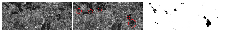
A typical result obtained with the proposed change detection technique is reported in Fig. 9 . The proposed approach gives a unified statistical thresholding procedure to perform change detection based on statistical features that have a known distribution under the no-change hypothesis. This approach is essentially non-parametric and is highly parallelizable.