|
|

|

|
| e-Pub |
Section: New Results
Epidermal cell layer thickness variability in Arabidopsis floral meristems
Participants : Gaël Michelin, Grégoire Malandain.
This work is made in collaboration with Yassin Refahi (Sainsbury Lab., University of Cambridge) and Jan Traas (ENS Lyon), within the Morphogenetics Inria Project Lab.
Flowers from the same species display a great robustness in their global shape and their developing stage can be theoretically identifiable to their size. The cells in epidermal (L1) and sub-epidermal (L2) layers of the floral meristem divide anticlinally, i.e. in a sideway fashion that ensures that L1 and L2 remain distinct. Thus a goodness-of-fit criterion on L1 and L2 layers is considered as an adequate registration quality measure in the inter-individual spatio-temporal registration framework developed in [8] .
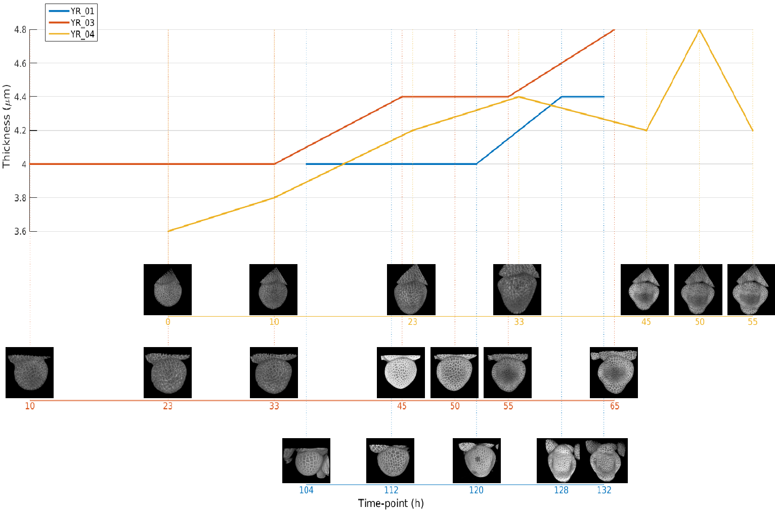
The aim of the present work is to investigate the variability of L1 layer thickness over development stages of an individual and between individuals. The study results may impact the way we process the inter-individual spatial registration. Therefore we measured the thickness distribution (histogram) of the L1 cells and we plotted the distribution of cells thickness (see figure 5 ) on images provided from three distinct floral meristems at acquisition time-points. Our results tend towards showing that L1 thickness increases over time non-uniformly, with a higher L1 thickness on sepals for advanced developing stages. We also observed an inter-individual thickness variability of about for developing floral meristems at close developing stages. Future investigations will consist in taking a larger set of data to assess our first observations, in providing a biological interpretation of these observations and in using this knowledge to propose a refined spatial registration method.