|
|

|

|
| e-Pub |
Section: New Results
Extraction and Analysis of the Vascular Network to Classify and Grade Kidney Tumors in Histological Imaging
Participants : Alexis Zubiolo, Eric Debreuve, Xavier Descombes.
This work is made in collaboration with Philippe Pognonec (Team TIRO, CEA/UNS), Damien Ambrosetti (Histopathology department, CHU Pasteur, Nice).
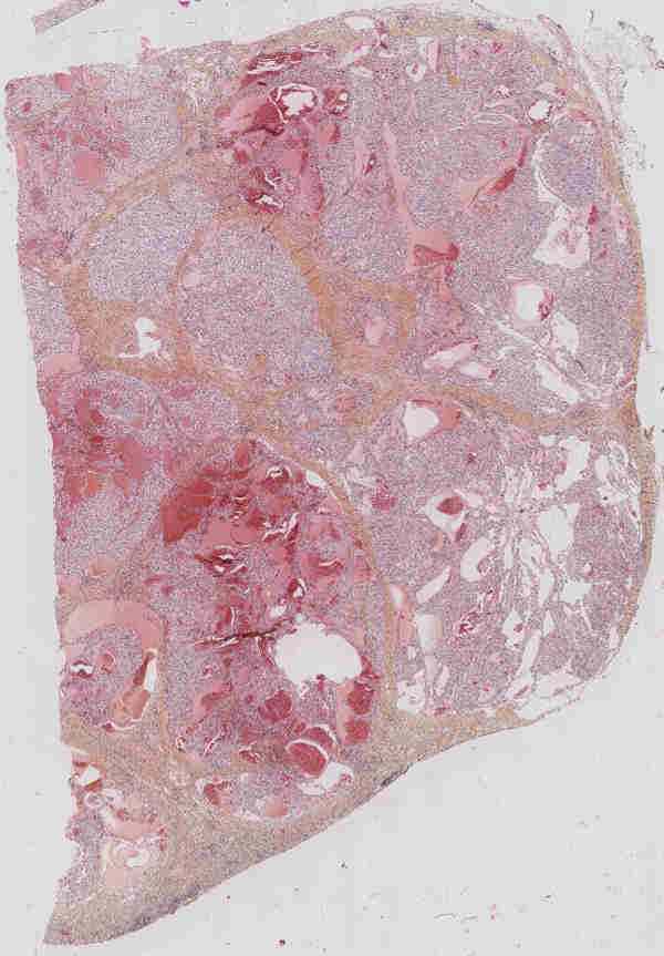
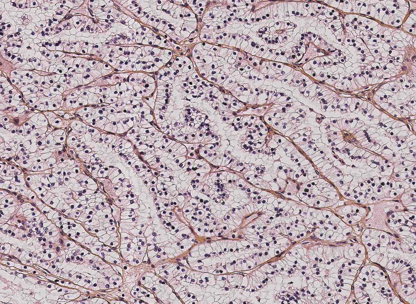
The renal carcinoma is the most frequent type of kidney cancer (between 90% and 95% of all cases). Twelve classes of carcinoma can be distinguished, among which the clear cell carcinoma (CCRCC) and the papillary carcinoma (PRCC) are the two most common (75% and 10% of the cases, respectively). After the carcinoma has been diagnosed, the tumor is ablated and prepared for histological examination (fixation, staining, slicing, observation with a microscope). Along with genetic tests and protein reactions, the histological study allows to classify and grade the tumor in order to make a prognosis and to take decisions for the subsequent patient treatment. Digital histology is a recent domain (routinely, histological slices are studied by MDs directly on the microscope). The pioneer works deal with the automatic analysis of cells. However, one crucial factor for carcinoma classification is the structure of the vascular network. Coarsely, CCRCC is characterized by a “fishnet” structure while the PRCC has a tree-like structure.
In this context, our goal was to extract the vascular network from a given histological slice, compute features of the underlying graph structure, and classify the tumor into CCRCC or PRCC based on these features. The histological images being huge (typically, 100k x 100k pixels), they must be split into tiles (with some overlap to ease the combination of results) and processed tile-wise. The first step is to combine the color channels so that the vessels are as highlighted as possible. Then, the vascular network is detected by a processing pipeline including tailored, Gabor-like filtering, thresholding, and extraction of the skeleton. Small gaps in the skeleton are filled and some pruning is performed. Finally, the skeleton is converted to a graph representation. Based on the medical interpretation procedure, we focused our analysis of the graph on the following elements: the number of terminal and junction nodes, and the terminal branches. We proposed to compute the ratio between the number of terminal nodes and the number of junctions (T/J ratio), and the average length of terminal branches. Both features seem to be adapted to classification, especially the T/J ratio which, on the fairly small database of cases we currently have, exhibits an average value 65% higher for PRCC.