|
|

|

|
| e-Pub |
Section: New Results
Hyperspectral Image Processing for Detection and Grading of Skin Erythema
Participant : Josiane Zerubia [contact] .
This work was carried out in collaboration with Ali Madooei (Simon Fraser University (https://www.sfu.ca/), Canada), Ramy Abdlaty (McMaster University (https://www.mcmaster.ca/), Canada), Lilian Doerwald-Munoz (Hamilton Health Sciences - General Hospital (http://www.hamiltonhealthsciences.ca/), Canada), Joseph Hayward (Hamilton Health Sciences - General Hospital, Canada), Mark Drew (Simon Fraser University, Canada) and Qiyin Fang (McMaster University, Canada).
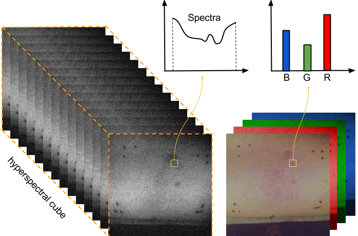
This study focused on detection and grading of skin erythema using hyperspectral image processing. The ultimate objective is to build a system for monitoring radiation response in individuals using hyperspectral imaging technology and image processing. The present project was to investigate the possibility of monitoring the degree of skin erythema. To this aim, we proposed an image processing pipeline and conducted controlled experiments to demonstrate the efficacy of the proposed approach for (1) reproducing clinical assessments, and (2) outperforming RGB imaging data (see Fig. 5). We combined the problem of erythema detection and grading into a multi-class classification problem where each pixel is classified as one of the four erythema classes or a non-erythema class. We used a weighted LDA (linear discriminant analysis) classifier to deal with noisy labels. Moreover, we devised pre-processing steps to deal with noisy measurements. We evaluate the system against the clinical assessment of an experienced clinician. We also compare the performance to that of using digital photography (instead of hyperspectral images) [9]. The results from this preliminary study are encouraging and indicate that hyperspectral image data contain relevant information, and indeed outperform imaging photography. In future, we want to extend the technique to further detect other skin responses to radiation (such as dry/moist desquamation, skin necrosis, etc.) and also to experiment with real patients undergoing radiotherapy.