|
|

|

|
| e-Pub |
Section: New Results
Modeling and identification of the sensory-motor system
Neuroplasticity and recovery in remote (sub)cortical structures following wide-awake surgery of infiltrative low-grade gliomas: investigation of fMRI and EEG signals by standard and nonlinear methods
Participants : Anthony Boyer, Jérémy Deverdun [CHU Montpellier] , Hugues Duffau [CHU Montpellier] , Emmanuelle Le Bars [CHU Montpellier] , Sofiane Ramdani [LIRMM] , David Guiraud, Nicolas Menjot de Champfleur [CHU Montpellier] , François Bonnetblanc.
Wide-awake surgery of brain tumour is used to optimize the resection of tumoral tissue. Postoperatively, patients show mild and temporary neurological deficits despite massive cerebral resections. Reasons for these impairments along with the compensation mechanisms operating within the cortex and subcortical structures are barely understood. The objective of this project is to reveal the remote effects of the tumour and its resection, to determine their nature measuring changes induced in functional Magnetic Resonance Imagery (fMRI) and electroencephalographic signals using standard and nonlinear methods.
In a first attempt to better understand the direct consequences of wide-awake surgery we focused on the thalamus insofar as, topologically, it is the largest input source and output target of the cortex. It plays a major role in corticosubcortical and corticocortical interactions and is expected to be heavily impacted by the tumour removal while being essential to the recovery process. Studying the thalamus, based on its very particular anatomical properties, could provide essential indications regarding the behaviour of cortical and subcortical centers.
We carried out Amplitude of Low-Frequency fluctuations and Regional Homogeneity analyses on resting state fMRI data before and after the tumour removal, including an original 24h postoperative acquisition. We intended to assess possible changes in spontaneous neuronal activity over time, characterizing different facets of slow-wave hemodynamic fluctuations. We particularly sought evidences of disrupted and atypical neuronal activity emerging within deafferented thalamic subterritories.
This work revealed significant alterations of neuronal activity within distinct thalamic territories, in accordance with its neuro-anatomo-functional organization. We showed a transient decrease of neuronal activity intensity and homogeneity within the ipsilesional thalamus directly related with the anatomical dee- and deafferentation induced by the neurosurgery and a concomitant increase of neuronal activity and temporal synchrony in homologous regions of the contralesional thalamus, leading to a significant interhemispheric imbalance during the immediate postoperative period. Evidences of diaschisis-like phenomenon primarily affecting higher order thalamic nuclei of the ipsilesional thalamus and the extensive involvement of the contralesional thalamus in the postoperative period promote the thesis of transient diaschisis-induced contralesional compensation for patients who underwent wide-awake surgery (Figure 12).
|

Understanding the effects of direct electrical stimulation of the brain during wide awake surgery
Participants : Marion Vincent, François Bonnetblanc, David Guiraud, Hugues Duffau, Mitsuhiro Hayashibe, Olivier Rossel.
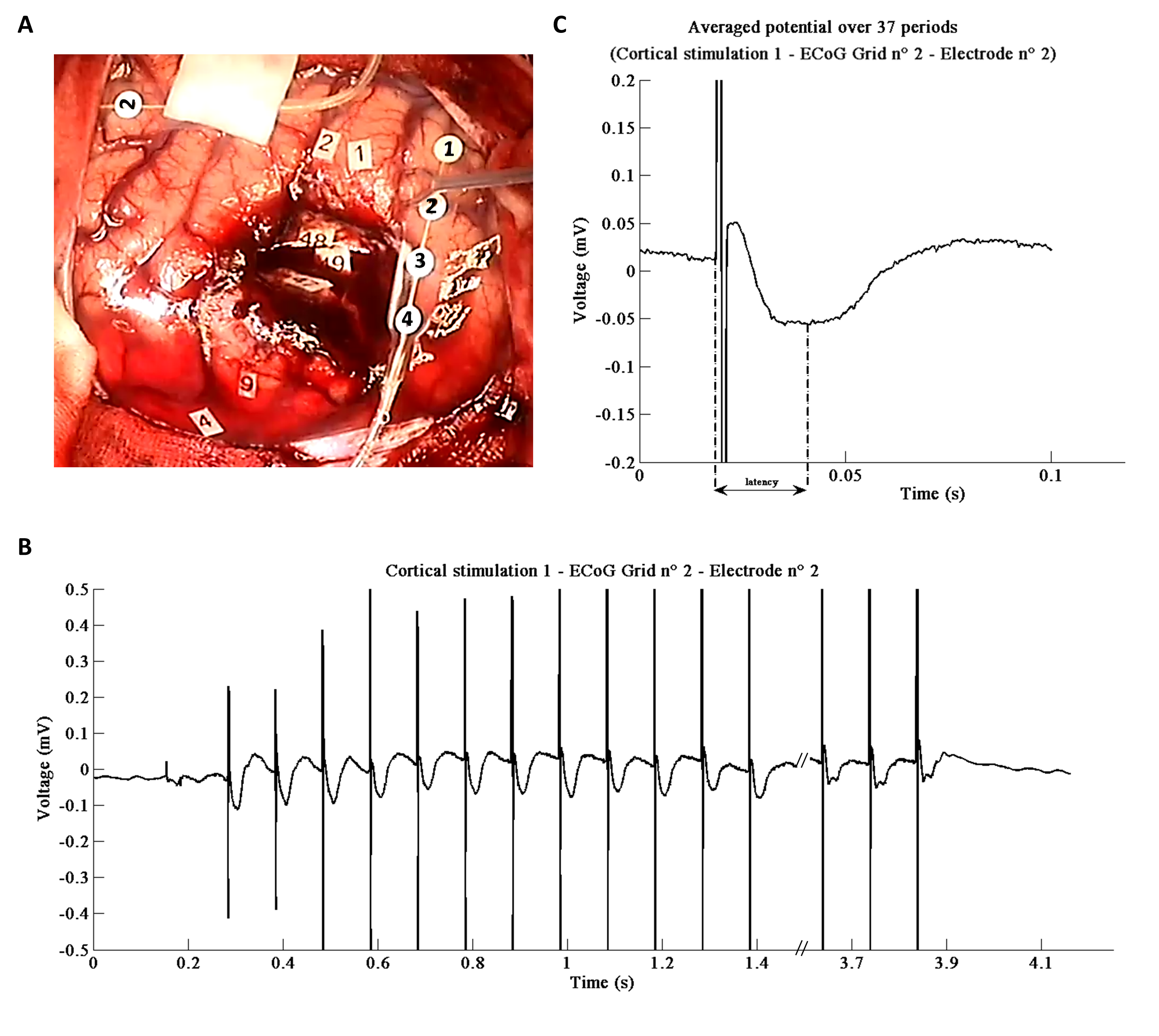
Real-time functional mapping of the brain combined with direct electrical stimulation (DES) has been widely recommended for the awake neurosurgery of slow-growing and infiltrative brain tumors, to guide the resection [53]. Intra-operative DES is generally applied at 60 Hz in Europe (50 Hz in some other countries) (biphasic stimuli, single pulse duration 1 ms, intensity from 2 to 6 mA under local anesthesia, and during 1 to 5 s). By generating transient perturbations, it allows the real-time identification of both cortical areas and sub-cortical white matter pathways that are essential for the function. Its use lowers the probability of resecting essential functional areas near or within the tumor. However, the electrophysiological effects of DES remain poorly understood, locally and at a more remote distance [36], [9].
The investigation of this topic requires the recording of evoked potential. DES can be used to probe the spatio-temporal connectivity and dynamics of short- or long-range networks in vivo and in real time when combined with electrophysiological recordings (e.g. electroencephalography (EEG) or electroencephalography (ECoG)). This approach has been used for pre-surgical planning of drug-resistant epileptic patients by using an ECoG grid implanted at the surface of the grey matter. Matsumoto et al. [55] sought to measure in vivo connectivity with DES (rather than studying its propagation) but observed that a low-frequency cortical application of DES (1 Hz, constant current, and alternating rectangular wave pulses of 0.3 ms, with an intensity around 10-12 mA) induces 'cortico-cortical' evoked potentials (CCEPs) around 10-50 ms after stimulation. These properties are incompatible with the detection of EPs during awake brain surgery, when DES is classically applied at 60 Hz due to stimulation artefacts. Conversely, 100 ms (i.e. a frequency of 10 Hz) seems to be a sufficient time-window that facilitates real-time averaging to detect these CCEPs for further on-line analysis of brain connectivity during the surgery.
In addition, in the studies mentioned above, ECoG signals were recorded in a classical common mode (CM) configuration, i.e. the signal was measured between each channel of interest and a reference electrode. Also, in all this literature, CCEPs were measured by averaging a large set of trials together. This off-line averaging actually prevents the use of ECoG recording to monitor the evoked potentials on-line. Recently, by lowering the DES frequency to 10 Hz and by using a differential recording mode (DM) for ECoG signals, in which the signal is measured between two adjacent electrodes, we were able to record for the first time on-line CCEPs easily with a standard current amplitude of stimulation (2 ms) and without averaging the data [41] (Figure 13.
Recording ECoG in a DM enabled increasing the focality and the signal to noise ratio of the raw data. Ongoing experiments on new patients corroborate the reproducibility of this protocol. This unusual way of recording ECoG could improve the spatial resolution of the recordings in the three dimensions (in surface and in depth). Moreover, this method was used under general anesthesia but could also be performed on-line during the awake surgery. It would enable the investigation of the connectivity and to probe directly rapid plastic changes of cortical excitability.
|
A study on the effect of electrical stimulation as a user stimuli for motor imagery classification in Brain-Machine Interface
Participants : Saugat Bhattacharyya, Maureen Clerc, Mitsuhiro Hayashibe.
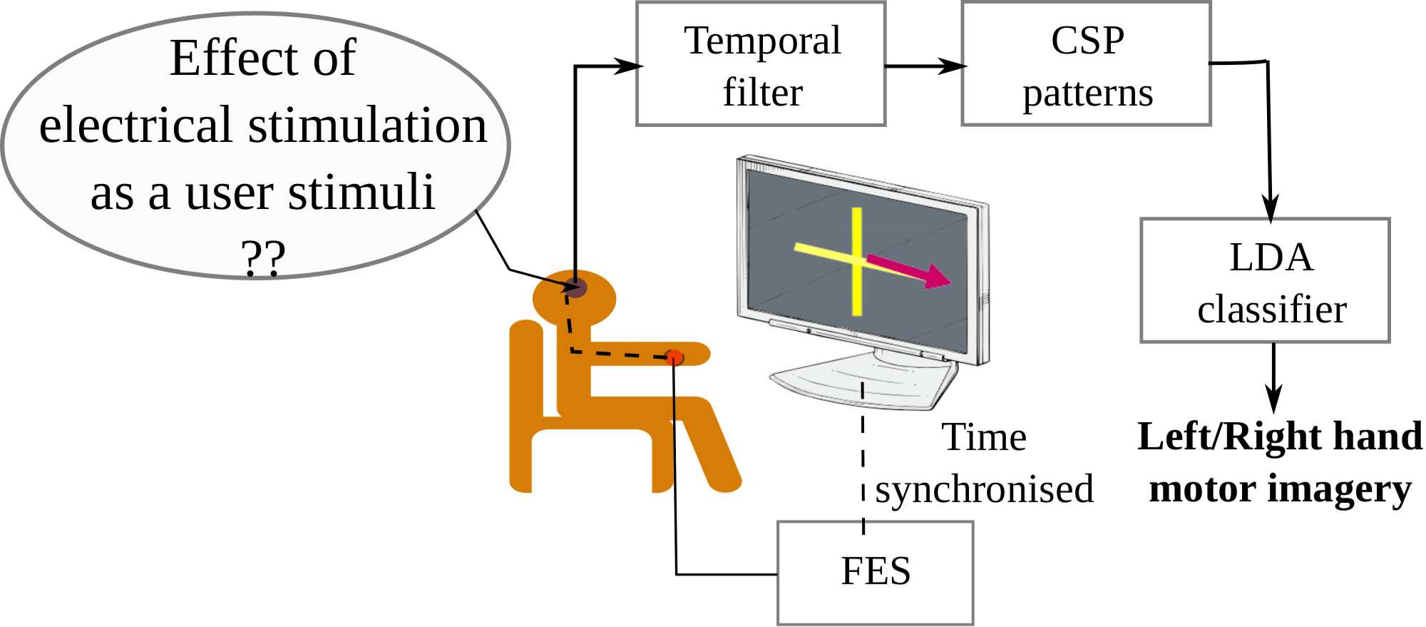
Functional Electrical Stimulation (FES) provides a neuroprosthetic interface to non-recovered muscle groups by stimulating the affected region of the human body. FES in combination with Brain-machine interfacing (BMI) has a wide scope in rehabilitation because this system directly links the cerebral motor intention of the users with its corresponding peripheral muscle activations. In this paper, we report the preliminary results of the effect of electrical stimulation during a motor imagery training task on healthy subjects and its comparison with visual stimuli.
The experiment designed for this work is divided into three sessions: only visual, only FES and both visual-FES stimuli. The sessions consist of instructing the subjects through a sequence of repetitive stimuli to execute the corresponding motor imagery task, which in our case, is left and right hand movement. The FES session is similar to the visual one except in place of the arrows, stimulation is directly induced in the fore-arm of the hand of interest, without providing any visual information. In the Visual-FES session, both the combined stimulations are time-synchronized to each other. After acquisition, the incoming raw EEG signal is band-pass filtered at 8-30 Hz. Then, common spatial filters (CSP) is applied to extract features relevant to left- and right-motor hand movement EEG signals. CSP is a spatial filter widely used in BMI because the spatial patterns contain highly discriminative features between two classes. In this study, we prepare the feature vectors using 6 spatial filters which is then transferred as inputs to a linear discriminant analysis (LDA) classifier. Finally, the classifier detects the corresponding motor intention of the subject, i.e., left and right motor movement. A block diagram of our experimental setup during Visual-FES session is illustrated in Fig. 14.
|
Classification results shows a significant rise in accuracy for 2 (of 3) subjects which suggest a positive influence of FES during motor imagery training of the subjects. It was noted that both the subjects had no previous experience on BMI, then they were not familiar with generating motor imagery with visual stimuli. Visual stimuli are the widely accepted form of motor training but the subject requires constant training to reach an optimal result. Based on the results of this study, we can infer that electrical stimulation can also be used for motor training and it can potentially provide better performance as it can make natural proprioceptive feedback related to motor performance than visual stimuli which requires user's recognition regarding the visual cue.
A Study on the Effect of Electrical Stimulation During Motor Imagery Learning in Brain-Computer Interfacing
Participants : Saugat Bhattacharyya, Maureen Clerc, Mitsuhiro Hayashibe.
Functional Electrical Stimulation (FES) stimulates the affected region of the human body thus providing a neuroprosthetic interface to non-recovered muscle groups. FES in combination with Brain-computer interfacing (BCI) has a wide scope in rehabilitation because this system can directly link the cerebral motor intention of the users with its corresponding peripheral mucle activations. Such a rehabilitative system would contribute to improve the cortical and peripheral learning and thus, improve the recovery time of the patients.
To date, in BCI experiments feedback is commonly provided to the subject by means of a visual medium. On observing the feedback, the subject would attempt to perform his task. It is an interesting notion if one includes electrical stimulation to help in augmenting the performance of the motor task at hand. Thus, in this paper, we report the preliminary results of the effect of electrical stimulation on the learning of the subject during a motor imagery training task on healthy subjects. Through this study, we aim at employing FES as a proprioceptive feedback to the subject to improve the learning of the subject both in terms of accuracy and time.
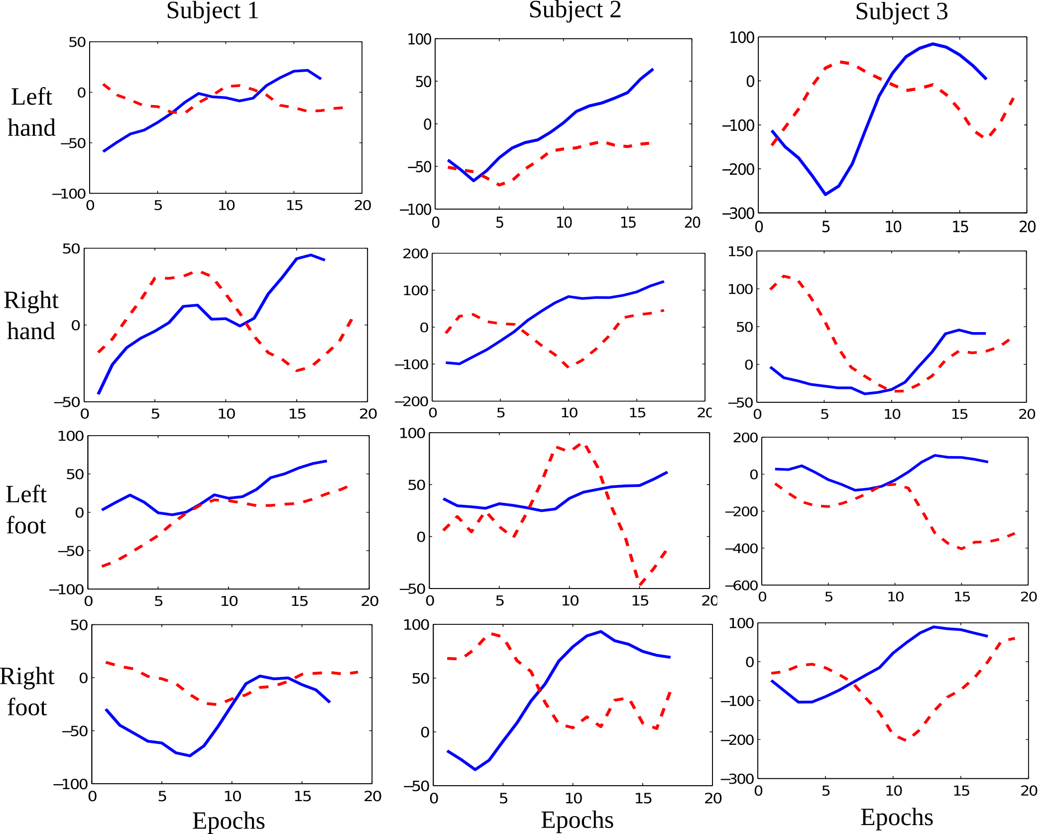
In this experiment the participants performed four motor tasks: left hand movement, right hand movement, left foot movement and right foot movement across 6 separate sessions. A session provides instructions to the participant through a sequence of visual cues to execute one of the four motor tasks and each visual cue is termed as `trial'. Further, for data analysis, each trial are separated into time windows, termed as epochs. Each session consists of a feedback session provided visually to the participant at each trial, quantified by the hyperplane distance of the decoder. Before the start of the experiment, the participants undergo a training session for decoder training and to acclimatize to the tasks. Common Spatial Patterns is employed as features which is given as inputs to the Linear Discriminant decoder. The decoder designed in this work is a 2-level hierarchy. The first level classifies between left and right motor imagery and the second level discriminates between hand and foot motor imagery. In 3 of the 6 sessions, surface electrical stimulation (ES) is transmitted to the subject during the feedback period to aid the participant in performing the task. Thus, in this paper, we named the ES induced sessions as FES sessions and the sessions with only visual feedback as VIS sessions.
We report the learning during FES and VIS session feedback for each trial. For this purpose, we measure the distance of the feature vector from the hyperlane for each epoch updated at every 0.125 seconds. We took this parameter to study the feedback effect because the larger the distance from the hyperplane, the higher is the confidence of the classifier to detect the right output. The average feedback curve for all the correctly classified trials of both FES (in blue) and VIS (in red) are shown in Fig. 15. From the curves we assume that greater the slope of the curve, faster is the learning demonstrated by the subject. Subject 1 demonstrates an increasing learning effect (greater slope) for FES feedback for all the limbs, except Right foot as compared to the VIS feedback. The figures for Subject 2 illustrates a more prominent learning effect during FES feedback and it is clearly differentiable for VIS feedback even though Subject 1 showcased a higher increase in accuracy across trials than Subject 2. It is also noted from the figures of both the subjects that VIS feedback has a frequent increasing and decreasing trend of the curve. Subject 3 had a decrease in accuracy during FES feedback as compared to the VIS feedback which can be validated from the figure that the discriminability between the FES and VIS feedback are not as prominent in comparison to the other subjects. We can infer from these results that the electrical stimulation had a positive influence during motor task learning and with an increase in sessions one can assume ES to provide a faster learning. The steady increase of learning during FES sessions can be attributed to the fact that the subjects reported to be more motivated to perform the tasks when an ES was provided and they felt the inclusion of ES helped in their imagination. On the other hand, during VIS sessions the subjects reported to lose motivation in-between the tasks.
|
NIRS-EEG joint imaging during transcranial direct current stimulation
Participants : Mehak Sood [IIT Hyderabad, India] , Pierre Besson [Euromov, UM] , Makii Muthalib [Euromov, UM] , Utkarsh Jindal [IIT Hyderabad, India] , Stéphane Perrey [Euromov, UM] , Anirban Dutta, Mitsuhiro Hayashibe.
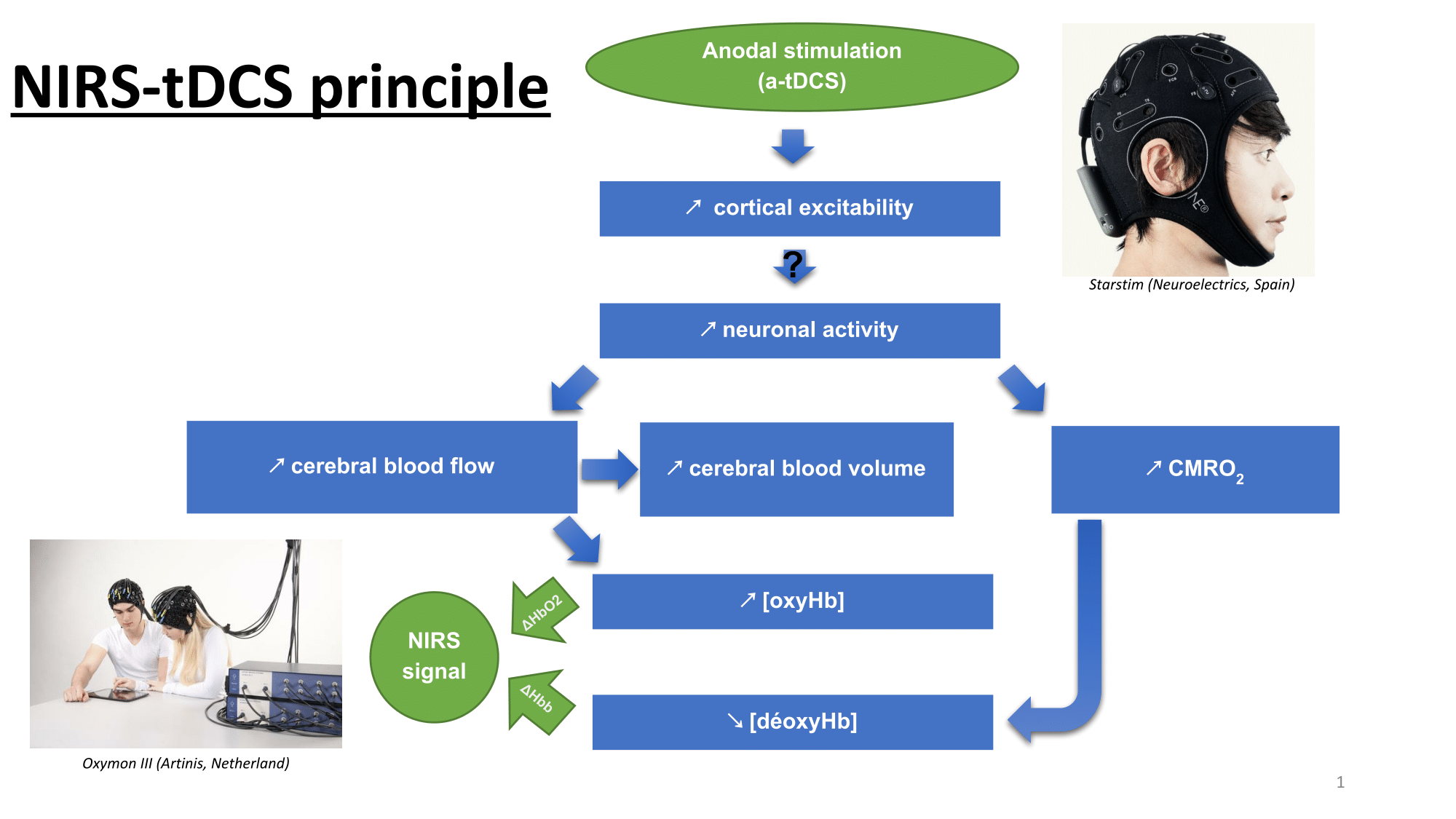
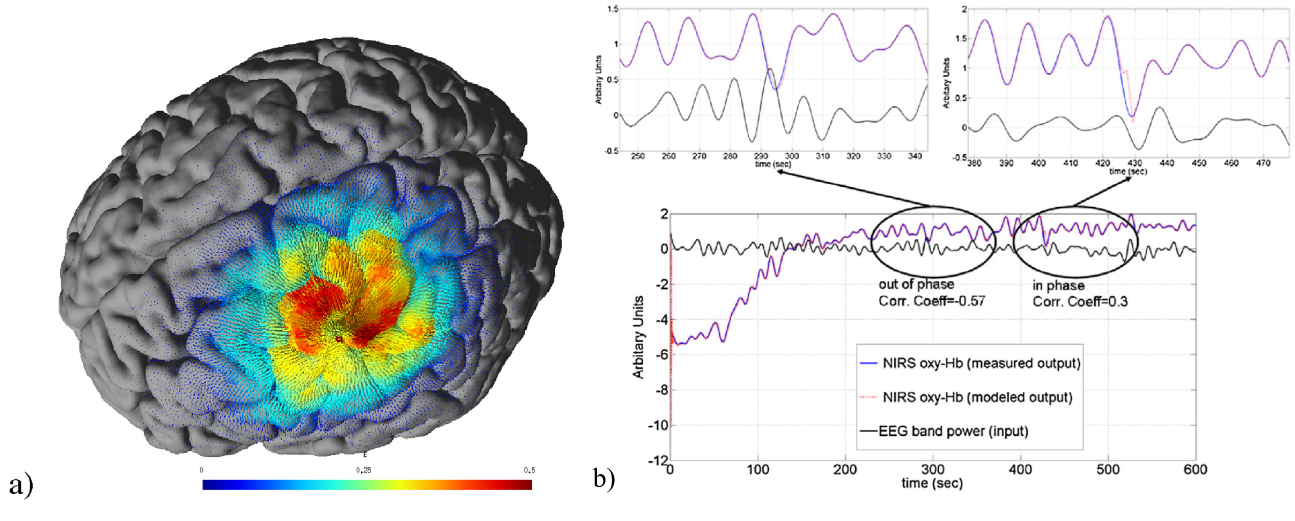
Transcranial direct current stimulation (tDCS) has been shown to perturb both cortical neural activity and hemodynamics during (online) and after the stimulation, however mechanisms of these tDCS-induced online and after-effects are not known. Here, online resting-state spontaneous brain activation may be relevant to monitor tDCS neuromodulatory effects that can be measured using electroencephalography (EEG) in conjunction with near-infrared spectroscopy (NIRS). We present a Kalman Filter based online parameter estimation of an autoregressive (ARX) model to track the transient coupling relation between the changes in EEG power spectrum and NIRS signals during anodal tDCS (2 mA, 10 min) using a 4x1 ring high-definition montage. Our online ARX parameter estimation technique using the cross-correlation between EEG band-power (0.5-11.25 Hz) and NIRS oxy-hemoglobin signal in the low frequency range was shown in 5 healthy subjects to be sensitive to detect transient EEG-NIRS coupling changes in resting-state spontaneous brain activation during anodal tDCS. Conventional sliding window cross-correlation calculations suffer a fundamental problem in computing the phase relationship as the signal in the window is considered time-invariant and the choice of the window length and step size are subjective. Here, Kalman Filter based method allowed online ARX parameter estimation using time-varying signals that could capture transients in the coupling relationship between EEG and NIRS signals. Our new online ARX model based tracking method allows continuous assessment of the transient coupling between the electrophysiological (EEG) and the hemodynamic (NIRS) signals representing resting-state spontaneous brain activation during anodal tDCS. It is supported by Franco-Indian Inria-DST project funding and by the LabEx NUMEV (ANR-10-LABX-20).
|
Is EMG a good signal to assess fatigure under FES in different stimulation modes?.
Participants : Willy Fagart, Robin Candau [EUROMOV] , Anthony Gelys [Propara Center] , Mitsuhiro Hayashibe, David Guiraud.
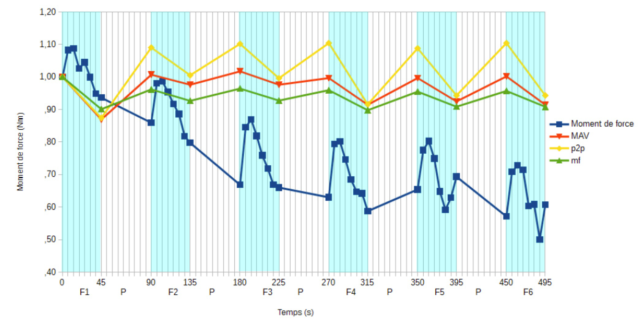
The study that we have undertaken aims to analyse the neuromuscular fatigue in 3 paralysed subjects with spinal cord injury and to find if there is a link between the torque and the EMG signal. 6 series of 8 trains of stimulation (30 Hz, 400s, 3 on / 2 off, in maximal intensity) were used to lead a muscular fatigue on the soleus muscle. At the beginning and the end of every series of stimulation, we measured the intermediate state of fatigue by evoking muscular twitch (1Hz, 400 s and of maximal intensity) on the 2 legs. The temporal component and frequency of electromyographic activity were analyzed. These values were correlated with the torque. At the end of the protocol of stimulation, the torque decreased on 5 legs on 6 (ranging from -39 % to -20 %, p<0,05). A polynomial of degres 2 relation was found between the torque and the peak to peak value of the EMG signal. Nevertheless this relation does not remain reliable in a clinical context with regard to the variability of the data. This variability represents the processes of potentiation of the electric and mechanical answer as well as the expression of the mechanisms which contribute to the muscular fatigue.
|
EleVANT project: a diagnostic evaluation of acute stroke by near infrared spectroscopy and transcranial direct current stimulation coupling.
Participants : Victor Vagné, Vincent Costalat, David Guiraud, Emmanuelle Le Bars, Stéphane Perrey [EUROMOV] , Olivier Rossel, Mitsuhiro Hayashibe.
Cerebral infarctions can now be treated with new techniques using intravenous thrombolysis and mechanical clearance. Their proven efficacy is directly correlated to the time lapse between the start of symptoms and initiation of treatment. Currently, a definitive diagnosis can only be made once the patient has realized a radiological imaging (CT scan or MRI) on a medical centre equipped with these expensive devices, thus enabling the medical team to initiate the appropriate treatment. Transit times during the pre-hospitalisation phase before diagnosis are therefore often longer and have the greatest negative impact on the patient's prognosis. The association of NIRS and tDCS enables recording modifications in cortical tissue oxygenation induced by electrostimulation. A case-control study demonstrated the capacity of near infra-red spectroscopy (NIRS), combined with transcranial direct current stimulation (tDCS) to diagnose established cerebral ischaemia. According to this study, the affected hemisphere with impaired circulation showed significantly less change in cerebral hemoglobin oxygenation than the healthy side in response to anodal tDCS. This preliminary study showed the feasibility of identifying the lesioned hemisphere in subacute stroke patient. In collaboration with Gui de Chauliac Hospital, I2FH and Euromov, the EleVANT project is aiming to prospectively evaluate the NIRS-tDCS technique in the diagnosis of acute cerebral ischaemia. This low cost technology could be used in a mobile way for the very early diagnosis of cerebral infarction and thus reduce treatment delays, opening the way to a new generation of diagnostic tools. A first NIRS-tDCS helmet prototype was developed to gather our Oxymon NIRS and Starstim tDCS devices, allowing good optodes-scalp and electrodes-scalp contact, while reducing both movement artifact and set-up time. This helmet was improved steps by steps as tests were done to attempt several parameters (among others electrodes location, amplitude and time of stimulation). A 4 NIRS optodes and 2 electrodes montage was retained to test and validate the proof of principle. Preliminary results are encouraging and need further investigation to be strongly validated.
Otherwise, as effects of anodal tDCS on hemodynamic response remain discussed, we'll proceed in parallel with the establishment of MRI protocols to attempt a validation of these effects.