|
|

|

|
| e-Pub |
Section: New Results
Multi-Angle TIRF reconstruction for studying the cell adhesion phenomenon
Participants : Emmanuel Soubies, Laure Blanc-Féraud, Sébastien Schaub.
This work is made in collaboration with Agata Radwanska and Ellen Van Obberghen-Schilling from Institut de Biologie Valrose (iBV) at Nice.
Understanding cell adhesion mechanism is of a major importance in biology for example in the context of tumoral angiogenesis (Process of blood vessels creation from existing ones.). However, this process occurs at the vicinity of the cell membrane within a layer of a few hundred nanometer making classical microscopy devices unable to image such biological structures due to their lack of resolution in the axial direction. An interesting alternative would be to use a multi-angle total internal reflection illumination together with numerical reconstruction algorithms in order to reach a nanoscale precision in the axial direction.
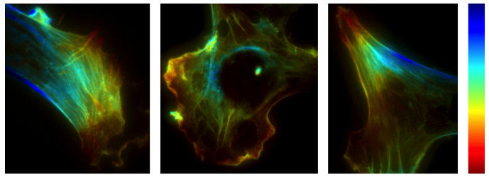
Following this idea, we made use of our previous work on MA-TIRF reconstruction to produce color-coded maps (see the example on Figure 1), with an axial resolution of 20 nm, of biological samples provided by Agata Radwanska and Ellen Van Obberghen-Schilling from the Institut de Biologie Valrose. The information obtained from the study of the reconstructed images have confirmed known behaviors of some proteins involved in the cell adhesion process allowing us, by this way, to complete the validation of our reconstruction method. Moreover, the 3D reconstructions have provided new information concerning the axial position of the observed biological proteins, information which was unavailable for previous studies conducted with other microscopy systems.