|
|

|

|
| e-Pub |
Section: New Results
White Blood Cells Segmentation and Classification in Bone Marrow Images
Participants : Mohammed Lamine Benomar, Xavier Descombes.
This work is made in collaboration with Chikh Amine and Mourtada Benazzouz from GBM Lab. (Tlemcen University). Our experiments were performed on an image database acquired in the Hemobiology service of the Tlemcen Hospital (Algeria).
The differential count of white blood cells (WBC) for medical diagnosis requires a careful observation in peripheral blood and bone marrow microscopic images in order to detect abnormal or suspicious cells. However, this process (screening) is time consuming, requiring concentration, experience and competence of the expert. The diagnosis depends on the correct recognition of cells. For that, computer analysis image system is required to automate the process in order to help experts, reduce the time and increase the accuracy. The main important steps in such systems are segmentation and classification of white blood cells.
The proposed approach to locate WBC in bone marrow microscopic smear could be divided into three main steps: pre-processing, segmentation and classification. The main concept of the segmentation and classification algorithm employed uses WBCs color, texture and morphological properties.
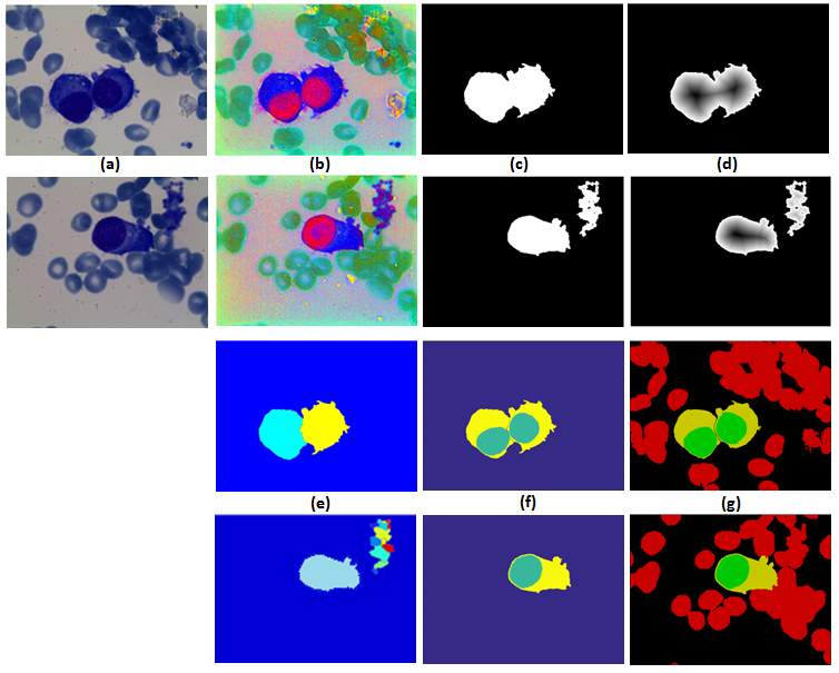
The first step is to reveal chromatic characteristics of the WBC by applying decorrelation stretch to multichannel RGB image, simple color transformation and Otsu thresholding to suppress background and most of the red blood cells. In the segmentation step, two techniques have been used which are Marker Controlled Watershed followed by MLE (Maximum Likelihood Estimator) to differentiate between WBC, the grouped red blood cells and artifacts using shape, color and texture features. Then Otsu thresholding based on HSL color space to separate WBC nucleus and cytoplasm (see Figure 5). Finally, white blood cells were classified into two categories related to the type of Myeloma, this step is based on features extraction and then applying a classifier.