|
|

|

|
| e-Pub |
Section: New Results
3D Coronary vessel tracking in x-ray projections
Participants : Emmanuelle Poulain, Grégoire Malandain.
This work is made in collaboration with Régis Vaillant (GE-Healthcare, Buc, France) and Nicholas Ayache (Inria Asclepios team).
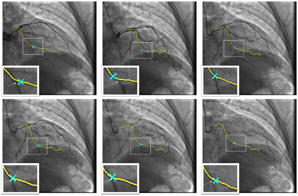
Percutaneous Coronary Intervention (PCI) is a minimally procedure which is used to treat coronary artery narrowing. During the guidewire navigation, the lesion is crossed and in some cases, the physician could benefit from a visual assessment of the coronary wall. The x-ray imaging interventional system used for per-operative guidance is not able to display this information mostly by lack of density resolution. On the contrary, Computed Tomography Angiography (CTA) is a modality which has the capability of capturing the characteristics of the vessel wall.
Fusing pre-operative CT angiography with per-operative angiographic and fluoroscopic images is thus considered by physicians as a potentially useful tool for improved guidance. To be adopted, this tool has required the development of tracking methods adapted to the deformations of the arteries caused by the cardiac motion. We have proposed a 3D/2D temporal tracking of one coronary vessel, based on a spline deformation, using pairings with a controlled 2D stretching or contraction along the paired curves and a preservation of the length of the 3D curve which corresponds to the anatomic propriety [8], [9]. Experiments were conducted on a database of 10 vessels from 5 distinct patients, with dedicated metrics assessing both the global registration and the local coherency of the position along the vessel. The proposed results demonstrate the efficiency of the proposed method, with an average standard deviation of 2 mm for the localization of landmarks (see Fig. 14).
|