|
|

|

|
| e-Pub |
Section: New Results
A Monte-Carlo approach for missing wedge restoration in cryo-electron tomography
Participants : Emmanuel Moebel, Charles Kervrann.
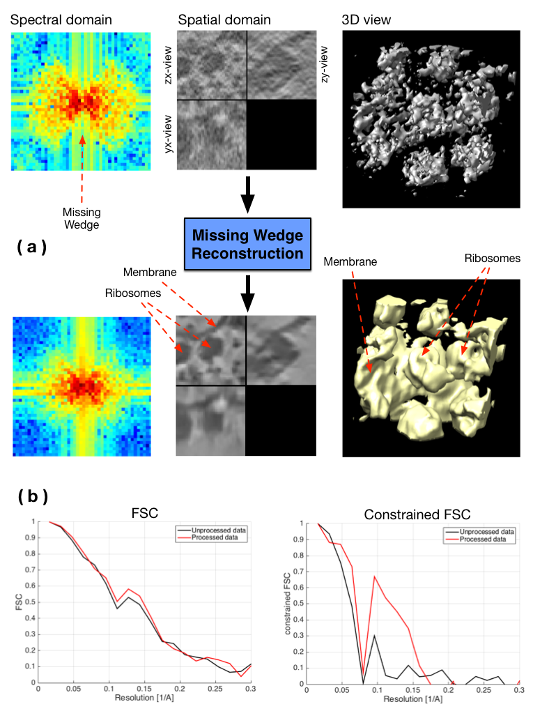
We investigated a Monte-Carlo approach to restore spectral information in the missing wedge (MW) in cryo-electron tomography (CET). The MW is known to be responsible for several types of imaging artifacts, and arises because of limited angle tomography: it is observable in the Fourier domain and is depicted by a region where Fourier coefficient values are unknown (see Fig. 3). The proposed computational method tackles the restoration problem by filling up the MW by iterating the two following steps: adding noise into the MW (step 1) and applying a denoising algorithm (step 2). The role of the first step is to propose candidates for the missing Fourier coefficients and the second step acts as a regularizer. Also, specific constraint is added in the spectral domain by imposing the known Fourier coefficients to be unchanged through iterations. We justified this approach in the Monte-Carlo simulation and Bayesian framework. In practice, different denoising algorithms (BM3D, NL-Bayes, NL-means...) can be applied. In our experiments, several transforms have been tested in order to apply the constraint (Fourier transform, Cosine transform, pseudo-polar Fourier transform). Convincing results have been achieved (see Fig. 3) using the Fourier Shell Correlation (FSC) as an evaluation metric.
Collaborators: Damien Larivière (Fondation Fourmentin-Guilbert),
Julio Ortiz (Max-Planck Institute, Martinsried, Germany).
|