|
|

|

|
| e-Pub |
Section: New Results
Brain-Computer Interfaces
Defining Brain-Computer Interfaces: A Human-Computer Interaction Perspective
Participants : Hakim Si Mohammed, Ferran Argelaguet, Anatole Lécuyer [contact] .
Regardless of the term used to designate them, Brain-Computer Interfaces (BCIs) are “Interfaces” between a user and a computer in the broad sense of the term. This paper aims to discuss how BCIs have been defined in the literature from the day the term was introduced by Jacques Vidal. In [32], from a Human-Computer Interaction perspective, we propose a new definition of Brain-Computer Interfaces as : "any artificial systems that directly converts brain activity into input of a computer process". As they are interfaces, such definition should not include the finality and objective of the system they are used to interact with. To illustrate this, we compared BCIs with other widely used Human-Computer Interfaces, and drew analogies in their conception and purpose.
This work was done in collaboration with the Inria LOKI team.
A conceptual space for EEG-based brain-computer interfaces
Participant : Anatole Lécuyer [contact] .
Brain-Computer Interfaces have become more and more popular these last years. Researchers use this technology for several types of applications, including attention and workload measures but also for the direct control of objects by the means of BCIs. In [7] we present a first, multidimensional feature space for EEG-based BCI applications to help practitioners to characterize, compare and design systems, which use EEG-based BCIs. Our feature space contains 4 axes and 9 sub-axes and consists of 41 options in total as well as their different combinations. In addition we present the axes of our feature space and we position our feature space regarding the existing BCI and HCI taxonomies. We also showed how our work integrates the past works, and/or complements them.
The use of haptic feedback in Brain-Computer Interfaces and Neurofeedback
Participants : Mathis Fleury, Anatole Lécuyer [contact] .
|
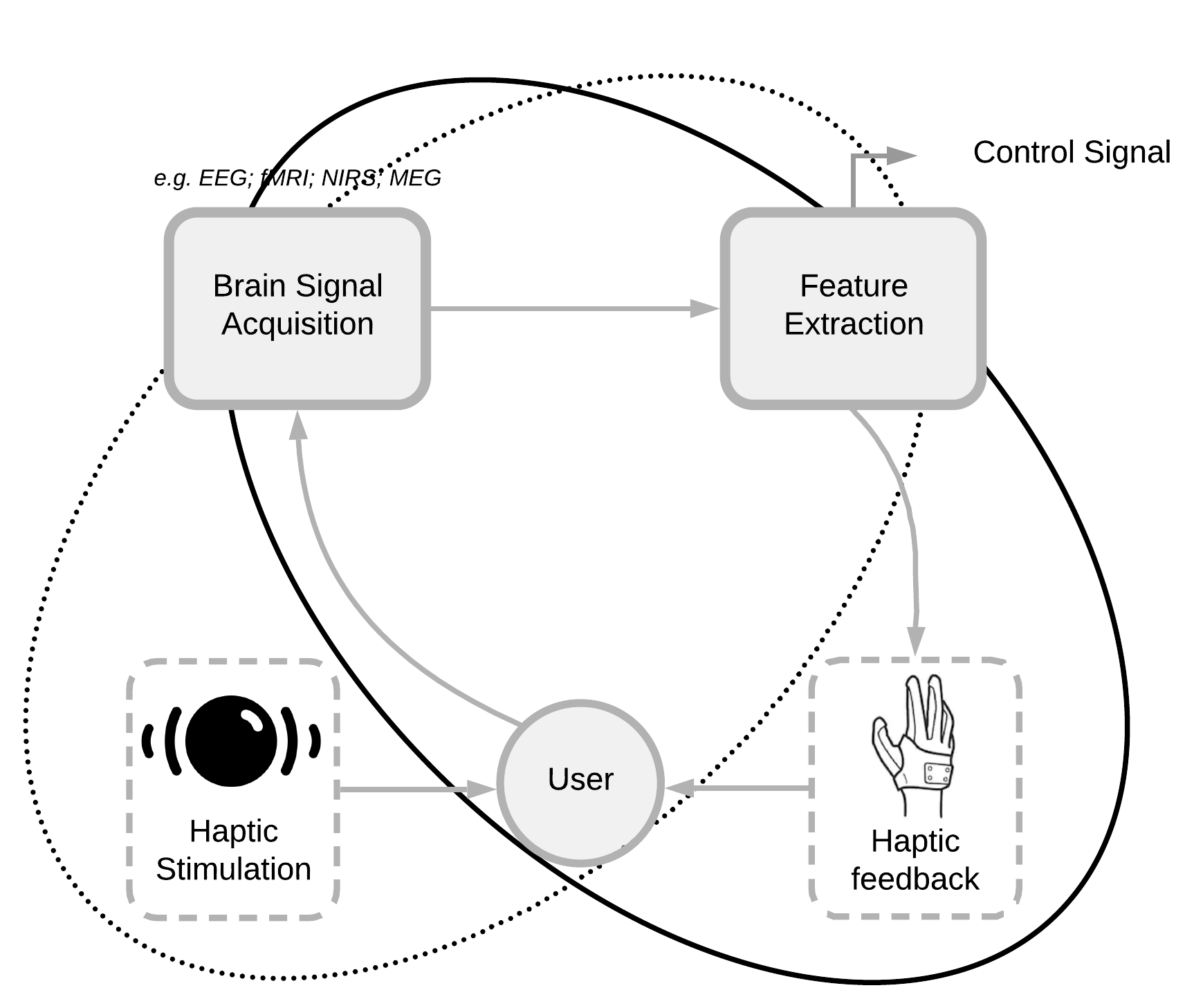
Neurofeedback (NF) and brain-computer interfaces are based on the recording of the cerebral activity associated with the requested task and the presentation of a feedback. The subject relies on the given feedback (visual, auditory or haptic) to learn and improve his mental strategy. It is therefore of crucial importance that it must be transmitted optimally. Historically, vision is the most used sensory modality in BCI/NF applications, but its use is raising potential issues. The more and more frequent use of haptic as a feedback modality reveals the limits of visual feedback; indeed, a visual feedback is not suitable in some cases, for individuals with an impaired visual system or during a mental motor imagery task (e.g. requiring a great abstraction). In such case, a haptic feedback would seem more appropriate. Haptic feedback has also been reported to be more engaging than visual feedback. This feedback could also contribute to close the sensory-motor loop. Haptic-based BCI/NF is a promising alternative for the design of the feedback and potentially improve the clinical efficacy of NF. In [38], [39] we have therefore surveyed the recent studies exploiting haptic feedback in BCI and NF.
This work was achieved in collaboration with the Inria EMPENN team.
Efficacy of EEG-fMRI Neurofeedback for stroke rehabilitation: a pilot study
Participants : Giulia Lioi, Mathis Fleury, Anatole Lécuyer [contact] .
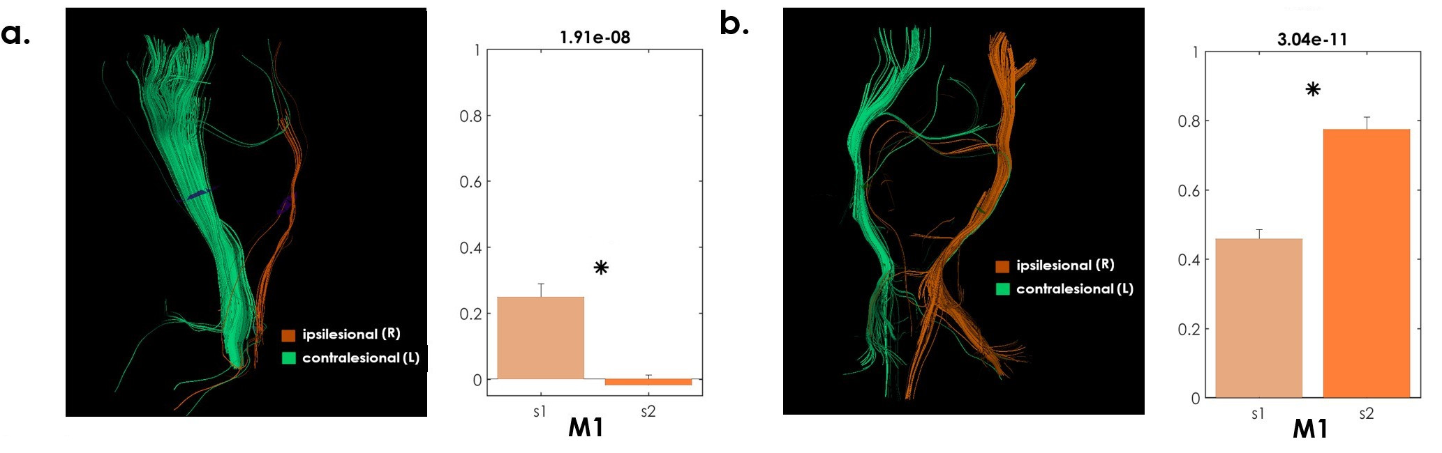
Recent studies have shown the potential of neurofeedback for motor rehabilitation after stroke. The majority of these NF approaches have relied solely on one imaging technique: mostly on EEG recordings. Recent study have gone further, revealing the potential of integrating complementary techniques such as EEG and fMRI to achieve a more specific regulation. In this exploratory work, multi-session bimodal EEG-fMRI NF for upper limb motor recovery was tested in four stroke patients.The feasibility of the NF training was investigated [41] with respect to the integrity of the cortico-spinal tract (CST), a well-established predictor of the potential for clinical improvement. Results indicated that patients exhibiting a high degree of integrity of the ipsilesional CST showed significant increased activation of the ipsilesional M1 at the end of the training. These preliminary findings confirm the critical role of the CST integrity for stroke motor recovery and indicate that this is importantly related also to functional brain regulation of the ipsilesional motor cortex.
This work was achieved in collaboration with Inria EMPENN team.
|
A multi-target motor imagery training using EEG-fMRI Neurofeedback
Participants : Giulia Lioi, Mathis Fleury, Anatole Lécuyer [contact] .
Upper limb recovery after stroke is a complex process. Recent studies have revealed the potential of neurofeedback training as an alternative or an aid to traditional therapies. Studies on cerebral plasticity and recovery after stroke indicate that premotor areas should be a preferred target for NF in the most severe patients while M1 stimulation may be more ef for patients with better recovery potential. Moreover, fMRI-NF studies (also on stroke patients) have shown that SMA is a robust correlate of motor imagery, while the activation of M1 is more dif to achieve, especially for short training sessions. Based on these results, in an exploratory work [13], we tested a dynamic NF training more strongly rewarding SMA activation in the NF training session and then increasing the M1 activation contribution in the NF session. We tested this novel approach on four stroke patients in a multisession bimodal EEG-fMRI NF training. To this end, we used an adaptive cortical region of interest (ROI) equal to a weighted combination of ipsilesional SMA and M1 activities and then varied the weights in order guide the patient training towards an improved activation of M1. Four chronic stroke patients with left hemiparesis participated to the study. The experimental protocol included an alternation of bimodal EEG-fMRI NF and unimodal EEG-only NF sessions. Preliminary results, on a short training duration, reveal the potential of a dynamic, multi-target/multimodal NF training approach.
This work was achieved in collaboration with Inria EMPENN team.
Bimodal EEG-fMRI Neurofeedback for upper motor limb rehabilitation
Participants : Giulia Lioi, Mathis Fleury, Anatole Lécuyer [contact] .
There is a growing interest in Neurofeedback or Brain computer interfaces for stroke rehabilitation. Integrating EEG and fMRI, two highly complementary imaging modalities, has potential to provide a more specific and efficient stimulation of motor areas. In this exploratory work [25], we tested the feasibility of a multi-session EEG-fMRI NF protocol on four chronic stroke patients, and its potential for upper-limb recovery. All the patients were able to upregulate their activity during NF training with respect to rest in the ipsilesional SMA and M1. Three over four patients showed a significant increase in ipsilesional M1 activation at the end of the protocol. Of these three individuals, two exhibited an increase in FMA-UE score. Preliminary results from this pilot study showed feasibility of bimodal EEG-fMRI in chronic stroke patients and indicated the potential of this training protocol for upper-limb recovery.
This work was achieved in collaboration with Inria EMPENN team.