|
|

|

|
| e-Pub |
Section: New Results
Morphological Analysis and Feature Extraction of Neurons from Mouse Cortices Multiscale 3D Microscopic Images
Participants : Alexis Zubiolo, Xavier Descombes, Eric Debreuve.
This work is jointly conducted with Kawssar Harb and Michèle Studer (iBV).
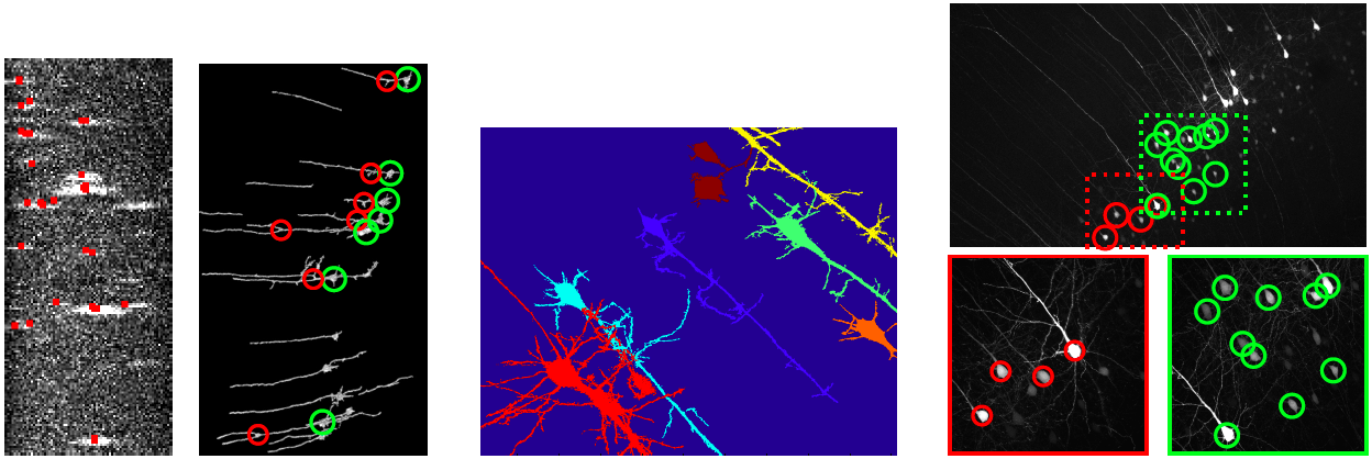
We propose a framework to analyze the morphology of mouse neurons in the layer V of the cortex from 3D microscopic images. We are given 8 sets of images, each of which is composed of a 10x image showing the whole neurons, and a few (2 to 5) 40x images focusing on the somas. The framework consists in segmenting the neurons on both types of images to compute a set of specific morphological features, and in establishing the correspondence between the neurons to combine the features we obtained, in a fully automatic fashion. On the 10x images, we use a multiple birth and cut algorithm to segment the sections of the apical dendrites. Merging these intersections provides the localization of the first branching of the apical dendrite (see Fig. 11 (left)). On the 40x images, we compute an hysteresis threshold to obtain a first segmentation (somas and dendrites starts) and apply iterative morphological operators to reconstruct the full dendrites (see Fig. 11 (middle)). The correspondence map between the two types of images is done using a bipartite graph matching model that associates each neuron configuration of a 40x image – a constellation – to a subset of neurons in the 10 image – the galaxy – (see Fig. 11 (right)).