|
|

|

|
| e-Pub |
Section: New Results
Whole-Slide Image Analysis of Renal Cell Carcinoma
Participants : Ana Rita Lopes Simoes, Eric Debreuve, Alexis Zubiolo, Xavier Descombes.
This work is jointly conducted with Thierry Pourcher, and Philippe Pognonec (TIRO, CEA-CAL-UNSA), and Damien Ambrosetti (CHU, Nice).
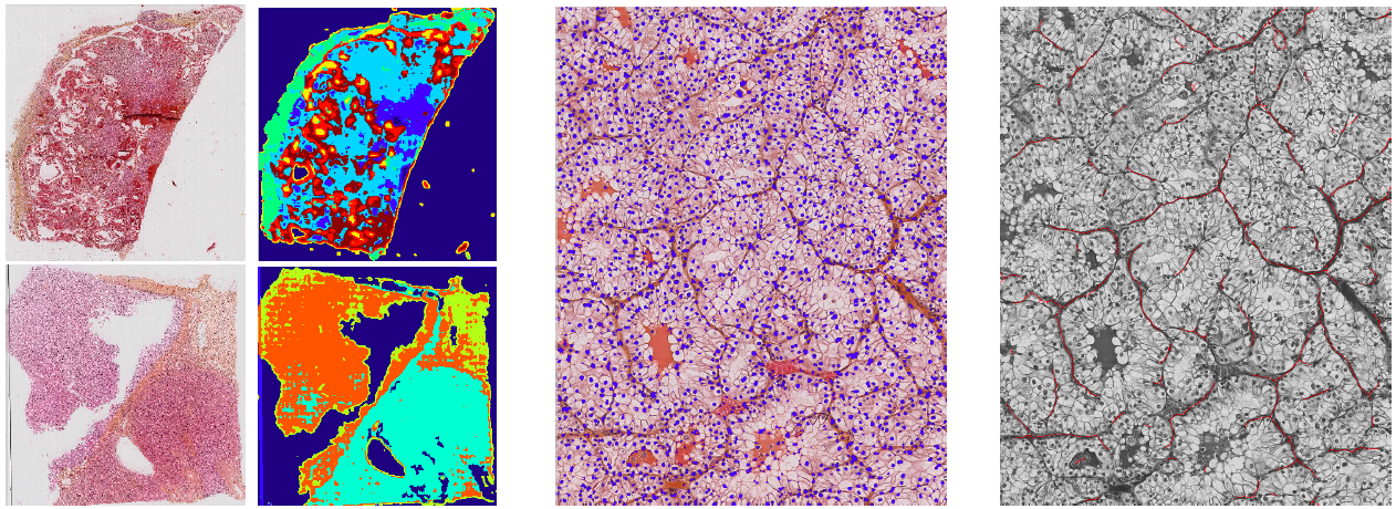
We study histology images of kidney cancer that present different subpopulations of cells (tumor, healthy tissue, stroma, fat, blood, ...). The goal is to analyze the images to help determine the cancer type and stage. Given the resolution of the images () that leads to very large images (around 100k100k pixels), a multiscale approach has been considered. At a larger scale, we focus on the cellular architecture and the vascular networks. Regions of interest (ROIs) have been detected with a pixelwise clustering based on neighborhood features (see Fig. 12 (left)). At a smaller scale, we extract more precise information from the cells (nucleus and cytoplasm sizes, shapes and colors, ...). The nuclei of the cells have been segmented using an Hessian determinant-based method (see Fig. 12 (middle)) which enables us to establish statistics about their size. Information on the vascular arborization has been extracted with a Frangi vesselness followed by a cleaning and gap filling post-processing (see Fig. 12 (right)).