Section:
New Results
Alterations of EEG rhythms and dynamics during motor preparation following wide-awake brain surgery
Participants :
Anthony Boyer, Sofiane Ramdani [LIRMM] , Hugues Duffau [CHU Montpellier] , Bénédicte Poulin-Charronnat [Université de Bourgogne] , David Guiraud, François Bonnetblanc.
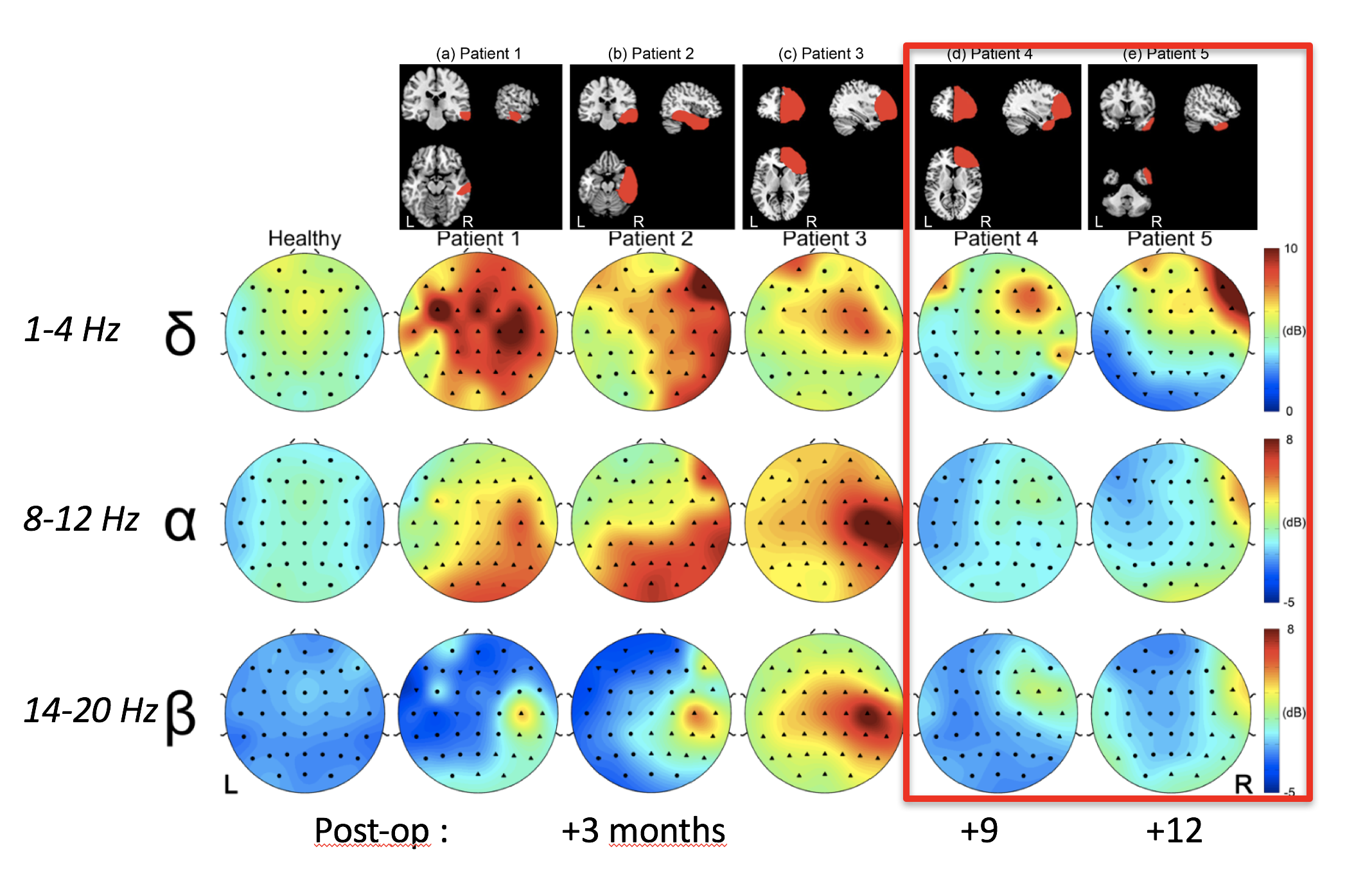
Awake brain surgery of tumour is used to optimize the resection of tumoral tissue. Postoperatively, patients show mild and temporary neurological deficits despite massive cerebral resections. Reasons for these impairments along with the compensation mechanisms operating within the cortex and subcortical structures are barely understood. The objective of this project is to reveal the remote effects of the tumour and its resection, to determine their nature measuring changes induced in functional Magnetic Resonance Imagery (fMRI) and electroencephalographic signals using standard and nonlinear methods. Recently, we focused on postoperative brain dynamics of patients, who underwent wide-awake surgery for Low grade gliomas (LGG). We analysed EEG data of 5 patients, who perfomed an ecological visuomanual task, comparing them to a control group of 8 healthy subjects (fig. 8). We used the motor preparation period to extract power features and phase space features to better characterise changes in EEG signal following surgery and subsequent functional reorganisation. The preparation period was chosen for its stationarity allowing analyses, which were not applicable during the ERP period. Our results clearly identify changes in postoperative brain dynamics of patients, who underwent wide-awake surgery. Both spectral and recurrence quantification analyses suggested imbalances between the injured and healthy hemispheres for patients, whether in terms of spectral power density or temporal structure of EEG signal. These investigations performed on the motor preparation period also provided important information regarding longitudinal recovery of brain dynamics. Although all patients in our study had very different tumours, both in size and location, it is interesting to note that the 2 patients, who underwent the experimental protocol respectively 9 and 12 months after surgery, showed more moderate alterations of spectral content and signal complexity independently of the lesion size. This may be seen as an indicator for EEG signal standardization in time and presumably a resumption of brain dynamics. These findings have potential clinical rehabilitation implications.
Figure
8. Topographical maps of spectral power: Maps illustrating the spatial distribution of the spectral power contained in a given frequency band over the scalp. We selected bands which showed significant peaks of spectral power for patients in comparison with control group (i.e. δ band: 1 - 4Hz, α/μ band: 9 - 12Hz and β band: 14 - 20Hz) and we then calculated the average power at each electrode for each band. Electrode locations are represented by a set of symbols: ● symbol is used when the corresponding spectral power falls within the 95% confidence interval estimated from healthy subjects. ▲ and ▼ symbols are respectively used when the power is either greater or smaller than the 95% confidence interval. α/μ band
|
|