Section:
New Results
Electrophysiological brain mapping: measuring evoked potentials induced by electrical stimulation and its physiological spreading in the human brain.
Participants :
Marion Vincent, François Bonnetblanc, David Guiraud, Hugues Duffau [CHU Montpellier] , Emmanuel Mandonnet [CHU Lariboisière, APHP] , Anthony Boyer.
Being able to change or inhibit the activity of a region or population of neurons in the brain is an essential approach in fundamental neuroscience, as it helps the researcher to determine the functional role of neurons. This approach is also important at a more applied level, for brain function mapping during neurosurgical procedures. It is well known that electrical stimulation (ES) affects neural activity by modifying the voltage gradient along the neuronal cell inducing depolarization or hyperpolarization of the membrane. When a current flows in tissues around neuronal cells, it can change their membrane potential and trigger an action potential. However, this general principle can be applied in vivo via several different settings and much is unknown about which neural elements are excited or inhibited locally and how this local perturbation spreads within the brain through physiological pathways [35]. We are now able to record different types of electrophysiological potentials that are evoked by ES in the human brain and we developed some basic methodological considerations required for their correct assessment [26] (fig.9). With our methodology, three different types of evoked potentials can now be measured during brain surgery in the operative room:
– Cortical evoked-potential (also called direct cortical response, DCR), when recording the cortex at the stimulation site,
– Cortico-axono-cortical evoked-potential, i.e. recording the cortex at a distant site from the stimulating site. These potentials are elicited by physiological propagation through white matter associative pathways from the locally stimulated area towards the distal area,
– Axono-cortical evoked potentials, when the cortex is distally recorded from a stimulation site within the white matter. These evoked potentials are technically difficult to observe. Their recording imposes important methodological considerations about the way they can be triggered and measured. In particular, proposed some factors potentially determining the generation of true cortico-axono-cortical evoked potentials, spreading from one stimulated cortical area to another distant one and passing through the white matter pathways.
Correctly measuring evoked potentials in the human brain induced by electrical stimulation is important in the clinical domain especially in the neurosurgical context. It remains challenging because of many pitfalls that can occur at the methodological level and few teams in the world are currently able to efficiently record these evoked potentials. Nevertheless, they can give strong real-time in vivo insights into the functional state and connectivity of a patient’s brain. In the next years measuring intraoperatively the evoked potentials with ES in the brain will be a new method for mapping the brain in vivo and in real time and taking into account the specificity of each patient’s brain.
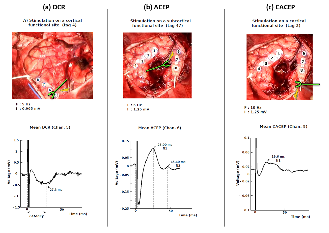
Figure
9. Example of mean evoked potentials induced by DES.
(a) Direct cortical responses (DCR) present one primary peak around 27 ms after the stimulation onset. They were measured on 4 patients on cortical sites distant of less than 2 cm from the cortical stimulation site.
(b) Axono-cortical evoked potentials (ACEP) presented a primary peak around 25 ms after DES, followed by a second peak 20 ms later. DES is applied subcortically, around 1.7 cm away from the recording site. ACEP were osberved on 2 patients.
(c) Cortico-axono-cortical evoked potentials (CACEP) were oberved in 1 patient. Cortical DES induced a one-peak waveform 14 to 35 ms after the artefact onset. CACEP were recorded on cortical site more than 2 cm away from the stimulation site.
|
|