|
|

|

|
| e-Pub |
Section: New Results
Detection and classification of neuronal extensions on fluorescence microscopy images: application to the study of metabolic diseases such as obesity or anorexia
Participants : Sarah Laroui, Eric Debreuve, Xavier Descombes.
This work is made in collaboration with Céline Cansell and Carole Rovere (IPMC, Sophia Antipolis).
The goal of this project is to classify 3D images of neuronal cells (astrocytes and microglia) into mice fed normally and mice fed with a high-fat diet (see Fig. 6). The distinction can be made in two different areas of interest of the hypothalamus: Median Eminence (EM) and Arcuate Nucleus (ARC).
|
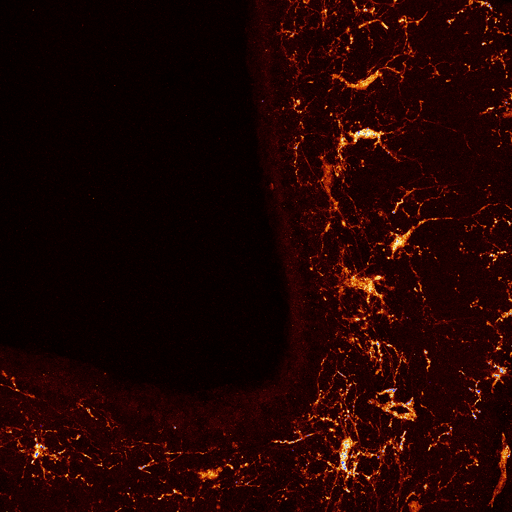
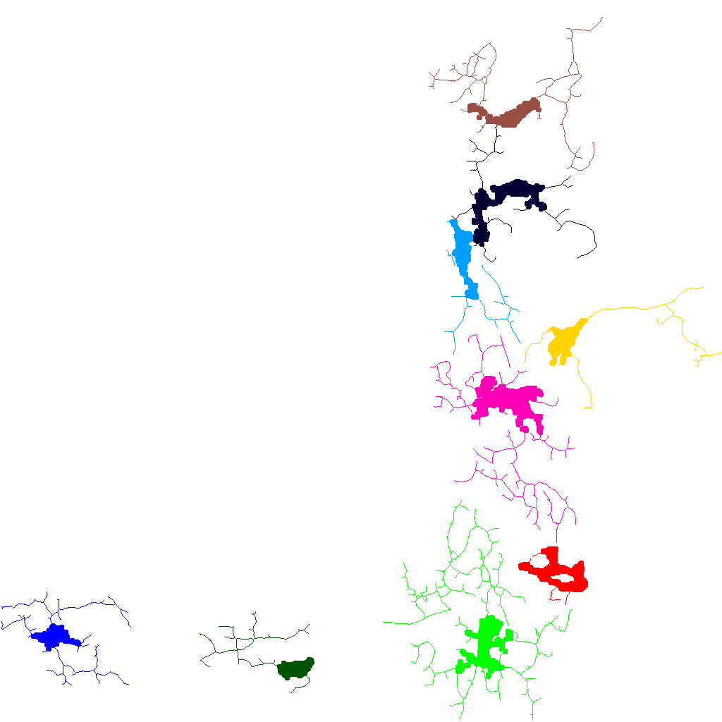
Astrocytes are perceived as networks. Our goal is to find out if there is a difference in the organization of these networks between the two areas of interest and between the two mouse models. Regarding inflammatory cells (microglias), we first segment each cell body and their extensions using a Frangi filter bank to enhance filamentous structures. This produces network pieces that must be joined to build one network per microglia. Thus, we connect filaments to soma and filaments to filaments using minimal paths (using an image-based, anisotropic metric) computed by dynamic programming. Finally, we extract geometrical and topological parameters such as the length and width of the extensions, the number of branches ... These parameters will be used for clustering microglia networks in order to identity the different populations.