|
|

|

|
| e-Pub |
Section: New Results
Comprehensive comparison of multi-labeled images
Participants : Gaël Michelin, Grégoire Malandain.
The data used for this work are courtesy of Yassin Refahi (Sainsbury Laboratory, Cambridge university) and Ulla-Maj Fiuza (CRBM, CNRS, Montpellier 1 & 2 university).
In the context of developmental biology, 3D+t microscopy imaging allows to quantitatively study the morphogenesis at the cellular level, but requires automated segmentation methods to handle the huge quantities of data. To minimize the necessary and tedious user interaction to correct unavoidable errors (3D images may have up to thousands of cells), it is desirable to improve such segmentation methods. This, in turn, motivates the need for a comprehensive evaluation methodology that will allow to automatically compare the outputs of two segmentation methods, not only in terms of cell border accuracy, but also in terms of cell detection.
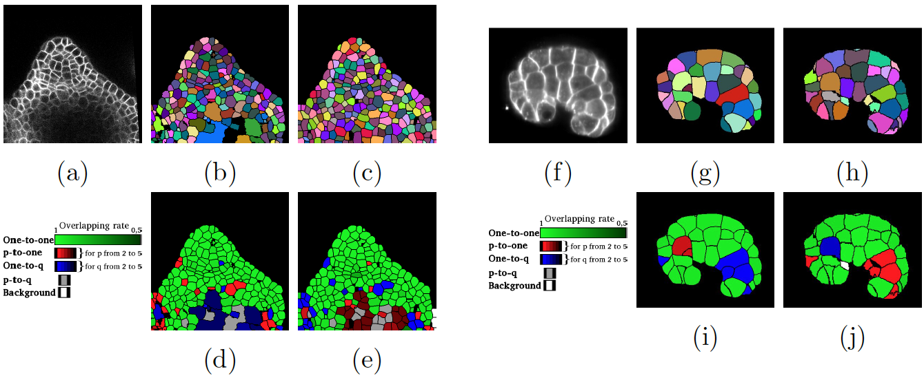
The aim of the present work is to propose such an original comprehensive segmentation comparison method that provides an objective way for multi-object segmentation comparison. This method enables to determine automatically a region-to-region correspondence map and provides asymmetric shape similarity indexes between two segmented images, with a robustness to potential region border variations. We illustrate the applicability of the proposed method with two examples in figure 10.