|
|

|

|
| e-Pub |
Section: New Results
Research axis 1: Medical Image Computing in Neuroimaging
Extraction and exploitation of complex imaging biomarkers involve an imaging processing workflow that can be quite complex. This goes from image physics and image acquisition, image processing for quality control and enhancement, image analysis for features extraction and image fusion up to the final application which intends to demonstrate the capability of the image processing workflow to issue sensitive and specific markers of a given pathology. In this context, our objectives in the recent period were directed toward 4 major methodological topics:
Diffusion imaging
L2 Similarity Metrics for Diffusion Multi-Compartment Model Images Registration
Participants : Renaud Hédouin, Olivier Commowick, Emmanuel Caruyer, Christian Barillot.
Diffusion multi-compartment models (MCM) allow for a fine and comprehensive study of the white matter microstructure. Non linear registration of MCM images may provide valuable information on the brain for example through population comparison. State-of-the-art MCM registration however relies on pairing-based similarity measures where the one-to-one mapping of MCM compartments is required. This approach leads to non differentiabilties or discontinuities, which may turn into poorer registration. Moreover, these measures are often specific to one MCM compartment model. We proposed [34] two new MCM similarity measures based on the space of square integrable functions, applied to MCM characteristic functions. These measures are pairing-free and agnostic to compartment types. We derived their analytic expressions for multi-tensor models and proposed a spherical approximation for more complex models. Evaluation was performed on synthetic deformations and inter-subject registration, demonstrating the robustness of the proposed measures.
Block-Matching Distortion Correction of Echo-Planar Images with Opposite Phase Encoding Directions
Participants : Renaud Hédouin, Olivier Commowick, Élise Bannier, Christian Barillot.
By shortening the acquisition time of MRI, Echo Planar Imaging (EPI) enables the acquisition of a large number of images in a short time, compatible with clinical constraints as required for diffusion or functional MRI. However such images are subject to large, local distortions disrupting their correspondence with the underlying anatomy. The correction of those distortions is an open problem, especially in regions where large deformations occur. We have proposed a new block-matching registration method to perform EPI distortion correction based on the acquisition of two EPI with opposite phase encoding directions (PED). It relies on new transformations between blocks adapted to the EPI distortion model, and on an adapted optimization scheme to ensure an opposite symmetric transformation. We have produced qualitative and quantitative results of the block-matching correction using different metrics on a phantom dataset and on in-vivo data. We have shown the ability of the block-matching to robustly correct EPI distortion even in strongly affected areas. This work has been published in IEEE Transactions on Medical Imaging [21].
Diffusion MRI processing for multi-compartment characterization of brain pathology
Participants : Renaud Hédouin, Olivier Commowick, Christian Barillot.
Diffusion weighted imaging (DWI) is a specific type of MRI acquisition based on the direction of diffusion of the brain water molecules. It allows, through several acquisitions, to model the brain microstructure, as white matter, which is significantly smaller than the voxel-resolution. To acquire a large number of images in a clinical setting, very-fast acquisition techniques are required as single-shot imaging. However these acquisitions suffer locally large distortions. We have proposed a block-matching registration method based on the acquisition of images with opposite phase-encoding directions (PED). This technique specially designed for Echo-Planar Images (EPI) robustly correct images and provides a deformation field. This field is applicable to an entire DWI series from only one reversed EPI allowing distortion correction with a minimal acquisition time cost. This registration algorithm has been validated both on phantom and on in vivo data and is available in our source medical image processing toolbox Anima. From these diffusion images, we are able to construct multi-compartments models (MCM) which can represent complex brain microstructure. Doing registration, averaging and atlas creation on these MCM images is required to perform studies and statistic analyses. We propose a general method to interpolate MCM as a simplification problem based on spectral clustering. This technique, which is adaptable for any MCM, has been validated on both synthetic and real data. Then, from a registered dataset, we performed a patient to population analysis at a voxel-level computing statistics on MCM parameters. Specifically designed tractography can also be used to make analysis, following tracks, based on individual anisotropic compartments. All these tools are designed and used on real data and contribute to the search of biomakers for brain diseases such as multiple sclerosis.
The challenge of mapping the human connectome based on diffusion tractography
Participant : Emmanuel Caruyer.
Tractography based on non-invasive diffusion imaging is central to the study of human brain connectivity. To date, the approach has not been systematically validated in ground truth studies. Based on a simulated human brain data set with ground truth tracts, we organized an open international tractography challenge, which resulted in 96 distinct submissions from 20 research groups. Here, we report the encouraging finding that most state-of-the-art algorithms produce tractograms containing 90 percent of the ground truth bundles (to at least some extent). However, the same tractograms contain many more invalid than valid bundles, and half of these invalid bundles occur systematically across research groups. Taken together, our results demonstrate and confirm fundamental ambiguities inherent in tract reconstruction based on orientation information alone, which need to be considered when interpreting tractography and connectivity results. Our approach provides a novel framework for estimating reliability of tractography and encourages innovation to address its current limitations [26].
Comparison of inhomogeneity distortion correction methods in diffusion MRI of the spinal cord
Participants : Haykel Snoussi, Emmanuel Caruyer, Christian Barillot.
Diffusion MRI (dMRI) is a modality that describes the geometry of neural architecture. Diffusion images suffer from various artifacts originating from subject and physiological motion, eddy currents and B0-field inhomogeneity. These can severely affect image quality particularly in the spine region. However, strategies exist to correct these distortions, including co-registration, point spread function, phase field map and reversed gradient polarity method (RGPM). We evalute various correction methods using RGPM which provides best results. More precisely, we compared Voss plus two other recent methods: Topup (FSL) and HySCO (ACID/SPM). This work was presented at the ESMRMB conference [38].
Arterial Spin Labeling:
Our contributions on this topic are illustrated in Fig. 2. Arterial Spin Labeling (ASL) enables measuring cerebral blood flow in MRI without injection of a contrast agent. Perfusion measured by ASL carries relevant information for patients suffering from pathologies associated with singular perfusion patterns.
|
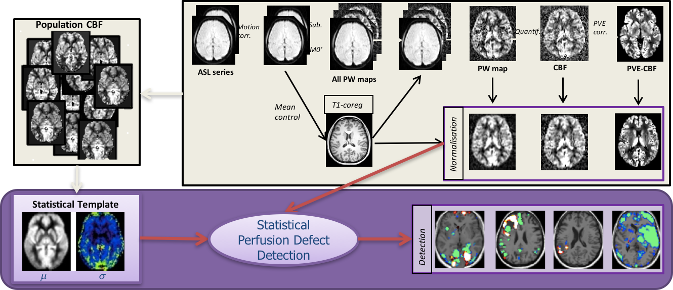
However this technique suffers from drawbacks such as low signal to noise ratio and poor resolution.
Patch-based super-resolution for arterial spin labeling MRI
Participants : Cédric Meurée, Pierre Maurel, Christian Barillot.
In this context, our contributions focused on a super resolution approach to reduce the influence of Partial Volume Effects (PVE) and obtain images close to the ones that would be acquired at a high resolution, but in a shorter scan duration. PVE are an important limitation of arterial spin labeling (ASL) acquisitions, impacting the validity of quantitative cerebral blood flow (CBF) estimations. This work consists of a super-resolution algorithm, which includes information of high resolution (HR) structural images to reconstruct HR CBF maps from low resolution ASL series, without increasing the acquisition time. Compared with nearest neighbor, trilinear and 3rd order spline interpolations, the proposed algorithm is found to generate a CBF image closer to the one obtained with a reference HR ASL acquisition. CBF calculations can therefore be improved by using this algorithm, which reduces the PVE [36].
Resting-state functional ASL
Participants : Corentin Vallée, Isabelle Corouge, Pierre Maurel, Christian Barillot.
We have started to work on resting-state functional ASL (rs-fASL). Rs-fASL in clinical daily practice and academic research stay discreet compared to resting-state BOLD. However, by giving direct access to cerebral blood flow maps, rs-fASL could lead to significant clinical subject scaled application as CBF can be considered as a biomarker in common neuropathology. As a new topic, we started by building a viable long sequence for rs-fASL. We take advantage of the long duration of the sequence to assess the link between overall quality of rs-fASL and duration of acquisition. To this end, we consider typical functional areas of the brain, and assess their quality compared to gold standards depending on the duration of acquisition. While some more work remain to be done, we tend to show there is an optimal duration of acquistion for rs-fASL. This work was submitted for the next ISMRM Conference.
Longitudinal atlas creation and brain development analysis
Participants : Antoine Legouhy, Olivier Commowick, Christian Barillot.
The study of brain development provides insights in the normal trend of brain evolution and enables early detection of abnormalities. We propose a method to quantify growth in three arbitrary orthogonal directions of the brain through linear registration. We introduce a 9 degrees of freedom transformation that gives the opportunity to extract scaling factors describing brain growth along those directions by registering a data base of subjects in a common basis. We apply this framework to create a longitudinal curve of scaling ratios along fixed orthogonal directions from 0 to 16 years highlighting anisotropic brain development. In pediatric image analysis, the study of brain development provides insights in the normal trend of brain evolution and enables early detection of abnormalities. Tools like longitudinal atlases allow to compute statistics on populations, understand brain variability at different ages to highlight changes in growth, shape, structure etc. We experimented different methods to perform longitudinal atlases. This work was submitted for the next ISMRM Conference.
Quantitative relaxation times estimation and processing:
The VisAGeS team has proposed new methodologies to exploit new relaxometry sequences, able to provide direct information on tissue properties (T1, T2, T2* relaxation times) and their alteration in diseases. Such sequences have a great potential in diagnostic and evolution study of patients suffering from various neurological diseases.
Gaining Insights Into Multiple Sclerosis Lesion Characteristics from Brain Tissue Microstructure Information: A Multi-Compartment T2 Relaxometry Approach:
Participants : Sudhanya Chatterjee, Olivier Commowick, Christian Barillot.
In addition to raw relaxation times, we have also studied other estimation methods able, from T2 relaxometry sequences, to estimate the fraction of myelin (myelin water fraction) inside each voxel, a quantity that may be largely impacted in neurological diseases. To this end, we have proposed new multi-compartment T2 estimation methods [42] with a new water three-compartment T2 model of tissue bounded water (free water, axons and cells, and myelin), using variable projection to make the estimation faster and more robust. Clinical trends and pathogenetic ways of onset and progression of Multiple Sclerosis (MS) in patients suggest that MS is a highly heterogeneous disease. MS is predominantly a White Matter (WM) disease, which is mainly composed of myelinated axons and neuroglia type cells. Demyelination and axonal loss characterize the condition of MS in a patient. However, they follow varying trends in patients. In this work, we propose a method in which T2 relaxometry data is used to obtain a quantitative brain tissue microstructure information. This information is then studied to check its corroborations with pathogenetic understanding of MS in literature [41].
Multi-Compartment T2 Relaxometry Model Using Gamma Distribution Representations: A Framework for Quantitative Estimation of Brain Tissue Microstructures:
Participants : Sudhanya Chatterjee, Olivier Commowick, Christian Barillot.
Advanced MRI techniques (e.g., d-MRI, MT, relaxometry etc.) can provide quantitative information of brain tissues. Image voxels are often heterogeneous in terms of microstructure information due to physical limitations and imaging resolution. Quantitative assessment of the brain tissue microstructure can provide valuable insights into neurodegenerative diseases (e.g., Multiple Sclerosis). In this work, we propose a multicompartment model for T2-Relaxometry to obtain brain microstructure information in a quantitative framework. The proposed method allows simultaneous estimation of the model parameters [42].
Multi-modal EEG and fMRI Source Estimation Using Sparse Constraints:
Participants : Saman Noorzadeh, Pierre Maurel, Christian Barillot.
In this work, a multi-modal approach is presented and validated on real data to estimate the brain neuronal sources based on EEG and fMRI. Combining these two modalities can lead to source estimations with high spatio-temporal resolution. The joint method is based on the idea of linear model already presented in the literature where each of the data modalities are first modeled linearly based on the sources. Afterwards, they are integrated in a joint framework which also considers the sparsity of sources. The sources are then estimated with the proximal algorithm. The results are validated on real data and show the efficiency of the joint model compared to the uni-modal ones. We also provide a calibration solution for the system and demonstrate the effect of the parameter values for uni- and multi-modal estimations on 8 subjects [37].