Section: New Results
Research axis 2: Applications in Neuroradiology and Neurological Disorders
Arterial Spin Labeling:
Participants : Jean-Christophe Ferré, Maia Proisy, Isabelle Corouge, Élise Bannier, Christian Barillot.
Arterial Spin Labeling is an attractive perfusion MRI technique due to its complete non-invasiveness. However it still remains confidential in clinical practice. Over the years, we have developed several applications to evaluate its potential in different contexts. In 2017, in the context of the MALTA project, we focused on the application of ASL to activation-fMRI. Functional Arterial Spin Labeling (fASL) has demonstrated its greater specificity as a marker of neuronal activity than the reference BOLD fMRI for motor activation mapping in healthy volunteers. Motor fASL was yet to be investigated in the context of tumors, under the assumption that fASL would be less sensitive to venous contamination induced by the hemodynamics remodeling in the tumor vicinity than BOLD fMRI. As the arterial transit time may be shortened in activation areas, we explored the ability of fASL to map the motor areas at different post-labeling delays (PLD) in healthy subjects and patient with brain tumor. As part of the PhD of Maia Proisy, we have also been working on processing and analyse MR perfusion images using arterial spin labeling in neonates and children for several purposes:
-
ASL and TOF-MRA are two totally non-invasive, easy-to-use MRI sequences for children in emergency settings. Hypoperfusion associated with homolateral vasospasm may suggest a diagnosis of migraine with aura (published in Cephalagia and presented in 3 congresses including RSNA)
-
Investigation of brain perfusion evolution between 6 month and 15 years using ASL sequence in order to provide reference values in this age range (Measurement of pediatric regional cerebral blood flow from 6 months to 15 years of age article under revision, presented in one national congress)
-
Work in Progress: ASL perfusion images in 20 neonates with hypoxic-ischemic encephalopathy that underwent MRI on day-of-life 3 and day-of-life 10.
Hybrid EEG-fMRI Neurofeedback:
Participants : Lorraine Perronnet, Marsel Mano, Élise Bannier, Mathis Fleury, Giulia Lioi, Christian Barillot.
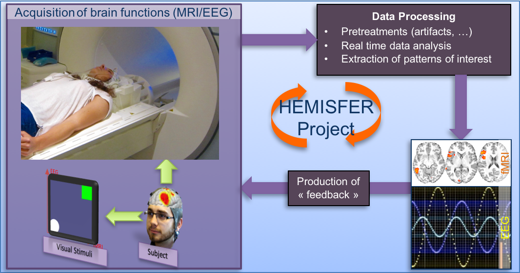
Over the last 4 years, we developed a whole new range of activities around hybrid EEG-MR imaging and neurofeedback for brain rehabilitation. We propose to combine advanced instrumental devices (Hybrid EEG and MRI platforms), with new man-machine interface paradigms (Brain computer interface and serious gaming) and new computational models (source separation, sparse representations and machine learning) to provide novel therapeutic and neuro-rehabilitation paradigms in some of the major neurological and psychiatric disorders of the developmental and the aging brain. We first performed a thorough state-of-the-art of Neurofeedback (NF) and restorative Brain Computer Interfaces (BCI) under EEG and fMRI modality as well as of EEG-fMRI integration, with a particular focus on applications in depression and motor rehabilitation. This enabled us to design a NF protocol based on motor imagery and compatible with EEG and fMRI. We implemented different types of feedback and compared for the first time the effects of unimodal EEG-NF and fMRI-NF versus bimodal EEG-fMRI-NF by looking both at EEG and fMRI activations. We also introduced a new feedback metaphor for bimodal EEG-fMRI-neurofeedback that integrates both EEG and fMRI signal in a single bi-dimensional feedback (a ball moving in 2D). The participants to this study were able to regulate activity in their motor regions in all NF conditions. Our results also suggest that that EEG-fMRI-neurofeedback could be more specific or more engaging than EEG-NF alone [31].
All the experiments were performed on the Neurinfo platform which is equipped with an EEG MR compatible 64-channel device in 2014 to perform joint EEG and BOLD or ASL fMRI. We developed, installed and successfully tested a hybrid EEG-fMRI platform for bimodal NF experiments. Our system is based on the integration and the synchronization of an MR-compatible EEG and fMRI acquisition subsystems. We developed two real-time pipelines for EEG and fMRI that handle all the necessary signal processing, the joint NF block that calculates and fuses the NF and a visualization block that displays the NF to the subject. The control and the synchronization of both subsystems with each other and with the experimental protocol is handled by the NF Control. Our platform showed very good real-time performance with various pre-processing, filtering, and NF estimation and visualization methods. Its modular architecture is easily adaptable to different experimental environments, and offers high efficiency for optimal real-time NF applications [27].
These developments came as part of the HEMISFER project which is conducted through a very complementary set of competences over the different teams involved (Visages Inserm U1228, HYBRID and PANAMA Teams from Inria/IRISA, EA 4712 team from University of Rennes I and ATHENA team from Inria Sophia-Antipolis). The overall principle of this project is illustrated in Fig. 3.
Multiple sclerosis:
Participants : Anne Kerbrat, Gilles Edan, Jean-Christophe Ferré, Benoit Combès, Olivier Commowick, Élise Bannier, Sudhanya Chatterjee, Haykel Snoussi, Emmanuel Caruyer, Christian Barillot.
The VisAGeS research team has a strong focus on applying the developed methodologies (illustrated in research axis 1) to multiple sclerosis (MS) understanding and the prediction of its evolution. Related to the EMISEP project on spinal cord injury evolution in MS, a first work investigated the magnetization transfer reproducibility across centers in the spinal cord and was accepted for presentation at ESMRMB [33]. Based on this work, a second work investigated the sensitivity of magnetization transfer to assess diffuse and focal burden in MS patients [43]. In parallel, methodological developments have addressed spinal cord diffusion data analysis, starting with a comparaison of several distortion correction methods [38].
Finally, we investigated myelin water fraction (MWF) estimation on multiple sclerosis and demonstrated in longitudinal studies [41] how these figures can be related with lesion evolution, paving the way towards myelin oriented MS evaluation of patient future evolution prediction (and thus treatment adaptation) and joint studies between different quantitative imaging modalities (e.g., diffusion).
Recovery imaging:
Participants : Isabelle Bonan, Stephanie Leplaideur, Élise Bannier, Jean-Christophe Ferré, Christian Barillot.
More common after a right hemispheric brain injury, misperception of body in space, impacting moves and posture is often associated with disturbance of spatial attention (behavioural symptoms of a failure in spontaneously reorienting attention to stimulus information in the left field). While different subjects use different references in their elaboration of spatial representation, body-centered coordinate systems are the most prevalent. As part of an fMRI substudy of a national research study on balance disorder rehabilitation, we investigated differences in activations during body-centered spatial tasks in corporeal and in extracorporeal space. Healthy controls and stroke patients were included in this fMRI sub study comprising 2 egocentric spatial tasks: perception of the midsagittal plane in extracorporeal space (straight-ahead task) and in corporeal space (longitudinal axis task). Results obtained on healthy control data were presented at the SOFMER conference and the journal paper is under review. For both tasks, cerebral activations largely dominated in the right hemisphere and essentially involved the right frontoparietal network. In addition, the straight-ahead task presented specific activations in the temporoparieto-insular cortex and thalamic areas. Patient data processing is ongoing in the context of an MD-PhD. In parallel, a master study investigated the brain structural connections between the cortical areas obtained from the fMRI study using diffusion MRI and the white matter query language.
White matter connectivity analysis in patients suffering from depression:
Participants : Julie Coloigner, Jean-Marie Batail, Jean-Christophe Ferré, Isabelle Corouge, Christian Barillot.
The mood depressive disorder (MDD) is a common chronically psychiatric disorder with an estimated lifetime prevalence reported to range from 10 percent to 15 percent worldwide. This disease is characterized by an intense dysregulation of affect and mood as well as additional abnormalities including cognitive dysfunction, insomnia, fatigue and appetite disturbance. Despite the extensive therapy options available for depression, up to 80 percent of patients will suffer from a relapse [1]. Consequently, exhibiting imaging biomarkers of this disease will support both a better understanding of the neural correlates underlying the depression, and a better diagnosis and treatment of individual depressed patients. Previous studies of structural and functional magnetic resonance imaging have reported several microstructural abnormalities in the prefrontal cortex, anterior cingulate cortex, hippocampus and thalamus [2]. These observations suggest a dysfunction of the circuits connecting frontal and subcortical brain regions, leading to a "disconnection syndrome" [3]. Given the small sample size used in the past studies, we proposed a more robust analysis using a larger cohort of patients suffering from depression, called LONGIDEP. The latter is a routine care cohort of patients suffering from mood depressive disorder who underwent a clinical evaluation, neuropsychological testing and brain MRI. The population sample consists of 125 patients suffering from depression and 65 healthy age and gender-matched, control subjects. A composite measure of medication load for each patient was assessed using a previously established method [4]. We investigated alterations of white matter integrity using a voxel-based analysis based on fractional anisotropy (FA) and the apparent diffusion coefficient (ADC) in patients with depression. Using graph theory-based analysis, we also examined white matter changes in the organization of networks in patients suffering from depression. Our findings provide robust evidence that the reduction of white-matter integrity in the interhemispheric connections and fronto-limbic neuronal circuits may play an important role in MDD pathogenesis. These results are consistent with an overall hypothesis that depression involves a disconnection of prefrontal, striatal, and limbic emotional areas.
Knowing and Remembering: Cognitive and Neural Influences of Familiarity on Recognition Memory in Early Alzheimer's Disease (EPMR-MA):
Participants : Pierre-Yves Jonin, Quentin Duché, Élise Bannier, Christian Barillot.
Inclusion of the 20 healthy participants in the "EPMR-MA" study (clinical trials ID NCT02492529) has been achieved, the inclusion phase will be achieved before 30th, december, 2017. Healthy controls data are pre-processed and the first analysis workflow proved promising, it should allow submitting a first paper at the beginning of 2018.
Semantic Dementia Imaging:
Participants : Jean-Christophe Ferré, Isabelle Corouge, Elise Bannier, Christian Barillot.
After demonstrating the relative preservation of fruit and vegetable knowledge in patients with semantic dementia (SD), we sought to identify the neural substrate of this unusual category effect. Nineteen patients with SD performed a semantic sorting task and underwent a morphometric 3T MRI scan. The grey-matter volumes of five regions within the temporal lobe were bilaterally computed, as well as those of two recently described areas (FG1 and FG2) within the posterior fusiform gyrus. In contrast to the other semantic categories we tested, fruit and vegetable scores were only predicted by left FG1 volume. We therefore found a specific relationship between the volume of a subregion within the left posterior fusiform gyrus and performance on fruits and vegetables in SD. We argue that the left FG1 is a convergence zone for the features that might be critical to successfully sort fruits and vegetables. We also discuss evidence for a functional specialization of the fusiform gyrus along two axes (lateral medial and longitudinal), depending on the nature of the concepts and on the level of processing complexity required by the ongoing task [28].


