Section: New Results
Lucid Dreaming in Narcolepsy
Participants : Pauline Daudet, Mario Chavez [Correspondant] , Smaranda Leu-Semenescu, Jean-Louis Golmard, Isabelle Arnulf.
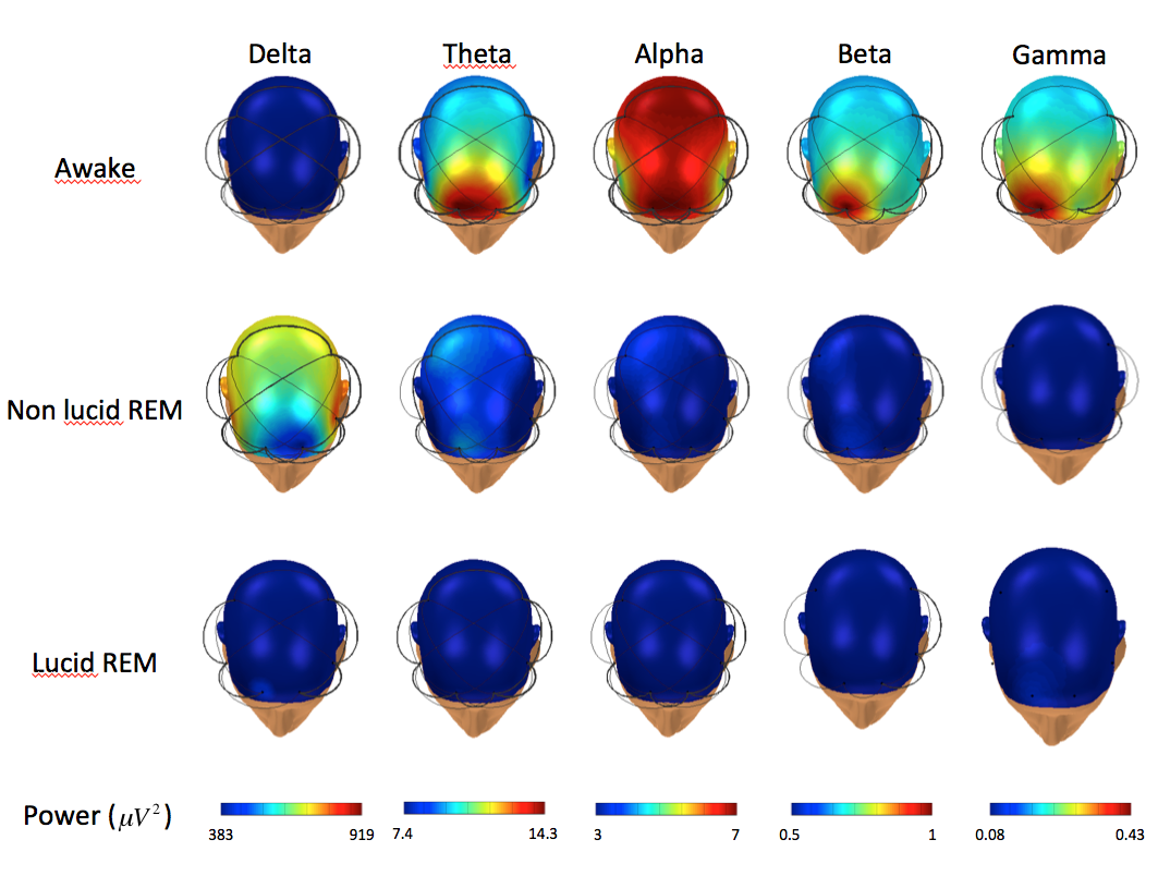
Lucid dreaming is the experience of being aware of dreaming while asleep and continuing to dream. Lucid dreams generally arise in REM sleep. Compared to non-lucid REM sleep, lucid REM sleep is associated with local frontal lobe EEG changes in the 40 Hz band, increased brain coherence, and increased activity on functional MRI in the bilateral precuneus, cuneus, parietal lobules, and prefrontal and occipito-temporal cortices, which may correspond to restored reflective consciousness. We decided to study the frequency and determinants of lucid dreaming in narcolepsy and to challenge patients' alleged ability to achieve lucid dreaming using sleep monitoring during nighttime and daytime sleep. Compared to 53 healthy controls, the 53 narcolepsy patients reported more frequent dream recall, nightmares and recurrent dreams. The frequency of cataplexy, hallucinations, sleep paralysis, dyssomnia, positivity, and the severity of sleepiness were similar in narcolepsy with and without lucid dreaming. The delta power in the electrode average, in delta, theta, and alpha powers in C4, and coherences between frontal electrodes were lower in lucid than non-lucid REM sleep in spectral EEG analysis. The duration of REM sleep was longer, the REM sleep onset latency tended to be shorter, and the percentage of atonia tended to be higher in lucid vs. non-lucid REM sleep; the arousal index and REM density and amplitude were unchanged. Our results suggest that narcoleptics have a high propensity for lucid dreaming without differing in REM sleep characteristics from people without narcolepsy. This also suggests that narcolepsy patients may provide useful information in future studies on the nature of lucid dreaming.
More details in [11]
|