

Keywords
Computer Science and Digital Science
- A3.3. Data and knowledge analysis
- A3.4. Machine learning and statistics
- A4.3. Cryptography
- A4.4. Security of equipment and software
- A4.8. Privacy-enhancing technologies
- A5.2. Data visualization
- A5.3. Image processing and analysis
- A5.4. Computer vision
- A5.6. Virtual reality, augmented reality
- A5.9. Signal processing
- A6.1. Methods in mathematical modeling
- A6.2. Scientific computing, Numerical Analysis & Optimization
- A6.3. Computation-data interaction
- A8.3. Geometry, Topology
- A9. Artificial intelligence
- A9.2. Machine learning
- A9.3. Signal analysis
- A9.6. Decision support
- A9.7. AI algorithmics
- A9.9. Distributed AI, Multi-agent
- A9.10. Hybrid approaches for AI
Other Research Topics and Application Domains
- B2.2. Physiology and diseases
- B2.3. Epidemiology
- B2.4. Therapies
- B2.6. Biological and medical imaging
- B2.6.1. Brain imaging
- B2.6.2. Cardiac imaging
- B2.6.3. Biological Imaging
1 Team members, visitors, external collaborators
Research Scientists
- Nicholas Ayache [Team leader, INRIA, Senior Researcher, HDR]
- Irene Balelli [INRIA, ISFP]
- James Benn [INRIA, Starting Research Position]
- Hervé Delingette [INRIA, Senior Researcher, HDR]
- Marco Lorenzi [INRIA, Researcher, HDR]
- Marta Nunez Garcia [INRIA, Starting Research Position]
- Xavier Pennec [INRIA, Senior Researcher, HDR]
- Maxime Sermesant [INRIA, Senior Researcher, HDR]
Post-Doctoral Fellows
- Safaa Al Ali [INRIA, from Oct 2022]
- Anna Calissano [INRIA]
- Amel Karoui [INRIA]
- Dimbihery Rabenoro [INRIA, until Apr 2022]
- Ghiles Reguig [Inria and startup MyDataModels, from Oct 2022]
PhD Students
- Hind Dadoun [UNIV COTE D'AZUR, 3IA Côte d'Azur]
- Gaetan Desrues [INRIA]
- Zhijie Fang [3IA COTE D'AZUR]
- Yann Fraboni [Accenture Labs Sophia Antipolis, CIFRE]
- Erwan Gaymard [ExactCure, CIFRE, from Dec 2022]
- Lisa Guzzi [Université Côte d'Azur, from Nov 2022, 3IA Côte d'Azur]
- Dimitri Hamzaoui [UNIV COTE D'AZUR]
- Josquin Harrison [INRIA]
- Etrit Haxholli [3IA COTE D'AZUR]
- Lucia Innocenti [3IA COTE D'AZUR, from Jun 2022]
- Florent Jousse [QuantificCare, CIFRE, until Apr 2022]
- Victoriya Kashtanova [3IA COTE D'AZUR]
- Huiyu Li [3IA COTE D'AZUR]
- Buntheng Ly [UNIV COTE D'AZUR, until Sep 2022]
- Elodie Maignant [INRIA]
- Morten Pedersen [INRIA]
- Santiago Smith Silva Rincon [UNIV COTE D'AZUR]
- Hari Sreedhar [UNIV COTE D'AZUR]
- Tom Szwagier [INRIA, from Oct 2022]
- Riccardo Taiello [3IA COTE D'AZUR]
- Yann Thanwerdas [INRIA, until Jun 2022]
- Paul Tourniaire [3IA COTE D'AZUR]
- Yingyu Yang [INRIA]
Technical Staff
- Florent Collot [Université de Bordeaux - IHU Liryc, Engineer, from Mar 2022]
- Francesco Cremonesi [INRIA, Engineer, from May 2022]
- Marie Deprez [INRIA, Engineer]
- Luis Gomes Pereira [INRIA, Engineer, from Apr 2022 until Feb 2022]
- Nicolas Guigui [Inria, Engineer, until Mar 2022]
- Mathilde Merle [Université de Bordeaux - IHU Liryc, Engineer, from Mar 2022]
- Marco Milanesio [UNIV COTE D'AZUR, Engineer]
- Mihaela Pop [INRIA, Engineer, until Jun 2022]
- Dimbihery Rabenoro [INRIA, Engineer, from May 2022 until Oct 2022]
- Jesus Jairo Rodriguez Padilla [INRIA, Engineer]
- Anaëlle Zanella [INRIA, Engineer, from Jul 2022]
Interns and Apprentices
- Tom Szwagier [INRIA, Intern, from Apr 2022 until Sep 2022, Master 2]
Administrative Assistant
- Isabelle Strobant [INRIA]
Visiting Scientists
- Federica Cruciani [University of Verona, Italy, from May 2022 until Sep 2022]
- Andreas Hansen [UNIV DTU, from Sep 2022]
- Hervé Lombaert [ ETS Montreal, until Feb 2022, Associate Professor of Computer Engineering]
- Alexander Panfilov [UNIV GAND, from Sep 2022 until Sep 2022]
External Collaborator
- Nicolas Cedilnik [inHEART]
2 Overall objectives
2.1 Description
Our long-term goal is to contribute to the development of what we call the e-patient (digital patient) for e-medicine (digital medicine).
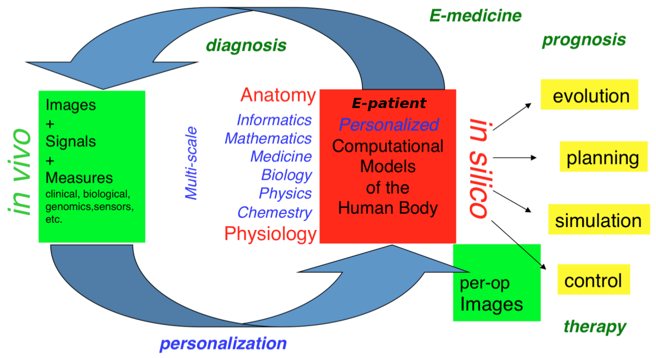
- the e-patient (or digital patient) is a set of computational models of the human body able to describe and simulate the anatomy and the physiology of the patient's organs and tissues, at various scales, for an individual or a population. The e-patient can be seen as a framework to integrate and analyze in a coherent manner the heterogeneous information measured on the patient from disparate sources: imaging, biological, clinical, sensors, ...
- e-medicine (or digital medicine) is defined as the computational tools applied to the e-patient to assist the physician and the surgeon in their medical practice, to assess the diagnosis/prognosis, and to plan, control and evaluate the therapy.
The models that govern the algorithms designed for e-patients and e-medicine come from various disciplines: computer science, mathematics, medicine, statistics, physics, biology, chemistry, etc. The parameters of those models must be adjusted to an individual or a population based on the available images, signals and data. This adjustment is called personalization and usually requires solving difficult inverse problems. The overall picture of the construction of the personalized e-patient for e-medicine was presented at the College de France through an inaugural lecture and a series of courses and seminars (fr), concluded by an international workshop.
2.2 Organisation
The research organization in our field is often built on a virtuous triangle. On one vertex, academic research requires multidisciplinary collaborations associating informatics and mathematics to other disciplines: medicine, biology, physics, chemistry ... On a second vertex, a clinical partnership is required to help defining pertinent questions, to get access to clinical data, and to clinically evaluate any proposed solution. On the third vertex, an industrial partnership can be introduced for the research activity itself, and also to transform any proposed solution into a validated product that can ultimately be transferred to the clinical sites for an effective use on the patients.
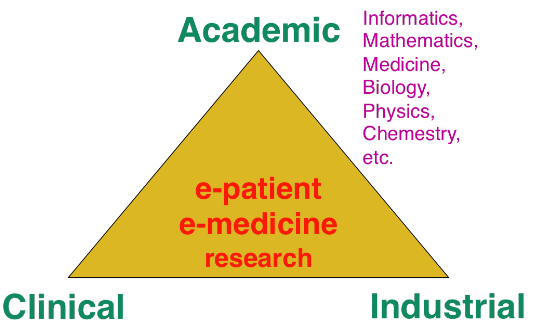
Keeping this triangle in mind, we choose our research directions within a virtuous circle: we look at difficult problems raised by our clinical or industrial partners, and then try to identify some classes of generic fundamental/theoretical problems associated to their resolution. We also study some fundamental/theoretical problems per se in order to produce fundamental scientific advances that can help in turn to promote new applications.
3 Research program
3.1 Introduction
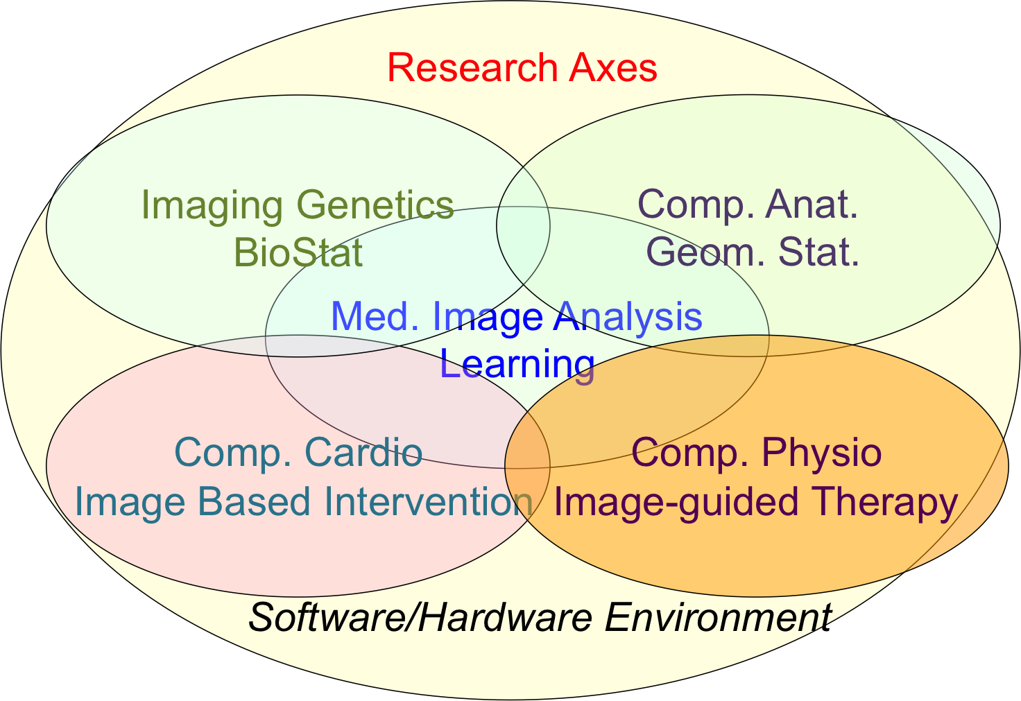
Our research objectives are organized along 5 scientific axes:
- Biomedical Image Analysis & Machine Learning
- Imaging & Phenomics, Biostatistics
- Computational Anatomy, Geometric Statistics
- Computational Physiology & Image-Guided Therapy
- Computational Cardiology & Image-Based Cardiac Interventions
For each scientific axis, we introduce the context and the long term vision of our research.
3.2 Biomedical Image Analysis & Machine Learning
The long-term objective of biomedical image analysis is to extract, from biomedical images, pertinent information for the construction of the e-patient and for the development of e-medicine. This relates to the development of advanced segmentation and registration of images, the extraction of image biomarkers of pathologies, the detection and classification of image abnormalities, the construction of temporal models of motion or evolution from time-series of images, etc.
In addition, the growing availability of very large databases of biomedical images, the growing power of computers and the progress of machine learning (ML) approaches have opened up new opportunities for biomedical image analysis.
This is the reason why we decided to revisit a number of biomedical image analysis problems with ML approaches, including segmentation and registration problems, automatic detection of abnormalities, prediction of a missing imaging modality, etc. Not only those ML approaches often outperform the previous state-of-the-art solutions in terms of performances (accuracy of the results, computing times), but they also tend to offer a higher flexibility like the possibility to be transferred from one problem to another one with a similar framework. However, even when successful, ML approaches tend to suffer from a lack of explanatory power, which is particularly annoying for medical applications. We also plan to work on methods that can interpret the results of the ML algorithms that we develop.
3.3 Imaging & Phenomics, Biostatistics
The human phenotype is associated with a multitude of heterogeneous biomarkers quantified by imaging, clinical and biological measurements, reflecting the biological and patho-physiological processes governing the human body, and essentially linked to the underlying individual genotype. In order to deepen our understanding of these complex relationships and better identify pathological traits in individuals and clinical groups, a long-term objective of e-medicine is therefore to develop the tools for the joint analysis of this heterogeneous information, termed Phenomics, within the unified modeling setting of the e-patient.
To date the most common approach to the analysis of the joint variation between the structure and function of organs represented in medical images, and the classical -omics modalities from biology, such as genomics or lipidomics, is essentially based on the massive univariate statistical testing of single candidate features out of the many available. This is for example the case of genome-wide association studies (GWAS) aimed at identifying statistically significant effects in pools consisting of up to millions of genetics variants. Such approaches have known limitations such as multiple comparison problems, leading to underpowered discoveries of significant associations, and usually explain a rather limited amount of data variance . Although more sophisticated machine learning approaches have been proposed, the reliability and generalization of multivariate methods is currently hampered by the low sample size relatively to the usually large dimension of the parameters space.
To address these issues this research axis investigates novel methods for the integration of this heterogeneous information within a parsimonious and unified multivariate modeling framework. The cornerstone of the project consists in achieving an optimal trade-off between modeling flexibility and ability to generalize on unseen data by developing statistical learning methods informed by prior information, either inspired by "mechanistic" biological processes, or accounting for specific signal properties (such as the structured information from spatio-temporal image time series). Finally, particular attention will be paid to the effective exploitation of the methods in the growing Big Data scenario, either in the meta-analysis context, or for the application in large datasets and biobanks.
Federated learning in multi-centric studies. The current research scenario is characterised by medium/small scale (typically from 50 to 1000 patients) heterogeneous datasets distributed across centres and countries. The straightforward extension of learning algorithms successfully applied to big data problems is therefore difficult, and specific strategies need to be envisioned in order to optimally exploit the available information. To address this problem, we focus on learning approaches to jointly model clinical data localized in different centres. This is an important issue emerging from recent large-scale multi-centric imaging-genetics studies in which partners can only share model parameters (e.g. regression coefficients between specific genes and imaging features), as represented for example by the ENIGMA imaging-genetics study, led by the collaborators at University of Southern California. This problem requires the development of statistical methods for federated model estimation, in order to access data hosted in different clinical institutions by simply transmitting the model parameters, that will be in turn updated by using the local available data. This approach is extended to the definition of stochastic optimization strategies in which model parameters are optimized on local datasets, and then summarized in a meta-analysis context. Finally, this project studies strategies for aggregating the information from heterogeneous datasets, accounting for missing modalities due to different study design and protocols. The developed methodology finds important applications within the context of Big Data, for the development of effective learning strategies for massive datasets in the context of medical imaging (such as with the UK biobank), and beyond.
3.4 Computational Anatomy, Geometric Statistics
Computational anatomy is an emerging discipline at the interface of geometry, statistics and image analysis which aims at developing algorithms to model and analyze the biological shape of tissues and organs. The goal is not only to establish generative models of organ anatomies across diseases, populations, species or ages but also to model the organ development across time (growth or aging) and to estimate their variability and link to other functional, genetic or structural information. Computational anatomy is a key component to support computational physiology and is evidently crucial for building the e-patient and to support e-medicine.
Pivotal applications include the spatial normalization of subjects in neuroscience (mapping all the anatomies into a common reference system) and atlas to patient registration to map generic knowledge to patient-specific data. Our objectives will be to develop new efficient algorithmic methods to address the emerging challenges described below and to generate precise specific anatomical model in particular for the brain and the heart.
The objects of computational anatomy are often shapes extracted from images or images of labels (segmentation). The observed organ images can also be modeled using registration as the random diffeomorphic deformation of an unknown template (i.e. an orbit). In these cases as in many other applications, invariance properties lead us to consider that these objects belong to non-linear spaces that have a geometric structure. Thus, the mathematical foundations of computational anatomy rely on statistics on non-linear spaces.
Geometric Statistics aim at studying this abstracted problem at the theoretical level. Our goal is to advance the fundamental knowledge in this area, with potential applications to new areas outside of medical imaging. Beyond the now classical Riemannian spaces, we aim at developping the foundations of statistical estimation on affine connection spaces (e.g. Lie groups), quotient and stratified metric spaces (e.g. orbifolds and tree spaces). In addition to the curvature, one of the key problem is the introduction of singularities at the boundary of the regular strata (non-smooth and non-convex analysis).
A second objective is to develop parametric and non-parametric dimension reduction methods in non-linear space. An important issue is to estimate efficiently not only the model parameters (mean point, subspace, flag) but also their uncertainty. We also want to quantify the influence of curvature and singularities on non-asymptotic estimation theory since we always have a finite (and often too limited) number of samples. A key challenge in developing such a geometrization of statistics will not only be to unify the theory for the different geometric structures, but also to provide efficient practical algorithms to implement them.
A third objective is to learn the geometry from the data. In the high dimensional but low sample size (small data) setting which is the common situation in medical data, we believe that invariance properties are essential to reasonably interpolate and approximate. New apparently antagonistic notions like approximate invariance could be the key to this interaction between geometry and learning.
Beyond the traditional statistical survey of the anatomical shapes that is developed in computational anatomy above, we intend to explore other application fields exhibiting geometric but non-medical data. For instance, applications can be found in Brain-Computer Interfaces (BCI), tree-spaces in phylogenetics, Quantum Physics, etc.
3.5 Computational Physiology & Image-Guided Therapy
Computational Physiology aims at developing computational models of human organ functions, an important component of the e-patient , with applications in e-medicine and more specifically in computer-aided prevention, diagnosis, therapy planning and therapy guidance. The focus of our research is on descriptive (allowing to reproduce available observations), discriminative (allowing to separate two populations), and above all predictive models which can be personalized from patient data including medical images, biosignals, biological information and other available metadata. A key aspect of this scientific axis is therefore the coupling of biophysical models with patient data which implies that we are mostly considering models with relatively few and identifiable parameters. To this end, data assimilation methods aiming at estimating biophysical model parameters in order to reproduce available patient data are preferably developed as they potentially lead to predictive models suitable for therapy planning.
Previous research projects in computational physiology have led us to develop biomechanical models representing quasi-static small or large soft tissue deformations (e.g. liver or breast deformation after surgery), mechanical growth or atrophy models (e.g. simulating brain atrophy related to neurodegenerative diseases), heat transfer models (e.g. simulating radiofrequency ablation of tumors), and tumor growth models (e.g. brain or lung tumor growth).
To improve the data assimilation of biophysical models from patient data, a long term objective of our research will be to develop joint imaging and biophysical generative models in a probabilistic framework which simultaneously describe the appearance and function of an organ (or its pathologies) in medical images. Indeed, current approaches for the personalization of biophysical models often proceed in two separate steps. In a first stage, geometric, kinematic or/ functional features are first extracted from medical images. In a second stage, they are used by personalization methods to optimize model parameters in order to match the extracted features. In this process, subtle information present in the image which could be informative for biophysical models is often lost which may lead to limited personalization results. Instead, we propose to develop more integrative approaches where the extraction of image features would be performed jointly with the model parameter fitting. Those imaging and biophysical generative models should lead to a better understanding of the content of images, to a better personalization of model parameters and also better estimates of their uncertainty.
3.6 Computational Cardiology & Image-Based Cardiac Interventions
Computational Cardiology has been an active research topic within the Computational Anatomy and Computational Physiology axes of the previous Asclepios project, leading to the development of personalized computational models of the heart designed to help characterizing the cardiac function and predict the effect of some device therapies like cardiac resynchronisation or tissue ablation . This axis of research has now gained a lot of maturity and a critical mass of involved scientists to justify an individualized research axis of the new project Epione, while maintaining many constructive interactions with the 4 other research axes of the project. This will develop all the cardiovascular aspects of the e-patient for cardiac e-medicine.
The new challenges we want to address in computational cardiology are related to the introduction of new levels of modeling and to new clinical and biological applications. They also integrate the presence of new sources of measurements and the potential access to very large multimodal databases of images and measurements at various spatial and temporal scales.
4 Application domains
The main applications of our research are in the field of healthcare and more precisely the domain of digital medicine and biomedical data analysis. The axes of research presented above are related to many branches of medicine including cardiology, oncology, urology, neurology, otology, pneumology, radiology, surgery, dermatology, nuclear medicine. Within those branches, the applications cover the following different stages of medicine : prevention, diagnosis, prognosis, treatment.
5 Highlights of the year
5.1 Distinctions
- Nicholas Ayache was elected a Fellow of the Asia-Pacific Artificial Intelligence Association on 14 December 2022.
5.2 Awards
- Yann Thanwerdas was awarded the first ATSI prize of the EDSTIC Doctoral school of Université Côte d'Azur for his PhD work on "Riemannian and stratified geometries of covariance and correlation matrices" under the supervision of Dr Xavier Pennec.
- Yann Fraboni obtained the Best Paper Award at the FL-IJCAI’22 for his work A General Theory for Client Sampling in Federated Learning41.
- Huiyu Li’s paper Data Stealing Attack on Medical Images: Is it Safe to Export Networks from Data Lakes?34 received the Best Paper Award at the 3rd MICCAI Workshop on Distributed, Collaborative and Federated Learning. The article is co-authored with N. Ayache and H. Delingette.
- Tom Szwagier, ranked 2nd at the ICLR 2022 Computational Geometry & Topology challenge.
5.3 Special Events
- N. Ayache was a keynote speaker at Global Congress on AI in Montreal in May 2022, and a keynote speaker at the Inserm Conference on the History of Medical Imaging in Sept 2022. He was invited on the French Radio "La matinale d'Europe 1" on the topic of AI for medicine, on 25 Nov 2022.
6 New software and platforms
6.1 New software
6.1.1 CardiacSegmentationPropagation
-
Keywords:
3D, Segmentation, Cardiac, MRI, Deep learning
-
Functional Description:
Training of a deep learning model which is used for cardiac segmentation in short-axis MRI image stacks.
- Publication:
-
Authors:
Qiao Zheng, Hervé Delingette, Nicolas Duchateau, Nicholas Ayache
-
Contact:
Qiao Zheng
6.1.2 CardiacMotionFlow
-
Keywords:
3D, Deep learning, Cardiac, Classification
-
Functional Description:
Creation of a deep learning model for the motion tracking of the heart, extraction of characteristic quantities of the movement and shape of the heart to classify a sequence of cine-MRI cardiac images in terms of the types of pathologies (infarcted heart, dilated , hypertrophied, abnormality of the right ventricle).
-
Authors:
Qiao Zheng, Hervé Delingette, Nicholas Ayache
-
Contact:
Qiao Zheng
6.1.3 MedINRIA
-
Keywords:
Visualization, DWI, Health, Segmentation, Medical imaging
-
Scientific Description:
MedInria aims at creating an easily extensible platform for the distribution of research algorithms developed at Inria for medical image processing. This project has been funded by the D2T (ADT MedInria-NT) in 2010, renewed in 2012. A fast-track ADT was awarded in 2017 to transition the software core to more recent dependencies and study the possibility of a consortium creation.The Empenn team leads this Inria national project and participates in the development of the common core architecture and features of the software as well as in the development of specific plugins for the team's algorithm.
-
Functional Description:
MedInria is a free software platform dedicated to medical data visualization and processing.
- URL:
-
Contact:
Olivier Commowick
-
Participants:
Maxime Sermesant, Olivier Commowick, Théodore Papadopoulo
-
Partners:
HARVARD Medical School, IHU - LIRYC, NIH
6.1.4 GP-ProgressionModel
-
Name:
GP progression model
-
Keywords:
Data modeling, Data visualization, Data integration, Machine learning, Biostatistics, Statistical modeling, Medical applications, Evolution, Brain, Uncertainly, Uncertainty quantification, Alzheimer's disease, Probability, Stochastic models, Stochastic process, Trajectory Modeling, Marker selection, Health, Statistic analysis, Statistics, Bayesian estimation
-
Functional Description:
Disease progression modeling (DPM) of Alzheimer's disease (AD) aims at revealing long term pathological trajectories from short term clinical data. Along with the ability of providing a data-driven description of the natural evolution of the pathology, DPM has the potential of representing a valuable clinical instrument for automatic diagnosis, by explicitly describing the biomarker transition from normal to pathological stages along the disease time axis.
In this software we reformulate DPM within a probabilistic setting to quantify the diagnostic uncertainty of individual disease severity in an hypothetical clinical scenario, with respect to missing measurements, biomarkers, and follow-up information. The proposed formulation of DPM provides a statistical reference for the accurate probabilistic assessment of the pathological stage of de-novo individuals, and represents a valuable instrument for quantifying the variability and the diagnostic value of biomarkers across disease stages.
This software is based on the publication:
Probabilistic disease progression modeling to characterize diagnostic uncertainty: Application to staging and prediction in Alzheimer's disease. Marco Lorenzi, Maurizio Filippone, Daniel C. Alexander, Sebastien Ourselin Neuroimage. 2019 Apr 15,190:56-68. doi: 10.1016/j.neuroimage.2017.08.059. Epub 2017 Oct 24. HAL Id : hal-01617750 https://hal.archives-ouvertes.fr/hal-01617750/
-
Release Contributions:
- New interface and output - Completely based on Pytorch
- URL:
- Publication:
-
Contact:
Marco Lorenzi
-
Participant:
Marco Lorenzi
6.1.5 Music
-
Name:
Multi-modality Platform for Specific Imaging in Cardiology
-
Keywords:
Medical imaging, Cardiac Electrophysiology, Computer-assisted surgery, Cardiac, Health
-
Functional Description:
MUSIC is a software developed by the Asclepios research project in close collaboration with the IHU LIRYC in order to propose functionalities dedicated to cardiac interventional planning and guidance. This includes specific tools (algorithms of segmentation, registration, etc.) as well as pipelines. The software is based on the MedInria platform.
- URL:
-
Contact:
Maxime Sermesant
-
Participants:
Florent Collot, Mathilde Merle, Maxime Sermesant
-
Partner:
IHU- Bordeau
6.1.6 SOFA
-
Name:
Simulation Open Framework Architecture
-
Keywords:
Real time, Multi-physics simulation, Medical applications
-
Functional Description:
SOFA is an Open Source framework primarily targeted at real-time simulation, with an emphasis on medical simulation. It is mostly intended for the research community to help develop new algorithms, but can also be used as an efficient prototyping tool. Based on an advanced software architecture, it allows the creation of complex and evolving simulations by combining new algorithms with algorithms already included in SOFA, the modification of most parameters of the simulation (deformable behavior, surface representation, solver, constraints, collision algorithm etc.) by simply editing an XML file, the building of complex models from simpler ones using a scene-graph description, the efficient simulation of the dynamics of interacting objects using abstract equation solvers, the reuse and easy comparison of a variety of available methods.
-
News of the Year:
The new version v20.06 has been released including new elements on SoftRobots + ModelOrderReduction integration, in addition to an improved architecture and lots of cleans and bugfixes.
- URL:
-
Contact:
Hugo Talbot
-
Participants:
Christian Duriez, François Faure, Hervé Delingette, Stephane Cotin, Hugo Talbot, Maud Marchal
-
Partner:
IGG
6.1.7 geomstats
-
Name:
Computations and statistics on manifolds with geometric structures
-
Keywords:
Geometry, Statistic analysis
-
Scientific Description:
Geomstats is an open-source Python package for computations and statistics on manifolds. The package is organized into two main modules: “geometry“ and “learning“.
The module `geometry` implements concepts in differential geometry, and the module `learning` implements statistics and learning algorithms for data on manifolds.
The goal is to provide an easily accessible library for learning algorithms on Riemannian manifolds.
-
Functional Description:
GeomStats is a Python package that performs computations on manifolds such as hyperspheres, hyperbolic spaces, spaces of symmetric positive definite matrices and Lie groups of transformations. It provides efficient and extensively unit-tested implementations of these manifolds, together with useful Riemannian metrics and associated Exponential and Logarithm maps. The corresponding geodesic distances provide a range of intuitive choices of Machine Learning loss functions. The operations implemented in GeomStats are available with different computing backends such as numpy, autograd, pytorch, and tensorflow.
-
Release Contributions:
Major update of the library after the April 2022 Hachathon in Saint Raphael.
-
News of the Year:
Three hackathons were organized (Sophia-Antipolis, Saint Raphael, Paris), bringing together (open-source) contributors from several institutions and very diverse background. The ICLR 2022 Computational Geometry & Topology challenge 2022 was organized to promote Geomstats to the Machine learning community and two sessions to present the sofware were also organized at workshops in Heidelberg and Paris, further contributing to the diffusion of the project.
GeomStats received the affiliated project status from NUMFOCUS. GeomStats was selected for Google "Season of Docs 2022", having the (payed) contribution of a technical writer during six months. A R version (which wraps the Python library) has been released. A new module on information geometry has been developed. A new module on stratified spaces has been started. Several new manifolds, metrics and learning methods have been added. Major refactorings have been done or planned in order to increase the robustness, correctness and ease of use of the library (especially for unit-tests, backends, and geometry module).
- URL:
- Publications:
-
Contact:
Luis Pereira
-
Participants:
Nicolas Guigui, Xavier Pennec, Yann Thanwerdas, Nina Miolane, Luis Pereira, Anna Calissano, Elodie Maignant, Nina Miolane, Alice Le Brigant
-
Partners:
University of California Santa Barbara, Université Panthéon-Sorbonne
6.1.8 MC-VAE
-
Name:
Multi Channel Variational Autoencoder
-
Keywords:
Machine learning, Artificial intelligence, Medical applications, Dimensionality reduction, High Dimensional Data, Unsupervised learning, Heterogeneity
-
Scientific Description:
Interpretable modeling of heterogeneous data channels is essential in medical applications, for example when jointly analyzing clinical scores and medical images. Variational Autoencoders (VAE) are powerful generative models that learn representations of complex data. The flexibility of VAE may come at the expense of lack of interpretability in describing the joint relationship between heterogeneous data. To tackle this problem, this software extends the variational framework of VAE to introduce sparsity of the latent representation, as well as interpretability when jointly account for latent relationships across multiple channels. In the latent space, this is achieved by constraining the variational distribution of each channel to a common target prior. Parsimonious latent representations are enforced by variational dropout. Experiments on synthetic data show that our model correctly identifies the prescribed latent dimensions and data relationships across multiple testing scenarios. When applied to imaging and clinical data, our method allows to identify the joint effect of age and pathology in describing clinical condition in a large scale clinical cohort.
-
Functional Description:
This software implements the work published in the paper "Sparse Multi-Channel Variational Autoencoder for the Joint Analysis of Heterogeneous Data" presented at the conference ICML 2019 (Long Beach, California, USA).
The software extends classical variational autoencoders by identifying a joint latent code associated to heterogeneous data represented in different channels. The software is implemented in Python and is based on Pytorch. It can be applied to any kind of data arrays, and provides functions for optimisation, visualisation and writing of the modelling results.
-
Release Contributions:
First release
-
News of the Year:
Method presented in the International Conference on Machine Learning (ICML 2019).
- URL:
-
Contact:
Luigi Antelmi
-
Participants:
Luigi Antelmi, Marco Lorenzi, Nicholas Ayache
-
Partner:
CoBteK
6.1.9 SOFA-CardiacReduction
-
Keywords:
Simulation, 3D modeling, Model Order Reduction, Cardiac
-
Scientific Description:
Modification of a finite element deformation model : meshless approach and frame-based description, reduction in the number of affine degrees of freedom and integration points.
-
Functional Description:
This SOFA plugin is intented to build a reduced model for deformable solids (especially cardiac simulations).
-
Contact:
Gaetan Desrues
-
Participants:
Gaetan Desrues, Hervé Delingette, Maxime Sermesant
6.1.10 Fed-BioMed
-
Name:
A general software framework for federated learning in healthcare
-
Keywords:
Federated learning, Medical applications, Machine learning, Distributed Applications, Deep learning
-
Scientific Description:
While data in healthcare is produced in quantities never imagined before, the feasibility of clinical studies is often hindered by the problem of data access and transfer, especially regarding privacy concerns. Federated learning allows privacy-preserving data analyses using decentralized optimization approaches keeping data securely decentralized. There are currently initiatives providing federated learning frameworks, which are however tailored to specific hardware and modeling approaches, and do not provide natively a deployable production-ready environment. To tackle this issue, Fed-BioMed proposes an open-source federated learning frontend framework with application in healthcare. Fed-BioMed framework is based on a general architecture accommodating for different models and optimization methods
-
Functional Description:
The project is based on the development of a distributed software architecture, and the establishment of a server instance from which remote experiments are triggered on the clients sites. The software is distributed to the client's sites, allowing to run machine learning models on the local data. Model parameters are then trasmitted to the server for federated aggregation.
Fed-BioMed finds application in all projects based on the development of learning models for multi-centric studies.
-
Release Contributions:
Fed-BioMed is based on Python and Pytorch. The software was recently revised (under an ADT) to adopt the libraries Pygrid and Pysyft.
-
News of the Year:
The develpment is ongoing and Fed-BioMed is currently at the version 4.1. For up-to-date news refer to the official project's page: https://fedbiomed.gitlabpages.inria.fr/
- URL:
- Publication:
-
Authors:
Marco Lorenzi, Santiago Smith Silva Rincon, Marc Vesin, Tristan Cabel, Irene Balelli, Andréa Senacheribbe, Carlos Zubiaga Pena, Jonathan Levy
-
Contact:
Marco Lorenzi
6.1.11 EchoFanArea
-
Name:
delineation of the border of the fan in ultrasound
-
Keywords:
Ultrasound fan area, Deep learning, Statistics, Image processing
-
Functional Description:
This software allows the delimitation of the acquisition cone in ultrasound imaging and the inpainting of annotations (lines, characters) inside the cone. It allows both the perfect de-identification of the images but also to standardize the content of the images. It relies on a parametric probabilistic approach to generate a training dataset with region of interest (ROI) segmentation masks. This data will then be used to train a deep U-Net network to perform the same task in a supervised manner, thus considerably reducing the calculation time of the method, one hundred and sixty times faster. These images are then processed with existing filling methods to remove annotations present within the signal area.
- URL:
- Publication:
-
Authors:
Hind Dadoun, Hervé Delingette, Nicholas Ayache, Anne-Laure Rousseau
-
Contact:
Hind Dadoun
-
Partner:
Nhance
6.1.12 ProMFusion
-
Keywords:
Image segmentation, Data fusion
-
Functional Description:
Code related to the paper "Robust Fusion of Probability Maps" by Benoît Audelan, Dimitri Hamzaoui, Sarah Montagne, Raphaële Renard-Penna and Hervé Delingette. The proposed approach allows to fuse probability maps in a robust manner with a spatial regularization of the consensus.
- URL:
- Publication:
-
Contact:
Benoit Audelan
-
Participants:
Benoit Audelan, Hervé Delingette, Dimitri Hamzaoui
6.1.13 SimulAD
-
Name:
Disease progression modeling for clinical intervention simulation
-
Keywords:
Data modeling, Clinical analysis, Clinical trial simulator, Alzheimer's disease, Pytorch, Variational Autoencoder, Ordinary differential equations
-
Scientific Description:
Recent failures of clinical trials in Alzheimer’s Disease underline the critical importance of identifying optimal intervention time to maximize cognitive benefit. While several models of disease progression have been proposed, we still lack quantitative approaches simulating the effect of treatment strategies on the clinical evolution. In this work, we present a data-driven method to model dynamical relationships between imaging and clinical biomarkers. Our approach allows simulating intervention at any stage of the pathology by modulating the progression speed of the biomarkers, and by subsequently assessing the impact on disease evolution.
-
Functional Description:
A machine-learning framework allowing to simulate the impact of intervention on the long-term progression of imaging and clinical biomarkers from collections of healthcare data
-
Contact:
Marco Lorenzi
-
Participants:
Clément Abi Nader, Marco Lorenzi, Nicholas Ayache, Philippe Robert
-
Partner:
Université Côte d'Azur (UCA)
6.1.14 SOFA-CardiacReduction
-
Keywords:
Simulation, 3D modeling, Model Order Reduction, Cardiac
-
Functional Description:
This SOFA plugin is intented to build a reduced model for deformable solids (especially cardiac simulations).
-
Contact:
Hervé Delingette
7 New results
7.1 Medical Image Analysis & Machine Learning
7.1.1 AI-based real-time diagnostic aid for abdominal organs in ultrasound
This work has been supported by the French government, through the 3IA Côte d’Azur Investments in the Future project managed by the National Research Agency (ANR) with the reference number ANR-19-P3IA-0002.
Keywords:
Participants: Hind Dadoun [Correspondant], Anne-Laure Rousseau, Hervé Delingette, Nicholas Ayache.
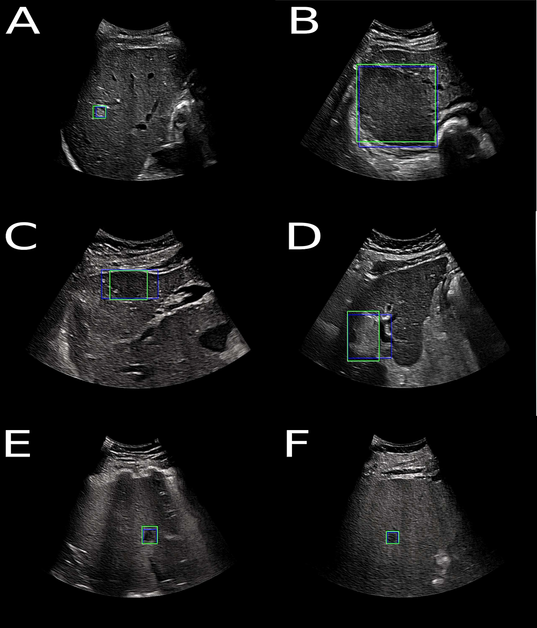
We developed a framework for the detection, localization, and characterization of focal liver lesions in B-mode ultrasound images on a curated dataset. The objective of this study was to compare the performance of expert physicians and non-expert caregivers to state-of-the-art methods with an ideal database. Malignant and benign lesions correctly identified by a Transformer based network DETR for the FLL characterization task are shown in Figure 4. This work has been published in Radiology: Artificial Intelligence 13.
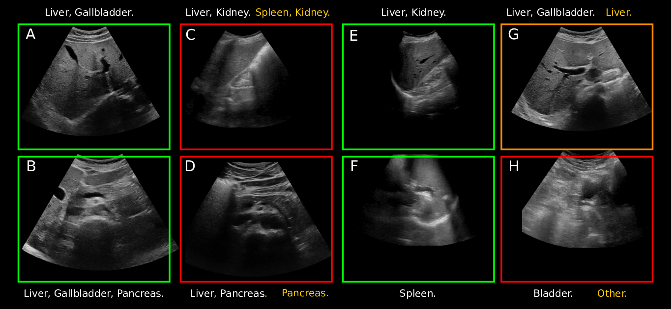
We then switched back to a database that more accurately reflects the reality of abdominal ultrasound analysis. We explored self-supervised and semi-supervised learning methods to leverage unlabeled data for abdominal organ classification in the presence of very few labeled data. Fig. 5 showcases images classified by the best performing model with four successful cases and four other cases where at least one label was misclassified or not detected. This work was submitted to a journal 51.
These works have been published in the thesis manuscript of H. Dadoun 45.
7.1.2 Machine Learning for MR-TRUS Prostate Image Fusion
This work has been supported by the French government, through the 3IA Côte d’Azur Investments in the Future project managed by the National Research Agency (ANR) with the reference number ANR-19-P3IA-0002.
Keywords:
Participants: Zhijie Fang [Correspondant], Hervé Delingette, Nicholas Ayache, Eric Gaudard, Paul Wiegel.
This is a work performed in collaboration with the Koelis Company (E. Gaudard and P. Wegiel)
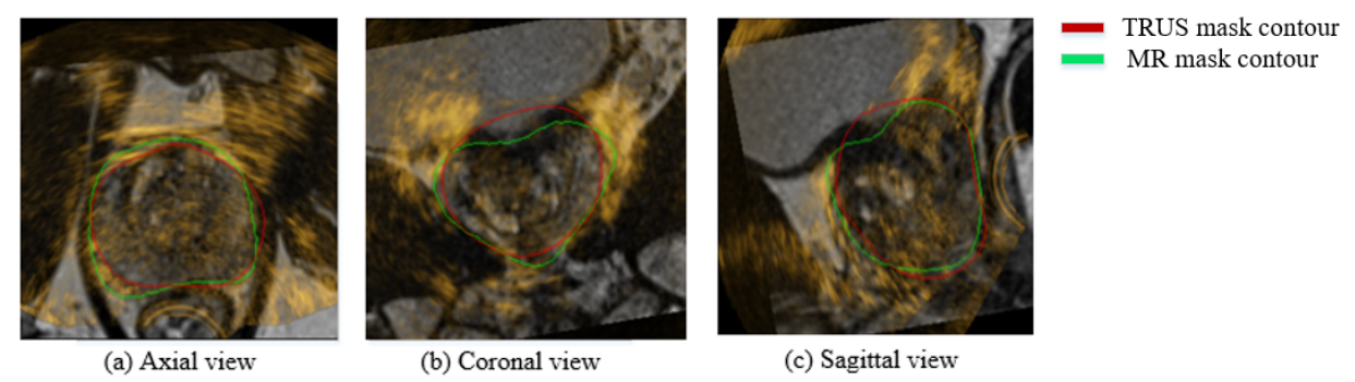
The use of pre-operative multiparametric MR images has considerably improved the sensitivity and specificity of the diagnosis of prostate tumors. Those MRI sequences are interpreted by a radiologist with a strong expertise in identifying the presence of potential cancer sites within the prostate. After the interpretation, the MRI scans are sent to a urologist for therapy planing. After their registration with per-operative ultrasound (US) images, MRI enables a high precision guidance of urologists of targeted biopsies (+/- 2.3 mm) arounf the suspicious regions of the prostate.
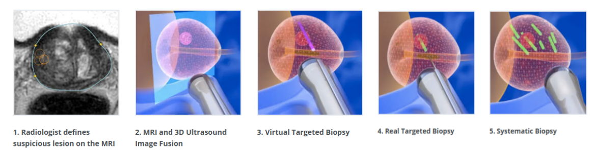
The objective of this thesis is to address the probabilistic registration of MR-TRUS prostate images.
7.1.3 AI-Based Diagnosis of Prostate Cancer from Multiparametric MRI
This work has been supported by the French government, through the 3IA Côte d'Azur Investments and UCA DS4H Investments in the Future project managed by the National Research Agency (ANR) with the reference numbers ANR-19-P3IA-0002and ANR-17-EURE-0004.
Keywords:
Participants: Dimitri Hamzaoui [Correspondant], Sarah Montagne, Raphaële Renard-Penna, Nicholas Ayache, Hervé Delingette.
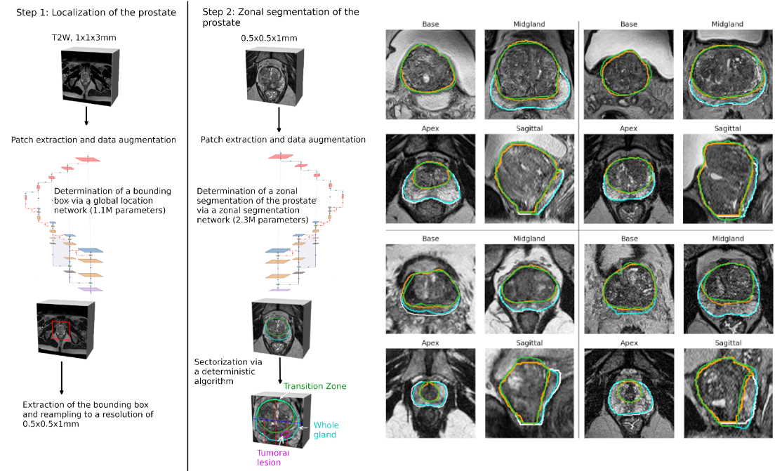
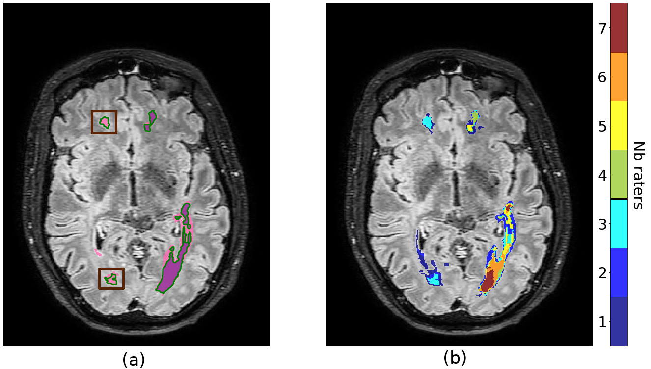
We proposed to automatically segment high-resolution T2 MR images of the prostate into peripherical / transition zones and anatomical sectors by using attention modules in a UNet network. The 2-step global framework is presented in Fig. 8. This method has been tested on two datasets : a public dataset and a private one from AP-HP segmented by up to 7 radiologists. When compared with a consensus segmentation, results were on par with previously published methods and segmentation capacities were comparable to an average performing radiologist. Zonal locations and sectorial positions of lesions annotated by radiologists were also preserved on the automated prostate segmentation 18.
We also compared three differents methods to measure prostate volumes, showing the efficiency and reliability of manual planimetry and ellipsoid methods 17.
7.1.4 MOrphologically-Aware Jaccard-Based ITerative Optimization (MOJITO) for Consensus Segmentation
This work has been supported by the French government, through the 3IA Côte d'Azur Investments and UCA DS4H Investments in the Future project managed by the National Research Agency (ANR) with the reference numbers ANR-19-P3IA-0002and ANR-17-EURE-0004.
Keywords:
Participants: Dimitri Hamzaoui [Correspondant], Sarah Montagne, Raphaële Renard-Penna, Nicholas Ayache, Hervé Delingette.
The aim of this method is to produce a consensus segmentation between several binary masks from different raters that is at the same time independent from background size (unlike the STAPLE algorithm) and that takes into account the global context of each structure (unlike the majority voting algorithm). To this end, we designed a method based on the Fréchet mean of the Jaccard distance. To speed up the algorithm, we used a heuristic method based on morphological distance maps and groups of raters, to process together voxels with the same properties. We compared extensively our method on three datasets 42 showing that it leads to consensus masks of intermediate size between Majority Voting and STAPLE and to different posterior probabilities than those methods. This work has been presented at the MICCAI workshop UNSURE 2022 42.
7.1.5 Data Stealing Attack when exporting trained network from datalakes
This project has been supported by the French government, through the 3IA Côte d’Azur Investments in the Future project managed by the National Research Agency (ANR) with the reference number ANR-19-P3IA-0002, and supported by the Inria Sophia Antipolis - Méditerranée, "NEF" computation cluster.
Keywords:
Participants: Huiyu Li [Correspondant], Nicholas Ayache, Hervé Delingette.
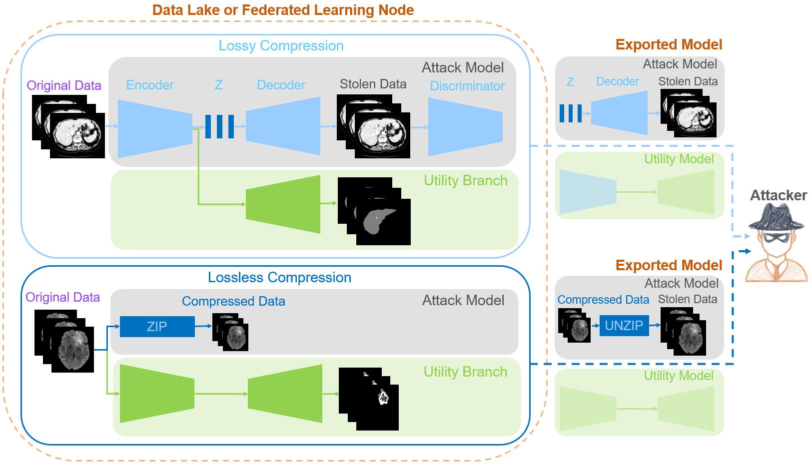
We introduce a new type of attack, the data stealing attack 34, allowing an attacker to recover training data from a remote data lake. The attack is based on losseless or lossy image compression techniques to reduce the size of the exported network and sharing part of the encoder with a utility network solving image segmentation task.
7.1.6 Mixed supervision and Deep Learning for the classification and localization of tumors in whole-slide images
This work has been supported by the French government, through the 3IA Côte d’Azur Investments in the Future project managed by the National Research Agency (ANR) with the reference number ANR-19-P3IA-0002.
Keywords:
Participants: Paul Tourniaire [Correspondant], Hervé Delingette, Nicholas Ayache, Marius Ilié, Paul Hofman.
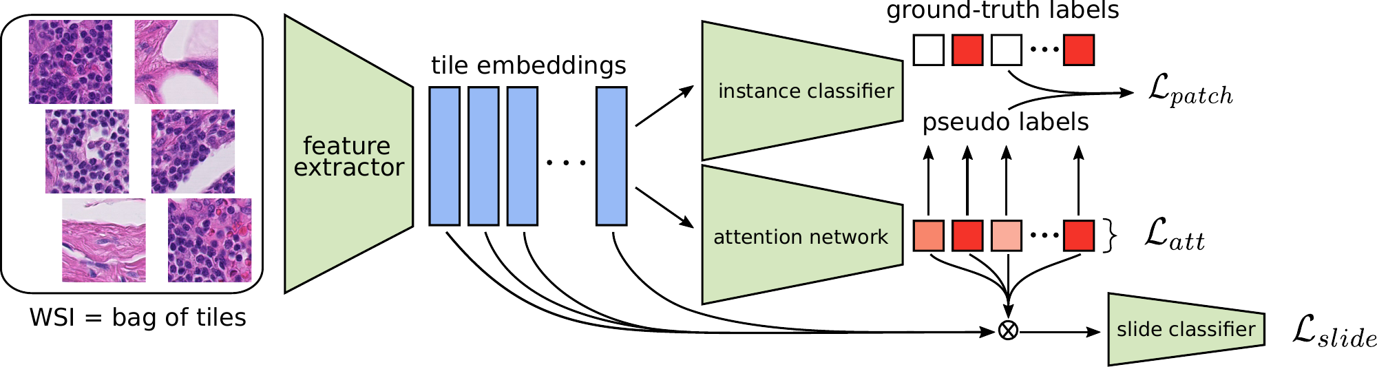
- Introduction of Mixed Supervision in the Clustering-constrained Attention Multiple Instance Learning (CLAM) model for whole-slide image tumor classification and localization (Figure 11).
- The attention scores are constrained thanks to a new loss function.
- The tile-level classifier is trained using both true and pseudo labels.
- Histological subtyping of pulmonary neuroendocrine tumors in whole-slide images 19.
7.1.7 Predicting PET-derived demyelination from multimodal MRI in multiple sclerosis
This work is done in collaboration with the Aramis-Project team of Inria in Paris and the researchers at the Brain and Spinal Cord Institute (ICM) located in Paris.
Keywords:
Participants: Theodore Soulier [MD, PhD student], Myriam Hamzaoui [PhD student], Marius chmidt-Mengin, Nicholas Ayache, Olivier Colliot, Bruno Stankoff.
We try to analyze PET-derived demyelination from multiparametric MRI, following the pioneering work of Wen Wei who defended his PhD in 2020. We also revisit the detection and analysis of MS lesions in brain MRIs 26.
7.2 Imaging & Phenomics, Biostatistics
7.2.1 Statistical Learning of Heterogeneous Data in Large-Scale Clinical Databases
Acknowledgments: IDEX UCAJEDI MNC3 Project (ANR-15-IDEX-01); 3IA (ANR-19-P3IA-0002); the OPAL infrastructure from Université Côte d'Azur.
Keywords: Ordinary Differential Equations; Generative models; Alzheimer's Disease; Disease Progression Modelling.
Participants: Clement Abi Nader [Correspondent], Nicholas Ayache, Philippe Robert, Marco Lorenzi.
Alzheimer's disease is a neurodegenerative disorder whose dynamics still remain partially unknown. The aim of this work is to develop computational models to better understand Alzheimer's disease progression, based on the analysis of imaging and clinical data. Lately, a collaboration with the LANVIE laboratory from the Geneva University Hospitals (HUG) has allowed to test the developed model in a clinical context.
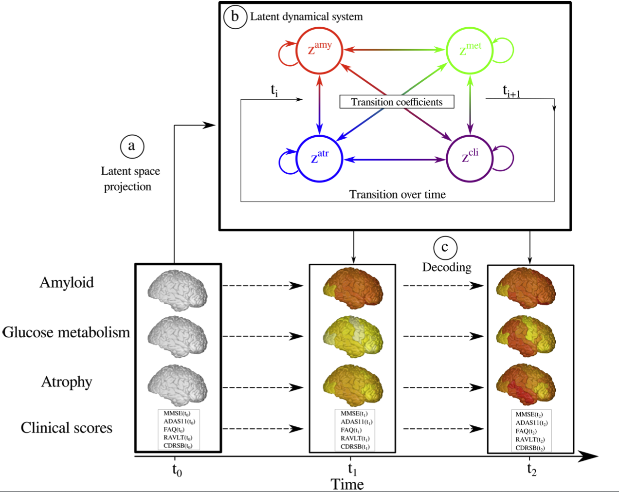
- Development of SimulAD, a statistical model for describing the evolution of Alzheimer's disease (Figure 12).
- Assessing the validity and generalization of SimulAD, by applying it on a new cohort from the Geneva Memory Center (GMC) 6.
7.2.2 Development of Genome-to-Phenome association methods using biologically inspired constraints
This work was funded by the grant ANR PARIS-15087 and ANR MITOMICS 2022000174.
Keywords:
Participants: Marie Deprez [Correspondant], Julien Moreira, Maxime Sermesant, Marco Lorenzi.
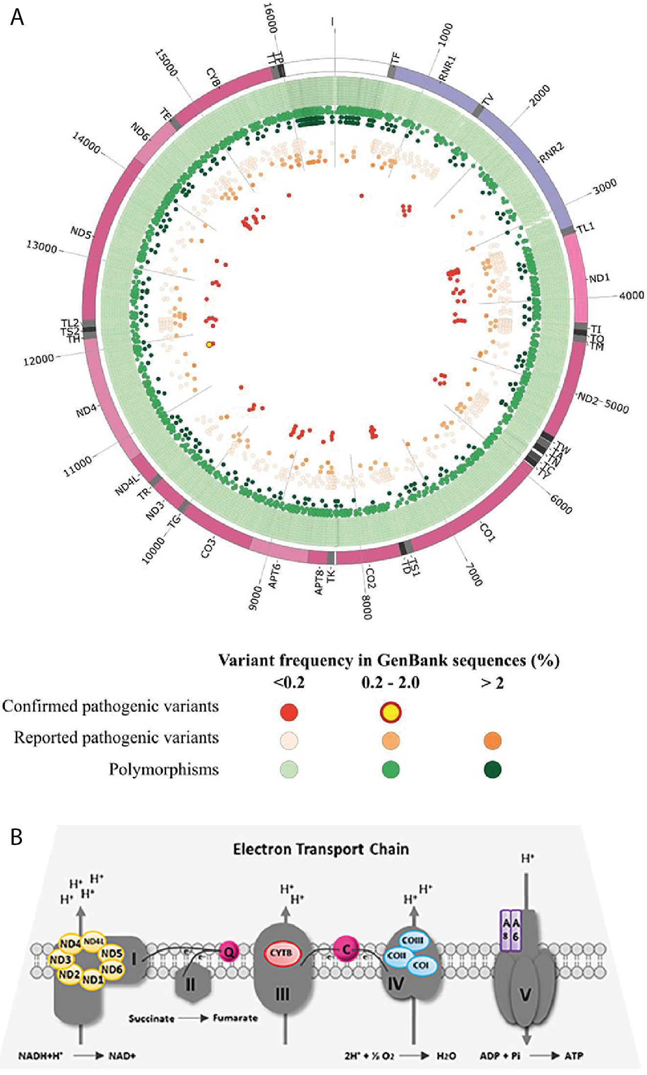
In the study of rare mitochondrial diseases, many analysis have been performed on mitochondrial DNA to identify rare variants and evaluate their pathogenicity. Yet, only a small number of studies focus on the co-occurences between multiple mitoDNA variants and/or nuclear variants and their impact on the pathological traits. Our participation in the MITOMICS project extends our previous work on the joint analysis of genomic and phenotypic data to uncover the genetics underpinning complex pathological traits.
This project is developed following multiple steps :
- Understanding the biological differences between mitochondrial and nuclear DNA, Figure 13.A. Mitochondrial DNA is circular, present in multiple copies and its maintenance is controlled both by itself and nuclear DNA.
- Study the biological / functional constraints that can be used for the prioritization of mtDNA variants, Figure 13.B. Including variants in protein-coding genes, but also in tRNAs and rRNAs.
- Some administrative work was also required to approve data security and management due to the specific sensibility of genomic data.
7.2.3 Exploring latent dynamical models for failure prediction in time-series of high-dimensional and heterogeneous data
This work was funded by 3IA Côte d'Azur.
Keywords:
Participants: Etrit Haxholli [Correspondant], Marco Lorenzi.
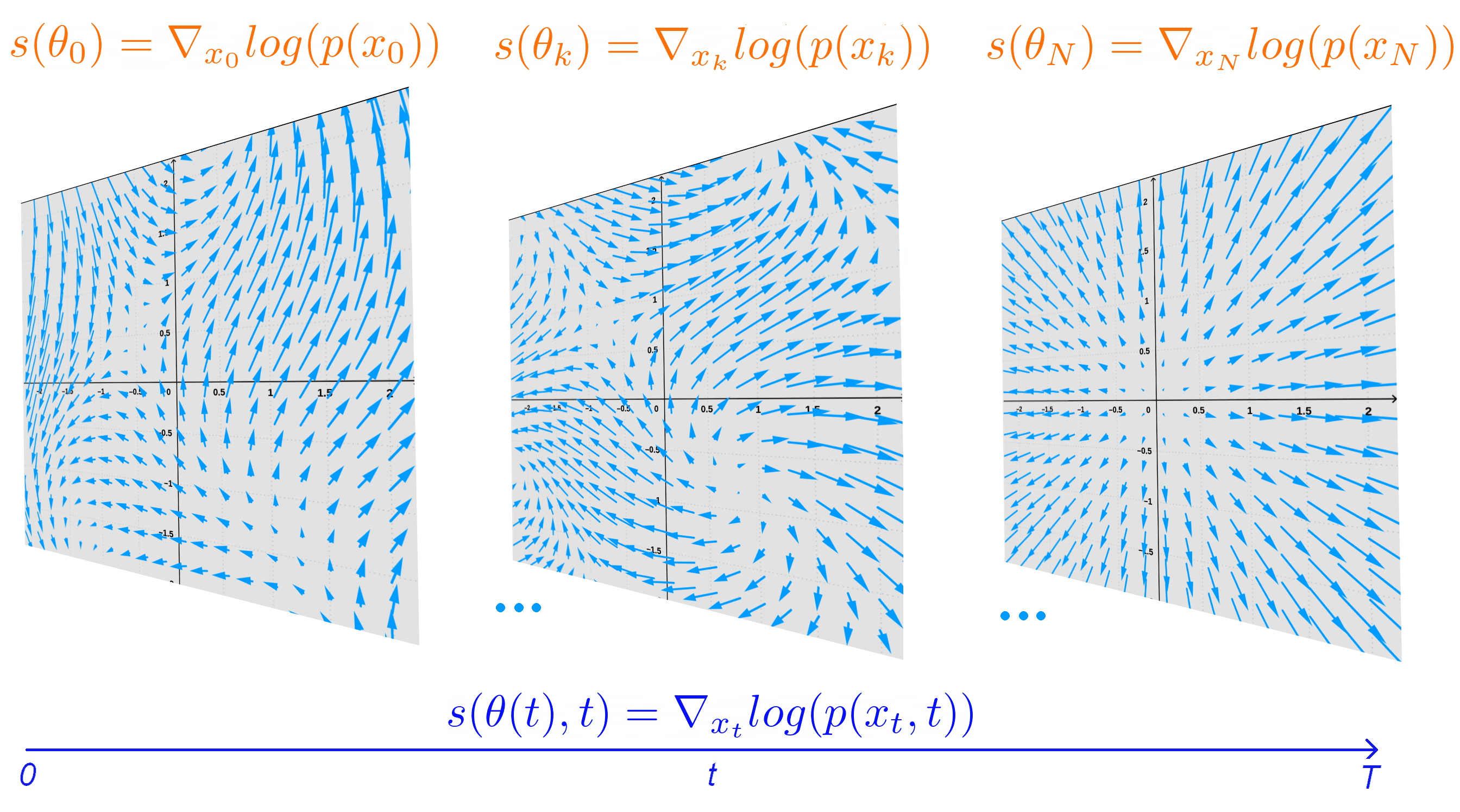
Normalizing flows provide a theoretical framework for normality quantification via density estimation, while diffusion probabilistic models (DPMs) define and efficiently train a continuous normalizing flow (CNF). Our contributions to the aforementioned fields include:
- We augment CNFs, give a closed form of their Jacobian using our continuous generalization of the chain rule, while ensuring that such transformations remain diffeomorphisms. This modification increases the flexibility of CNFs, as proven by the improved performance 54.
- In DPMs, steps in the reverse diffusion process can be optimized independently from each-other. We exploit this property, and show that by parallel training we can substantially reduce the training time and enhance performance (Figure 14).
- Under certain assumptions, we provide theoretical guarantees that the tail of a given marginal distribution coincides with the thickest tail of distributions defined on the range of the marginalized out variables. The algorithm we devised from this result (cross-tail estimation) boosts the estimation speed and quality of tail thickness 55.
7.3 Computational Anatomy & Geometric Statistics
7.3.1 Theoretically grounded computational tools for manifolds
This work was funded by the ERC grant Nr. 786854 G-Statistics.
Keywords:
Participants: Nicolas Guigui [Correspondant], Nina Miolane [UC Santa-Barbara, USA], Xavier Pennec.
Non-linear data naturally belong to manifolds and Riemannian geometry is the natural framework to compute on these manifolds. Provide a practically convenient computational framework that is rooted in the basic theory of Riemannian geometry is the challenge that we takle with the open-source Python package GeomStats. The package is organized into a geometry module, that implements the concepts of differential geometry, and a leraning module implementing generic statistics and learning algorithms for data on manifolds. Designing classes and their organization goes thus well beyond a simple implementation work: this is appealing to the mathematical theory of categories applied to geometric structures. This search for the ideal structure is also grounding the bases of our research in geometric statistics. We have published this year a 100 page chapter in Foundations & Trends in Machine Learning introducing the basic theory of Riemannian Geometry and Geometric Statistics illustrated by its implementation in Geomstats 14. This important companion paper to the geomstat library details for instance how principled algorithms can be designed on Riemannian quotient manifolds. A second example was provided by our theoretical and numerical analysis of discrete parallel transport methods with ladders 15, 44, that is both illustrated and backing up the thorough implementation in geomstats. More information about the Geostats software library itself is given in the software section.
7.3.2 The Measurement and Analysis of Shape: An Application of hydrodynamics and probability theory
This work was funded by the ERC grant Nr. 786854 G-Statistics.
Keywords:
Participants: James Benn [Correspondant], Stephen Marsland [Victoria University of Wellington, NZ].
A de Rham -current can be viewed as a map (the current map) between the set of embeddings of a closed -dimensional manifold into an ambient -manifold and the set of linear functionals on differential -forms. We demonstrate that, for suitably chosen Sobolev topologies on both the space of embeddings and the space of -forms, the current map is continuously differentiable, with an image that consists of bounded linear functionals on -forms. Using the Riesz representation theorem we prove that each -current can be represented by a unique co-exact differential form that has a particular interpretation depending on .
Embeddings of a manifold can be thought of as shapes with a prescribed topology. Our analysis of the current map provides us with representations of shapes that can be used for the measurement and statistical analysis of collections of shapes. We consider two special cases of our general analysis and prove that: (1) if then closed, embedded, co-dimension one surfaces are naturally represented by probability distributions on the ambient manifold; and (2) if then closed, embedded, one-dimensional curves are naturally represented by fluid flows on the ambient manifold. In each case we outline some statistical applications using an and metric, respectively 11.
7.3.3 Graph Alignment Exploiting the Spatial Organisation Improves the Similarity of Brain Networks
This work was funded by the ERC grant Nr. 786854 G-Statistics.
Keywords:
Participants: Anna Calissano [Correspondant], Samuel Deslauriers-Gauthier, Theodore Papadopoulo, Xavier Pennec.
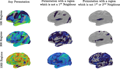
Can the structural and functional properties of a specific brain region of an atlas be assumed to be the same across subjects? In 49 we addressed this question by looking at the network representation of the brain, with nodes corresponding to brain regions and edges to their structural relationships. We perform graph matching on a set of control patients and on parcellations of different granularity to measure the connectivity misalignment between regions. The graph matching is unsupervised and reveals interesting insight on local misalignment of brain regions across subjects as shown in 15.
7.3.4 The Currents Space of Graphs
This work was funded by the ERC grant Nr. 786854 G-Statistics.
Keywords:
Participants: James Benn [Correspondant], Anna Calissano, Stephen Marsland [Victoria University of Wellington, NZ], Xavier Pennec.
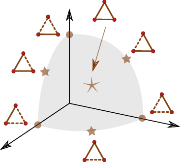
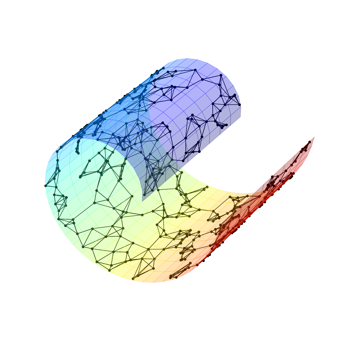
We defined in 48 a novel embedding space for binary undirected graphs, the Currents Spaces of Graphs. We represent simple graphs on vertices by 1-forms over a complete graph . It is shown that these 1-forms lie on a hypersphere in the Hilbert space of all 1-forms and the round metric induces a natural distance metric on graphs. The metric itself reduces to a global spherical Jaccard-type metric which can be computed from edge-list data alone. The structure of the graph space embedding for 3 vertices is illustrated in Fig.16. Lastly, we describe the global structure of the 1-forms representing graphs and how these can be exploited for the statistical analysis of a sample of graphs
7.3.5 Statistical modeling of face morphology for dermatology and plastic surgery
This work was supported by the ANRT Cifre contract 2019/0101 with QuantifiCare & Inria and by the French government through the 3IA Côte d'Azur Investments ANR-19-P3IA-0002 managed by the National Research Agency.
Keywords:
Participants: Florent Jousse [Correspondent], Xavier Pennec, Hervé Delingette, Arnaud Bletterer, Matilde Gonzalez.
We developed statistical modeling methods of the face to improve the automatic evaluation and follow-up of dermatological and cosmetic treatments. We first register a template mesh to the original 3D scans. In order to account for the different mesh connectivity and topological changes in face expressions such as mouth or eyes opening / closing, we propose a new Gaussian Process Morphable Models kernel based on geodesic distances that allow more flexible and realistic deformations. Our registration formulation also uses weighted least squares to select areas such as hair that do not need to be registered.
Then, we built a statistical shape model to quantify the skin sagging on the face. We use Partial Least Square Regression (PLSR) to find a linear relationship between the skin sagging score and geometric features on the face surface. Our jawline sagging model is easily interpretable and achieves the same performances as a human rater. The visualization of the PLSR latent variables shows that the model has captured jawline sagging deformations that are coherent with the jawline sagging used by the physicians.
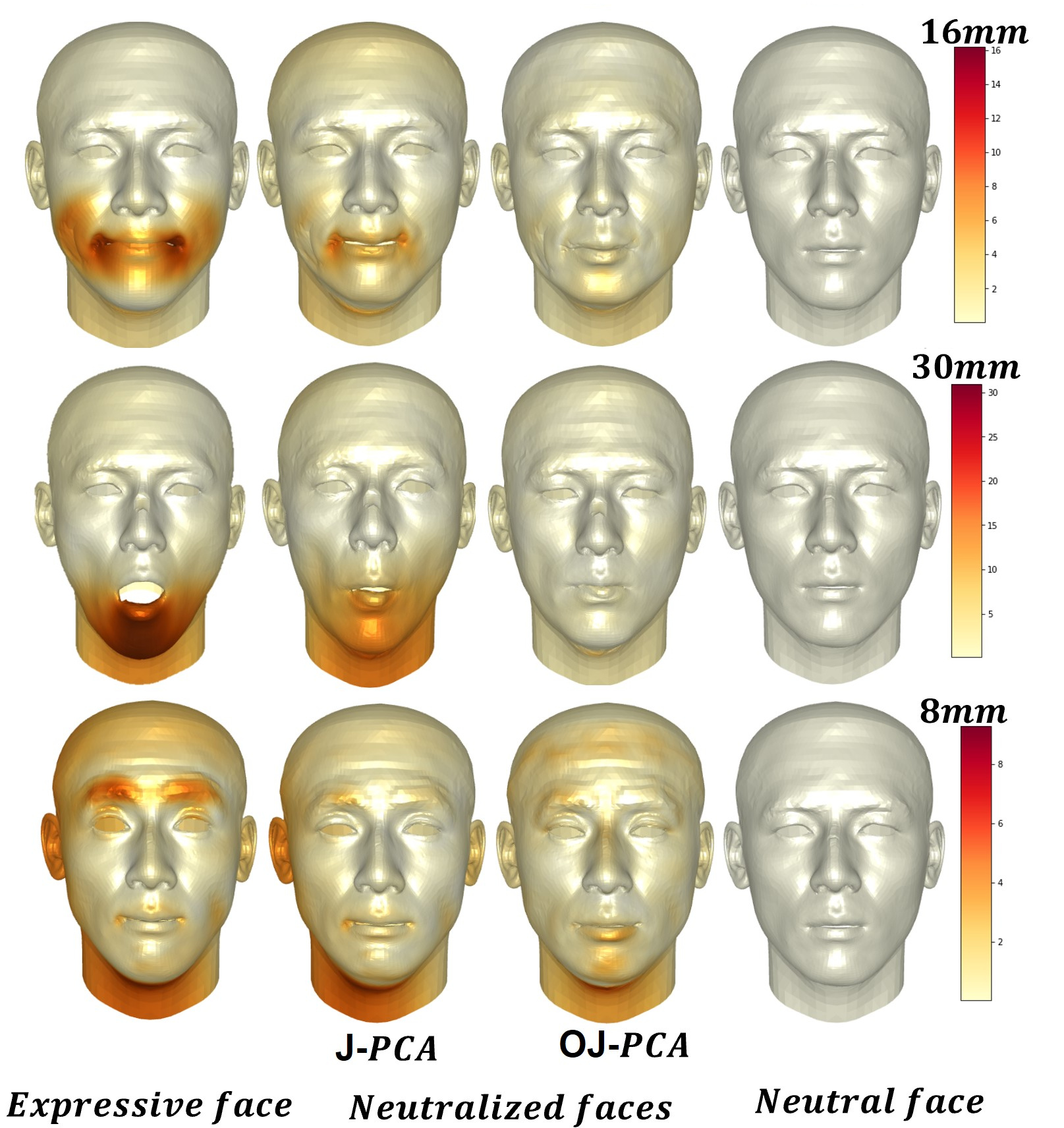
Finally, we propose to use a morphable face model to neutralize facial expressions from 3D face scans. A key point is to disantangle the facial expression from the person's identity. To this end, we propose a new simple method penalizing the non-orthogonality of the expression and identity subspaces. The effect of our quasi-orthogonality is most effective on large amplitude facial expressions, such as opening the mouth, where we can show very convincing facial expression neutralizations (see Fig.17). This work was publish in the thesis manuscript of F. Jousse46.
7.3.6 Manifold learning using neighbor graphs
This work was funded by the ERC grant No 786854 G-Statistics.
Keywords:
Participants: Elodie Maignant [Correspondant], Xavier Pennec, Alain Trouvé [ENS Paris-Saclay].
In this work, we review popular manifold learning methods relying on the notion of neighbor graph (Laplacian Eigenmaps, Isomap, LLE, t-SNE) with a particular focus on Locally Linear Embedding (LLE). This includes
- A deeper analysis of the different methods 60.
- Comparative studies on symptomatic and shape analysis examples.
- The generalisation of the methods to manifold-valued data.
7.3.7 Tangent phylogenetic PCA
This work was funded by the ERC grant No 786854 G-Statistics.
Keywords:
Participants: Morten Pedersen [Correspondant], Stefan Sommer [Univ. of Copenhagen, DK], Xavier Pennec.
Phylogenetic PCA (p-PCA) is a well-known version of PCA for observations that are leaf nodes of a phylogenetic tree. The method works on Euclidean data, but in evolutionary biology there is a need for applying it to data on manifolds, particularly shape-trees. We provide in 36 a generalization of p-PCA to data lying on Riemannian manifolds, called Tangent p-PCA. Figure 19 illustrates step 1 of our method, consisting of estimating the unknown root node of the phylogenetic tree.
7.3.8 A geometric framework for asymptotic inference of principal subspaces in PCA
This work was funded by the ERC grant No 786854 G-Statistics.
Keywords:
Participants: Dimbihery Rabenoro [Correspondant], Xavier Pennec.
In 56, we develop an asymptotic method for testing hypothesis on the set of all linear subspaces arising from PCA and for constructing confidence regions for this set. This procedure is derived from intrinsic estimation in each Grassmannian, endowed with a structure of Riemannian manifold, to which each of these subspaces belong.
7.3.9 Wrapped Gaussian Process Regression on Riemannian Manifolds
This work was funded by the ERC grant No 786854 G-Statistics.
Keywords:
Participants: Tom Szwagier [Correspondant], Arthur Pignet.
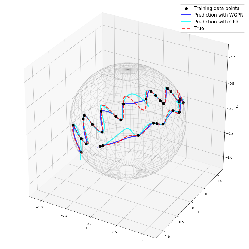
This work focuses on the implementation in the python library Geomstats of wrapped Gaussian process regression on Riemannian manifolds. This work won the second prize from the ICLR Computational Geometry &Topology Challenge associated to the ICLR 2022 Workshop on Geometrical and Topological Representation Learning. The challenge and its submissions are summarized in the white paper 23 and the code is available on the challenge repository.
7.3.10 Geometries of covariance and correlation matrices
This work was funded by the ERC grant Nr. 786854 G-Statistics and ANR UCAJEDI ANR-15-IDEX-01.
Keywords:
Participants: Yann Thanwerdas [Correspondant], Xavier Pennec.
In 28, 47, we investigated families of Riemannian and stratified geometries on covariance and correlation matrices.
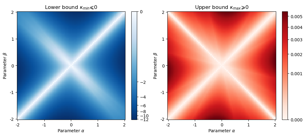
For SPD matrices, we use the principles of deformed metrics and balanced metrics to introduce Mixed-Power-Euclidean metrics which encompass the Euclidean, affine-invariant, log-Euclidean and BKM metrics. We also relate these MPE metrics to the -divergences of Information Geometry and we compute the curvature (see bounds on Figure 21). This work was published in 28. Furthermore, we characterized all the continuous metrics invariant by O(n) by means of three multivariate continuous functions, that encompass the classical metrics (log-Euclidean, BKM, power-Euclidean) for which we detail all the basic Riemannian operations 27.
On the space of full-rank correlation matrices, we showed that the curvature of the recently introduced quotient-affine metric is of non-constant sign and unbounded from above, which makes this geometry practically very complex. We introduce computationally more convenient Hadamard or even log-Euclidean metrics, along with their geometric operations. To recover the lost invariance under permutations, I define two new permutation-invariant log-Euclidean metrics, one of them being invariant under a natural involution on full-rank correlation matrices 29, 58.
For positive semi-definite symmetric (PSD) matrices, we studied the stratified Riemannian structure of the Bures-Wasserstein distance. A notable contribution was to compute the injection domain within each stratum and to characterize the length-minimizing curves between the strata of different ranks 57.
7.4 Computational Cardiology & Image-Based Cardiac Interventions
7.4.1 Causal data analysis of in-silico trials
This work has received funding from the European Union’s Horizon 2020 research and innovation programme under grant agreement No 101016496 (SimCardioTest).
Keywords:
Participants: Safaa Al Ali [Correspondant], Irene Balelli, Maxime Sermesant.
Within the context of SimCardioTest project, and specifically the "Data science & In-silico trials" workpackage (cf 22):
- Following the work of Llopis-Lorente et al. (2020) entitled In Silico Classifiers for the Assessment of Drug Proarrhythmicity, we propose to apply bayesian approaches & variational auto-encoder models in order to evaluate the tested set of biomarkers used to characterize drug-induced proarrhythmic risk in a probabilistic manner, and then develop an in-silico risk classifier.
- Next, we will investigate the explainability of the obtained results: causal discovery/inference & machine learning will play a major role.
7.4.2 Personalisation of cardiac models for the prediction of cardiac resynchronisation therapy response
This work has been supported by the French government through the National Research Agency (ANR) Investments in the Future 3IA Côte d’Azur (ANR-19-P3IA-000) and by Microport CRM funding.
Keywords:
Participants: Gaëtan Desrues [Correspondant], Serge Cazeau [Microport CRM], Thierry Legay [Microport CRM], Delphine Feuerstein [Microport CRM], Maxime Sermesant.
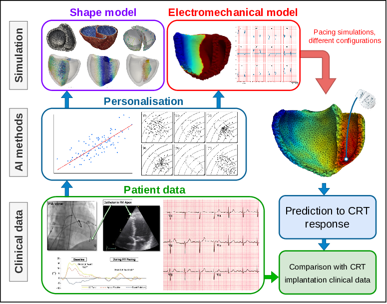
Patient-specific 3D electromechanical models can help in improving patient selection, therapy optimisation and interventional guidance. The aim of this project is to study the response to cardiac resynchronisation therapy, with the help of personalised cardiac models, see Figure 23. To this end, the following work has been realised:
- electrophysiological personalisation and stimulation experiments were compared to patient data undergoing CRT, presented at the EHRA 2022 conference,
- implementation of the cardiac mechanical model in the SOFA framework (anisotropy, hyperelastic properties of the tissue, sarcomere contraction, Lagrangian-based haemodynamic model, pericardium collision model),
- participation to the SOFA week 2022, annual workshop that gathers researchers from the SOFA community to present the latest developments around the software,
- parameters estimation for the cardiac mechanics using an AI-based surrogate model, presented at the SophIA summit 2022 conference,
- demonstration related to the patient-specific cardiac simulation pipeline, SimCardioTest 2022 workshop.
7.4.3 Predicting the risk of stroke from cardiac imaging
This work is funded by ERACoSysMed PARIS Project with Simula (Norway) and Hamburg University Hospital (Germany), and is in collaboration with the IHU Liryc of Bordeaux (France) and the Pompeu Fabra University in Barcelona (Spain).This work has been supported by the French government, through the 3IA Côte d’Azur Investments in the Future project managed by the National Research Agency (ANR) with the reference number ANR-19-P3IA-0002, as well as the IHU LIRYC with the reference number ANR-10-IAHU-04.
Keywords:
Participants: Josquin Harrison [Correspondant], Hubert Cochet [IHU Liryc, Bordeaux, France], Oscar Camara Rey [Pompeu Fabra University, Barcelona, Spain], Maxime Sermesant.
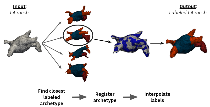
We aim at studying meshes of the Left Atrium for the prediction of Thrombosis in Atrial Fibrillation conditions, in order to predict the risk of stroke.
- We performed automatic labelling of pulmonary veins and appendage, leveraging on diffeomorphic registration (see fig.24)
- We extracted 80 markers on the labelled mesh and perform a statistical analysis on a cohort of patients.
- Using mesh processing and computational anatomy, we generated new dynamic meshes for a haemodynamic study on relevant markers
7.4.4 Deep Learning for Model Correction in Cardiac Electrophysiological Imaging
PhD funding from 3IA Côte d'Azur
Keywords:
Participants: Victoriya Kashtanova [Correspondant], Mihaela Pop, Maxime Sermesant, Andony Arrieula [Inria, Bordeaux], Mark Potse [Inria, Bordeaux], Ibrahim Ayed [LIP6, Paris], Patrick Gallinari [LIP6, Paris].
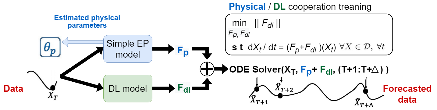
We present a fast physics-based deep learning framework to learn cardiac electrophysiology dynamics from data. It is based on the integration of a low-fidelity physical model and a learning component implemented here via neural networks (see Figure 25). We show experimentally in 43, 33, that this framework can reproduce the complex dynamics of the transmembrane potential and correctly identify the relevant physical parameters, even when only partial measurements are available.
The code and an example of the data used for APHYN-EP training are available on the official project page.
7.4.5 Interpratable Prediction of Post-Infarct Ventricular Arrhythmia using Graph Convolutional Network
Part of this work has been supported by the French Government, through the National Research Agency (ANR) 3IA Côte d'Azur (ANR-19-P3IA-0002), IHU Liryc (ANR- 10-IAHU-04), Université Côte d'Azur and Ecole Doctorale Sciences et Technologies de l'Information et de la Communication (EDSTIC).
Keywords:
Participants: Buntheng Ly [Correspondant], Sonny Finsterbach, Marta Nuñez-Garcia, Pierre Jaïs, Damien Garreau, Hubert Cochet, Maxime Sermesant.
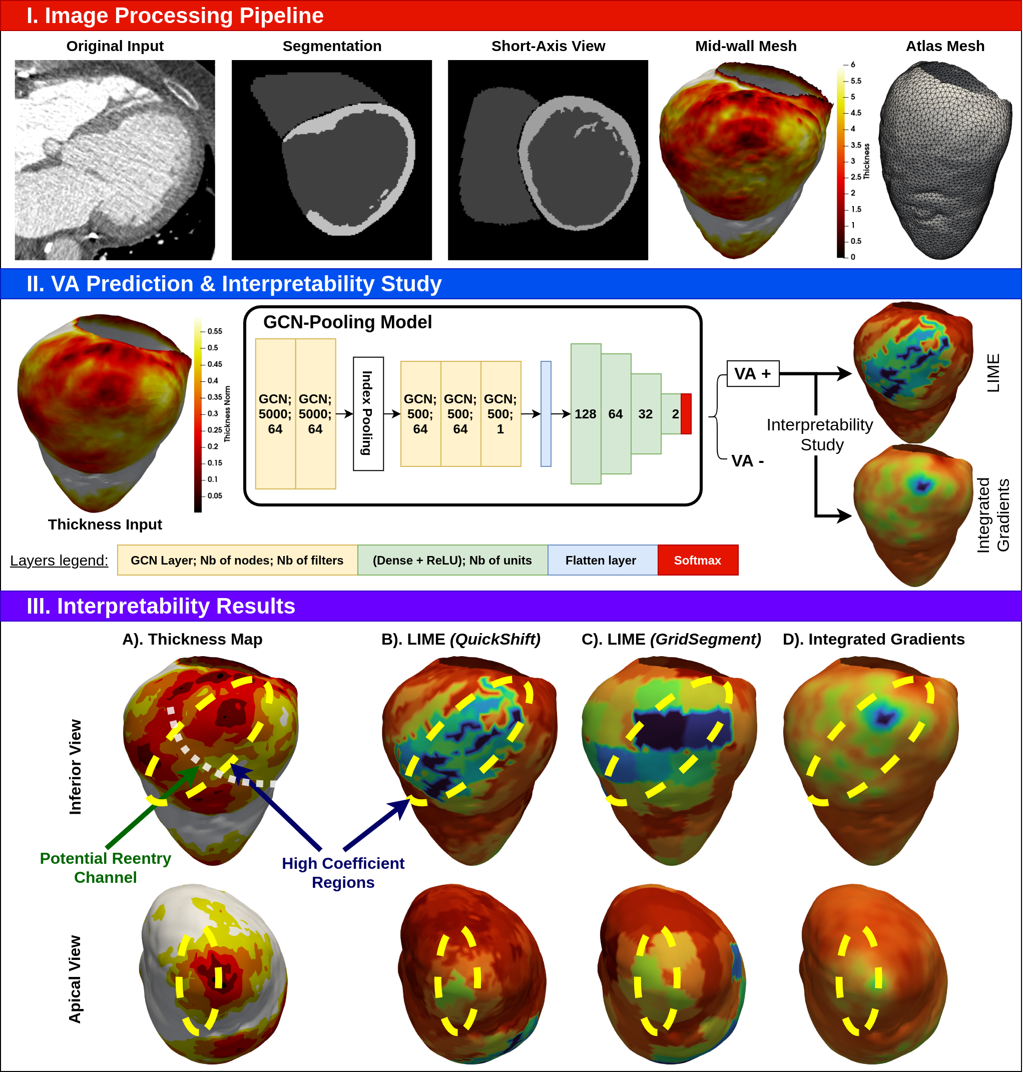
We proposed a fully automatic pipeline for the prediction of ventricular arrhythmia (VA) based of the left ventricular (LV) myocardial thinning observed in cardiac CT images 35. This pipeline leverages on the graph neural network (GCN-Pooling) and the interpretability methods (LIME & integrated gradients) to allow robust VA prediction, as well as the weakly supervised localisation of the arrhythmogenic substrate, Figure 26.
7.4.6 Automated CT quantification in presence of metal artifacts: wall thickness, and fat and calcification volume.
Keywords:
Participants: Marta Nuñez-Garcia [Correspondant], Buntheng Ly, Hubert Cochet, Maxime Sermesant.
-
Cardiac remodelling after myocardial infarction, including LV wall thinning, calcification and fat remodelling, is often responsible for Ventricular Arrhythmia. Artifacts caused by metallic implants such as implantable cardioverter defibrillators, artificial heart valves, etc. hamper automated tissue classification from CT images. We proposed an automated pipeline for the quantification of LV wall thickness, fat and calcification volume in big CT datasets affected by metal artifacts. Figure 27. 24.
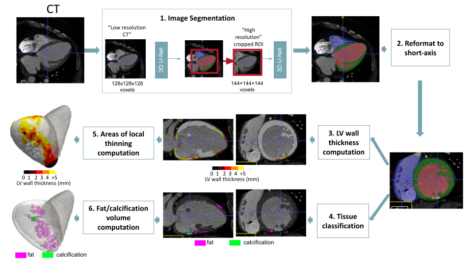
Automated CT quantification pipeline: 1. Image segmentation using the concatenation of 2 U-Nets; 2. Reformat to short-axis view to enable accurate wall thickness computation and data visualization in the preferred interpretation plane; 3. LV wall thickness computation; 4. Tissue classification (fat, calcification or healthy tissue). Estimation of optimal image-specific intensity thresholds using Gaussian Mixture Models, and distance maps to segment and exclude metal artifacts; 5. Scar size computation (abnormally thin LV wall); 6. Fat and calcification volume computation. Figure 27: Automated CT quantification pipeline: 1. Image segmentation using the concatenation of 2 U-Nets; 2. Reformat to short-axis view to enable accurate wall thickness computation and data visualization in the preferred interpretation plane; 3. LV wall thickness computation; 4. Tissue classification (fat, calcification or healthy tissue). Estimation of optimal image-specific intensity thresholds using Gaussian Mixture Models, and distance maps to segment and exclude metal artifacts; 5. Scar size computation (abnormally thin LV wall); 6. Fat and calcification volume computation. This lead to a new software for Automated fat and calcification quantification from CT images affected by metal artifacts.
7.4.7 Personalisation of electromechanical models in the context of cardiac resynchronization therapy.
This work has received funding from the European Union’s Horizon 2020 research and innovation programme under grant agreement No 101016496 (SimCardioTest).
Keywords:
Participants: Jairo Rodríguez Padilla [Correspondant], Gaëtan Desrues, Serge Cazeau, Thierry Legay, Delphine Feuerstein, Romano Setzu, Guilhem Faure, Maxime Sermesant.
In the context of CRT, we are currently working on the personalisation of our 3D electromechanical model in order to simulate what is observed in the clinic. This is, we start from a data base of patients that underwent CRT; with all this data (measurements from echocardiography and 12 lead ECG of patients) we generate a digital twin of the heart. Then we calibrate our model in order to simulate the realistic electromechanical activity observed by the cardiologists in their daily practice. Figure 28 exhibits an example of a simulated case.
- Publication in peer reviewed journal 25.
- Presentation in BoostUrCAreer Doctoriales 2022, entitled "Modeling of electromechanical activity of the heart: an application to cardiac resynchronization therapy", June 10th, 2022, Nice, France.
- Instructor in summer school "Heart modeling and numerical simulation", 20-24 June, 2022, at IHU-Liryc, Bordeaux, France.
- Presentation in 9th World Congress of Biomechanics, Taipei (July 10th - July 14th) 2022, entitled "Electromechanical output as function of action potential duration: a cardiac personalized simulation study."

7.4.8 Unsupervised Echocardiography Registration through Patch-based MLPs and Transformers
This project has been supported by the French government, through the 3IA Côte d’Azur Investments in the Future project managed by the National Research Agency (ANR) with the reference number ANR-19-P3IA-0002, and supported by the Inria Sophia Antipolis - Méditerranée, "NEF" computation cluster.
Keywords:
Participants: Zihao Wang, Yingyu Yang [Correspondant], Maxime Sermesant, Herve Delingette.
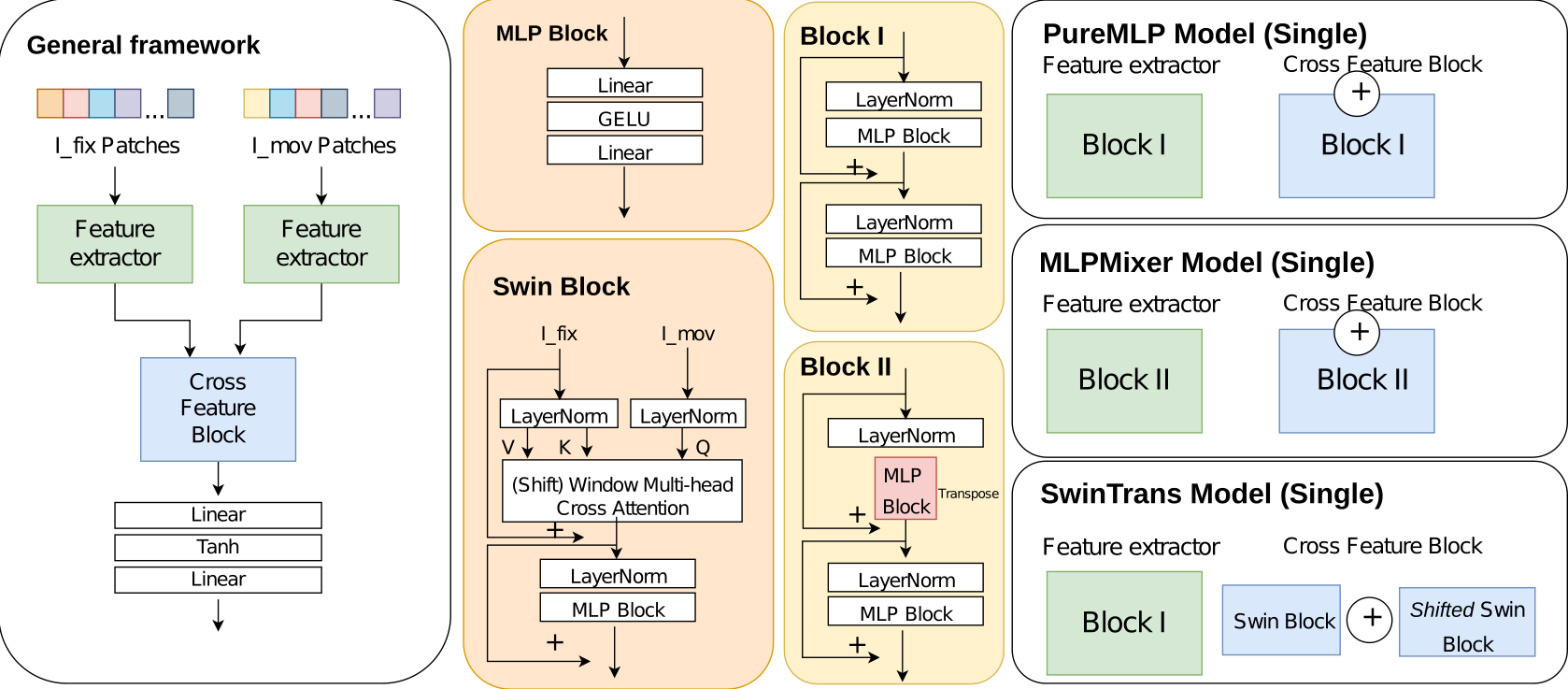
We proposed and compared three patch-based models with a popular CNN model. Our methods better preserve volume changes in terms of Jacobian determinants, thus generating robust registration fields with less unrealistic deformation. The results from experiments demonstrate that patch-based learning methods, with or without attention mechanisms, can lead to a high-quality unsupervised registration solution with adequate time and space complexity. Our codes are available on Gitlab and the work was published 39 in the STACOM'22 workshop.
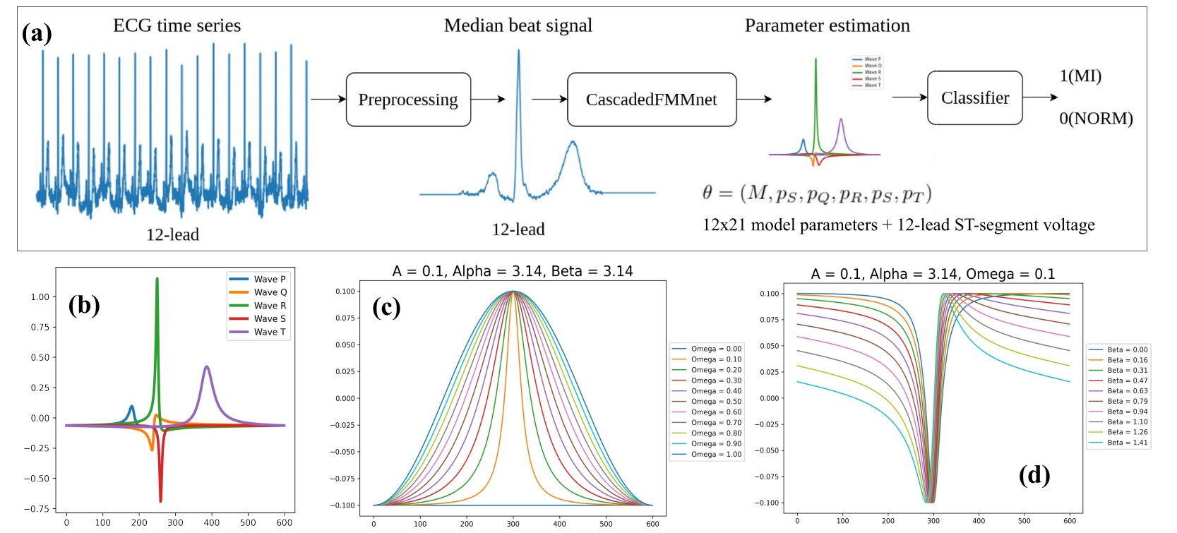
7.4.9 Explainable Electrocardiogram Analysis with Wave Decomposition: Application to Myocardial Infarction Detection
Keywords:
Participants: Yingyu Yang [Correspondant], Marie Rocher, Pamela Moceri, Maxime Sermesant.
In our study, we explore explainable ECG classification through explicit decomposition of single-beat (median-beat) ECG signal. In particular, every single-beat ECG sample is decomposed into five subwaves and each subwave is parameterised by a Frequency Modulated Moebius. Those parameters have explicit meanings for ECG interpretation. In stead of solving the optimisation problem iteratively which is time-consuming, we make use of an Cascaded CNN network to estimate the parameters for each single-beat ECG signal. Our preliminary results show that with appropriate position regularisation strategy, our neural network is able to estimate the subwave for P, Q, R, S, T events and maintain a good reconstruction accuracy (with R2 score 0.94 on test dataset of PTB-XL) in a unsupervised manner. Using the estimated parameters, we achieve very good classification and generalisation performance on myocardial infarction detection on four different datasets. The features of high importance are in accordance with clinical interpretations. Paper is accessible from HAL 40.
7.5 Multi-centric data and Federated Learning
7.5.1 Handling heterogeneity and missingness in Federated context
This project received financial support by the French government through the Agence Nationale de la Recherche (ANR), ref. num. ANR-19-CE45-0006.
Keywords:
Participants: Irene Balelli [Correspondant], Santiago Silva, Marco Lorenzi, Pierre-Alexandre Mattei [Inria, MAASAI Team], Aude Sportisse [Inria, MAASAI Team].
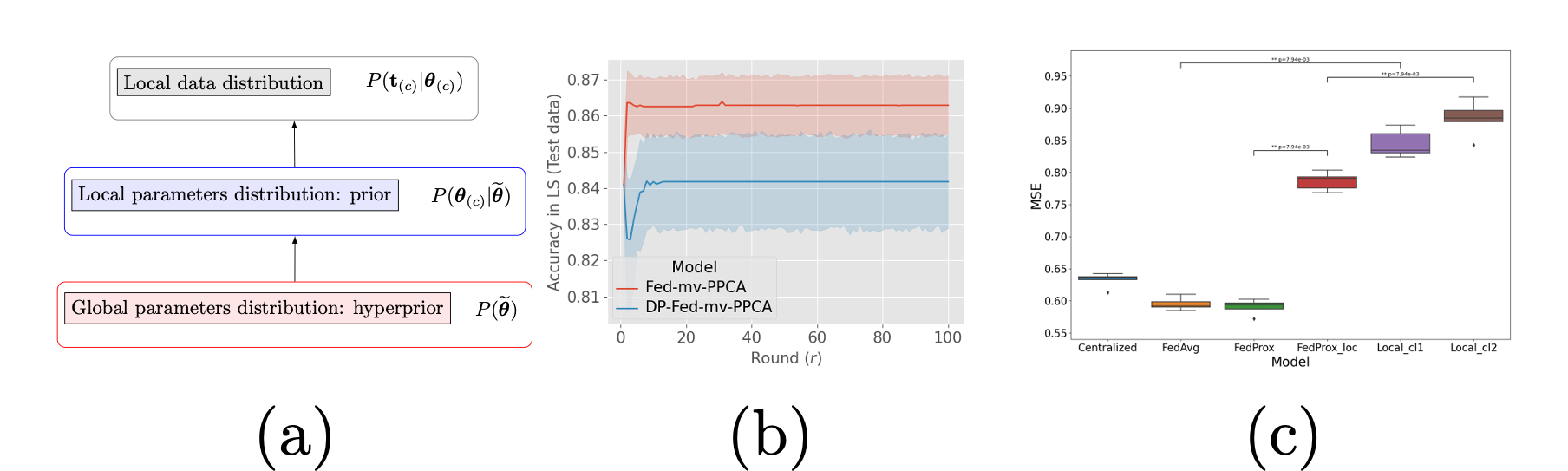
In 10 we addressed the issue of heterogeneous multi-views data assimilation across federated dataset through a novel Bayesian learning framework (Figure 31 (a)), which shows good reconstruction ability even in non-iid scenarios, allows to handle completely missing views, and provides an interpretable model of data variability and a tool for missing views imputation. In addition, our framework can be naturally coupled with differential privacy, ensuring theoretical privacy guarantees against data leakage (Figure 31 (b)).
We are further investigating the problem of missingness in a federated context, and we proposed a pipeline for data standardization and imputation of Missing At Random (MAR) entries in a federated context, based on the MIWAE method. We show improved performance in the imputation task when the model is trained coherently in a federated manner, with respect to local training and imputation (Figure 31 (c)).
- Bayesian hierarchical generative model for joint integration of multi-views heterogeneous decentralized data
- High quality data reconstruction even in the presence of missing views
- Formal privacy guarantees
- Handling of MAR data in a federated setting with MIWAE
- Improved robustness and generalizability with respect to local training
7.5.2 Real-world Deployment of Federated Learning in Biomedical Research Consortia with Fed-BioMed
Keywords:
Participants: Francesco Cremonesi [Correspondant], Sergen Cansiz, Yannick Bouillard, Marc Vesin, Marco Lorenzi.
Fed-BioMed is an open-source initiative aiming at enabling real-world deployment of federated learning in biomedical research. We investigated the architecture design of this framework to support real-world deployment in hospitals. Contrary to more generic approaches, Fed-BioMed's design is based on the specific requirements linked to its field of application:
- providing robust and easy-to-use tools for the governance of sensitive personal data and the federated training of ML models on these data;
- supporting seamless integration with biomedical data and healthcare interoperability standards, and supporting deployment in heterogeneous hospital infrastructures;
- maximising interactivity for data scientists and clinical researchers, enabling fast turnaround times for the development of ML algorithms;
- guaranteeing security from cyber attacks as well as gradient, model, and membership inference attacks.
Fed-BioMed is targeted at high trust, highly interactive research environments such as research consortia following the experiment workflow shown in Figure 32, but we also support deployment in a production mode. A publication is currently being redacted to outline the design space, goals, and current implementation of Fed-BioMed.
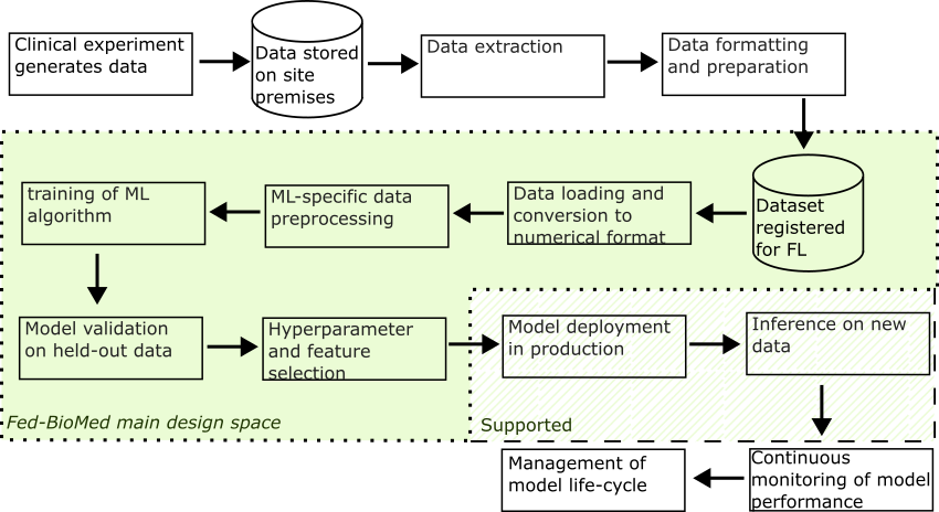
7.5.3 Reliability and Robustness of Federated Learning in Practical Applications
Keywords:
Participants: Yann Fraboni [Correspondant], Marco Lorenzi.
Federated learning (FL) enables a set of participants to jointly learn a global model without sharing their data.
- In 41, we introduce a novel formalization of FL aggregation scheme to prove the impact of a client sampling on FL convergence.
- We extend in 52 the aggregation scheme of 41 to account for asynchronous FedAvg, where the server creates a new global model whenever receiving a client's contribution.
- We develop in 53 SIFU, a novel unlearning framework for FL, that can measure a client's contribution in a trained model and unlearn it upon request with statistical guarantees.
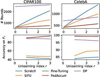
7.5.4 Multi-centric AI-based framework for prostate cancer patients management on active surveillance through robust federated learning
Project funded by 3IA Côte d'Azur
Keywords:
Participants: Lucia Innocenti [Correspondant], Marco Lorenzi, Michela Antonelli, Sebastien Ourselin.
Prostate cancer (PCa) is the second most frequently diagnosed cancer in men worldwide. Several studies have investigated AI-based computer-aided diagnosis (CAD) for PCa diagnosis, using medical images as input of AI systems. To ensure compliance with respect to data governance and privacy across centers we are relying on distributed learning.
- The final PhD goal is to develop a robust AI-based framework for AS patient selection and monitoring able to optimally adapt to the variability of real-world applications of AI in real clinical settings.
- The framework will be applied to a cohort of patients provided by the network of hospitals of Guy’s & St Thomas’ NHS Foundation Trust and by potential collaborating hospitals from Université Côte d’Azur.
- In this scenario, our first goal is to define the distributed environment. For this, we are working on an analysis of Multi-Centric AI-based frameworks in prostate segmentation applications, comparing FL with Consensus-Based ensembles.
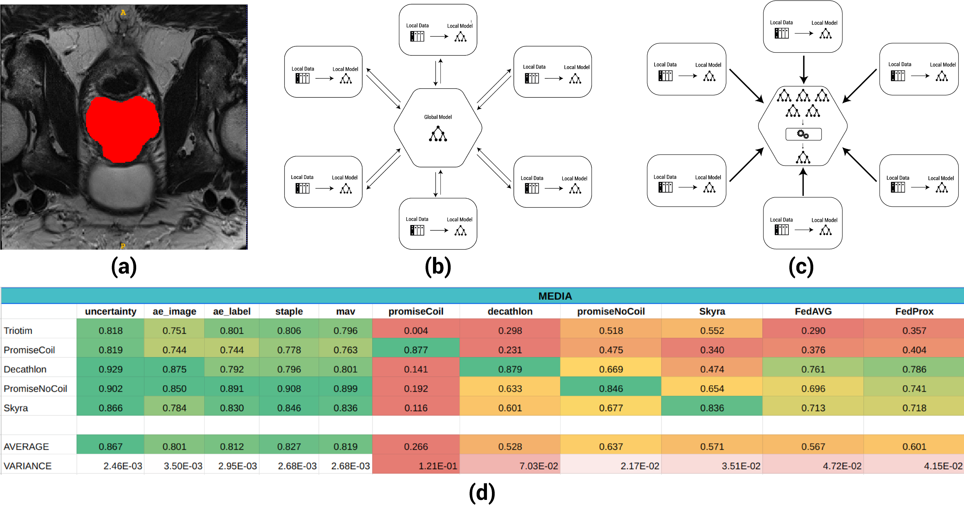
7.5.5 Federated ComBat (Fed-ComBat) for data harmonization on private decentralized data
H2020 / Marie Skłodowska-Curie Actions COFUND number 847579.
Keywords:
Participants: Santiago Silva [Correspondant], Marco Lorenzi, Andre Altmann.
Data harmonization is a crucial step in medical imaging data studies for removing bias between studies. Simultaneously, current concerns about patient's privacy and data sharing such as the GDPR and CCPA a paradigm shift is required towards secure and decentralized approaches for data modeling. ComBat is a widely used model that accounts for batch effects by relying on the estimation of fixed and random effects site-wise while preserving the covariate effects of interest (e.g., sex, age).
We propose a generalization on the ComBat model that allows to have a decentralized version relying on federated learning as well as the integration of nonlinear approaches for the preservation of covariate effects.
This work is in the process of being submitted as a journal publication in collaboration with the University College London (UCL).
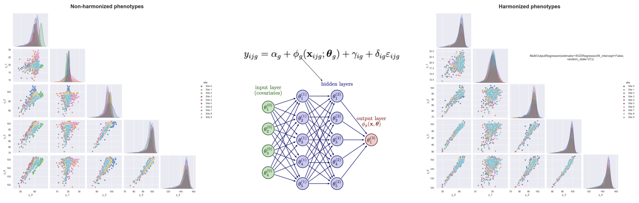
7.5.6 Data Structuration and Security in Large-Scale Collaborative Healthcare Data Analysis
This work has been supported by the French government, through the 3IA Côte d’Azur Investments.
Keywords:
Participants: Riccardo Taiello [Correspondant], Marco Lorenzi, Melek Önen, Olivier Humbert.
Our work aims to study the impact of privacy-preserving methods in Federated Learning for biomedical applications:
- We have proposed a privacy-preserving framework for medical image registration 37, Figure 36;
- We are implementing a Secure Aggregation scheme for Federated Learning in Fed-BioMed.
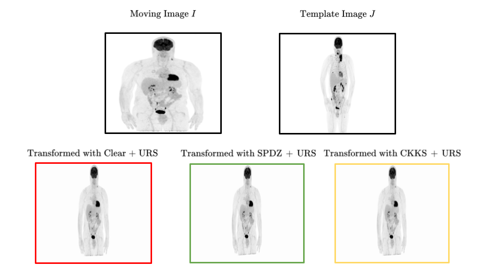
8 Bilateral contracts and grants with industry
8.1 Bilateral contracts with industry
8.1.1 Quantificare
Participants: Xavier Pennec, Hervé Delingette.
The company Quantificare is funding the PhD of Florent Jousse through a CIFRE grant, on the statistical analysis of shapes, deformations and appearance of anatomical surfaces for computer-aided dermatology and plastic surgery. The primary purpose is to model complex face deformations such as natural aging, facial expressions, surgical interventions and posture motions.
8.1.2 Koelis
Participants: Nicholas Ayache, Hervé Delingette.
The company Koelis participates in the thesis work of Zhijie Fang on the multimodal registration of MR and ultrasound images. The objective is to improve the accuracy the targeted biopsies inside the prostate.
8.1.3 Accenture Labs
Participants: Marco Lorenzi, Yann Fraboni.
Accenture Labs, Sophia Antipolis, France, is funding the CIFRE PhD work of Yann Fraboni, which aims at investigating the problem of bias and fairness in federated learning applications. Accenture also supports the development of the platform Fed-BioMed.8.1.4 MyDataModels
Participants: Marco Lorenzi.
MyDataModels, Sophia Antipolis, France, collaborates with Epione within the context of the postdoctoral project of Ghiles Reguig, aiming at developing a federated platform for the analysis of medical records in a network of hospitals in France.8.1.5 Microport CRM
Participants: Maxime Sermesant, Gaetan Desrues.
Microport CRM is a cardiac impantable device company, and is funding the PhD work of Gaëtan Desrues, which aims at using cardiac electromechanical modelling for cardiac resynchronisation therapy planning.8.1.6 ExactCure
Participants: Irene Balelli, Maxime Sermesant, Erwan Gaymard.
ExactCure, Nice, France, is funding the CIFRE PhD work of Erwan Gaymard, which aims at the development of novel methods for meta-modeling in pharmacometrics, to allow the co-integration of highly heterogeneous sources.8.1.7 Spin-off company inHEART
Participants: Maxime Sermesant.
inHEART1 is a spin-off of the Epione team and IHU Liryc founded in 2017. inHEART provides a service to generate detailed anatomical and structural meshes from medical images, that can be used during ablation interventions. inHEART received 2 awards, one from Aquitaine region and one i-LAB from the BPI. It raised 3.2 million euros in 2020. It currently employs 27 people. It is FDA and CE certified.
9 Partnerships and cooperations
9.1 International initiatives
9.1.1 International collaborations
-
Department of Computer Science, University of Copenhagen, DK
Xavier Pennec is collaborating with Pr. Stefan Sommer on stochastic and sub-Riemannian approached to statistics, in the framework of his ERC G-statistics and of the jointly advised PhD thesis of Morten Akhoj Pedersen.
-
DTU Compute, Technical University of Denmark, DK
Xavier Pennec and Anna Calissano are collaborating with the group of Aasa Feragen on neural networks in graph spaces, in particular in the context of the PhD of Andreas Hansen advised by A. Feragen at DTU. Maxime Sermesant is collaborating with Rasmus Paulsen on the ARTICHOKE project for arrhythmia prediction from imaging.
-
King's College London (KCL), London, UK
Marco Lorenzi is collaborator of the School of Biomedical Engineering & Imaging Sciences at King's College London. This collaboration focuses on the development of novel collaborative learning methods to improve prostate cancer detection in multicentric studies.
-
University College London (UCL), London, UK
Marco Lorenzi is collaborator of the COMputational Biology in Imaging and geNEtics (COMBINE) group within the Centre for Medical Image Computing (CMIC) of UCL. His collaboration is on the topic of spatio-temporal analysis of medical images and imaging-genetics, with special focus on brain imaging analysis and biomarker development.
-
Laboratory of Neuroimaging of Aging (LANVIE), Faculty of Medicine, Geneva University Hospitals (HUG)
Marco Lorenzi collaborates with the LANVIE laboratory led by Prof. Giovanni B. Frisoni. The collaboration consists in developing and translating novel approaches for disease progression modeling in neurodegenerative disorders, such as Alzheimer's disease.
-
Imaging Genetics Center (IGC),
University of Southern California (USC), CA, USA and Illinois Institute of Technology (IIT, IL, USA) Marco Lorenzi is currently collaborator of IGC and IIT for the investigation of the complex relationship between brain atrophy and genetics in Alzheimer's disease, in particular for demonstrating the effectiveness of multivariate statistical models in providing a meaningful description of the relationship between genotype and brain phenotype.
-
Laboratory of Physics of Fluids, University of Twente, NL
Herve Delingette is collaborating with Assistant Professor Guillaume Lajoinie, on the topics of Deep Learning for ultrasound imaging in the framework of the BoostUrCareer Cofund program and the thesis of Hari Sreedhar.
9.1.2 Participation in other International Programs
Project MDPARK, funded by Michael J. Fox Fundation
Participants: Marco Lorenzi.
-
Title:
Multimodal Dynamic Modeling and Prediction of Parkinsonian Symptom Progression
-
Partner Institution(s):
- Illinois Institute of Technology, USA
- Amsterdam University Medical Center, The Netherlands
- University of Southern California, USA
- Northwestern University School of Medicine, USA
- Date/Duration: 2022-2026
-
Additionnal info/keywords:
Connectomics, Disease models, Network analysis, Neuroimaging
9.2 International research visitors
9.2.1 Visits of international scientists
Hervé Lombaert
-
Status
Associate Professor of Computer Engineering
-
Institution of origin:
ETS Montreal
-
Country:
Canada
-
Dates:
Jan-Feb 2022
-
Context of the visit:
Collaboration with ETS Montreal, Canada
-
Mobility program/type of mobility:
Research stay
Alexander Panfilov
-
Status
Full Professor of biophysics
-
Institution of origin:
Ghent University
-
Country:
Belgium
-
Dates:
Sept. 2022
-
Context of the visit:
Collaboration with Ghent University, Belgium
-
Mobility program/type of mobility:
Research stay
Andreas Hansen
-
Status
PhD student
-
Institution of origin:
Technical University of Denmark
-
Country:
Denmark
-
Dates:
Oct-Dec 2022
-
Context of the visit:
Collaboration with A. Feragen's group at DTU.
-
Mobility program/type of mobility:
Research stay
Federica Cruciani
-
Status
PhD student
-
Institution of origin:
University of Verona
-
Country:
Italy
-
Dates:
May-Sept 2022
-
Context of the visit:
Collaboration with Prof. G. Menegaz, University of Verona.
-
Mobility program/type of mobility:
Research stay
9.3 European initiatives
9.3.1 H2020 projects
ERC G-Statistics
-
Title:
Geometric Statistics
-
Type:
ERC
-
Program:
H2020
-
Duration:
2018-2024
-
Inria contact:
Xavier Pennec
-
Coordinator:
Inria
-
Summary:
G-Statistics aims at exploring the foundations of statistics on non-linear spaces with applications in the Life Siences. Invariance under gauge transformation groups provides the natural structure explaining the laws of physics. In life sciences, new mathematical tools are needed to estimate approximate invariance and establish general but approximate laws. Rephrasing Poincaré: a geometry cannot be more true than another, it may just be more convenient, and statisticians must find the most convenient one for their data. At the crossing of geometry and statistics, G-Statistics aims at grounding the mathematical foundations of geometric statistics and to exemplify their impact on selected applications in the life sciences. So far, mainly Riemannian manifolds and negatively curved metric spaces have been studied. Other geometric structures like quotient spaces, stratified spaces or affine connection spaces naturally arise in applications. G-Statistics will explore ways to unify statistical estimation theories, explaining how the statistical estimations diverges from the Euclidean case in the presence of curvature, singularities, stratification. Beyond classical manifolds, particular emphasis will be put on flags of subspaces in manifolds as they appear to be natural mathematical object to encode hierarchically embedded approximation spaces. In order to establish geometric statistics as an effective discipline, G-Statistics will propose new mathematical structures and characterizations of their properties. It will also implement novel generic algorithms and illustrate the impact of some of their efficient specializations on selected applications in life sciences. Surveying the manifolds of anatomical shapes and forecasting their evolution from databases of medical images is a key problem in computational anatomy requiring dimension reduction in non-linear spaces and Lie groups. By inventing radically new principled estimations methods, we aim at illustrating the power of the methodology and strengthening the “unreasonable effectiveness of mathematics” for life sciences.
ERC ECSTATIC
-
Title:
Electrostructural Tomography – Towards Multiparametric Imaging of Cardiac Electrical Disorders
-
Type:
ERC
-
Program:
H2020
-
Duration:
2017 - 2022
-
Inria contact:
Maxime Sermesant
-
Coordinator:
U. Bordeaux
-
Summary:
Cardiac electrical diseases are directly responsible for sudden cardiac death, heart failure and stroke. They result from a complex interplay between myocardial electrical activation and structural heterogeneity. Current diagnostic strategy based on separate electrocardiographic and imaging assessment is unable to grasp both these aspects. Improvements in personalized diagnostics are urgently needed as existing curative or preventive therapies (catheter ablation, multisite pacing, and implantable defibrillators) cannot be offered until patients are correctly recognized. ECSTATIC aims at achieving a major advance in the way cardiac electrical diseases are characterized and thus diagnosed and treated, through the development of a novel non-invasive modality (Electrostructural Tomography), combining magnetic resonance imaging (MRI) and non-invasive cardiac mapping (NIM) technologies. The approach will consist of: (1) hybridising NIM and MRI technologies to enable the joint acquisition of magnetic resonance images of the heart and torso and of a large array of body surface potentials within a single environment; (2) personalising the inverse problem of electrocardiography based on MRI characteristics within the heart and torso, to enable accurate reconstruction of cardiac electrophysiological maps from body surface potentials within the 3D cardiac tissue; and (3) developing a novel disease characterisation framework based on registered non-invasive imaging and electrophysiological data, and propose novel diagnostic and prognostic markers. This project will dramatically impact the tailored management of cardiac electrical disorders, with applications for diagnosis, risk stratification/patient selection and guidance of pacing and catheter ablation therapies. It will bridge two medical fields (cardiac electrophysiology and imaging), thereby creating a new research area and a novel semiology with the potential to modify the existing classification of cardiac electrical diseases.
SimCardioTest
-
Title:
Simulation of Cardiac Devices & Drugs for in-silico Testing and Certification
- Link:
-
Duration:
2021 - 2024
-
Partners:
- Universidad Pompeu Fabra (UPF), Spain
- Institut national de recherche en Informatique et Automatique(Inria), France
- Virtual Physiological Human Institute for Integrative Biomedical Research vzw (VPH Institute), Belgium
- Sorin CRM SAS (Livanova), France
- ExactCure, France
- Simula Research Laboratory AS, Norway
- Université de Bordeaux (UBx), France
- Boston Scientific Scimed INC, United States
- Universitat Politecnica de Valencia (UPV), Spain
- InSilicoTrials Technologies S.P.A. (InSilicoTrials), Italy
-
Inria contact:
Maxime Sermesant
-
Coordinator:
Inria
-
Summary:
Despite massive investment in healthcare, huge R&D cost increase and regulatory pathway complexity hamper tremendously commercialisation of new devices & medicines, putting patient populations at risk of not receiving adequate therapy. At the same time, outside healthcare, computer modelling and simulation (CM&S) is precisely recognised to increase speed & agility while reducing costs of development. CM&S can create scientific evidence based on controlled investigations including variability, uncertainty quantification, and satisfying demands for safety, efficacy & improved access.
Cardiac modelling has dramatically gained maturity over the last decades, with personalisation to clinical data enabling validation. We selected a number of cardiac devices and medicines where CM&S is mature enough and that represent the most common cardiac pathologies, to demonstrate a standardised and rigorous approach for in-silico clinical trials.
SimCardioTest will bring a disruptive innovation by creating an integrated and secure platform standardising & bridging model simulations, in-silico trials, and certification support. This environment will go beyond the state-of-the-art in computational multi-physics & multi-scale personalised cardiac models. Diseased conditions and gender/age differences will be considered to overcome clinical trials limitations such as under-representation of groups (e.g. women, children, low socio-economic status). Advanced big data, visual analytics & artificial intelligence tools will extract the most relevant information.
It is critical that Europe demonstrates its capacity to leverage in-silico technology in order to be competitive in healthcare innovation. SimCardioTest exploitation aims at delivering a major economic impact on the European pharmaceutical and cardiac devices industry. It will accelerate development, certification and commercialisation, and will produce a strong societal impact contributing to personalised healthcare.
inEurHeart
-
Title:
inEurHeart: AI, Digital Twin & Clinical Trial for a Disruption in Catheter Ablation
- Link:
-
Duration:
2022 - 2024
-
Partners:
- Institut national de recherche en Informatique et Automatique(Inria), France
- Rotterdam University, Netherlands
- Inserm, France
- CHU Bordeaux, France
- inHEART, France
- Université de Bordeaux (UBx), France
-
Inria contact:
Maxime Sermesant
-
Coordinator:
Inria
-
Summary:
inEurHeart is an innovation project in Artificial Intelligence, Digital Twin & a Clinical Trial for a Disruption in Catheter Ablation for Ventricular Tachycardia, making ablation therapy accessible to most patients.
This project is a collaborative project between 5 organizations in France and Netherlands funded by EIT Health – the European Institute of Innovation and Technology, co-funded by the European Union. This project will exemplify how the academic-industrial relationships can be fostered and can lead to drastic changes in clinical practice.
EIT Health provides a unique opportunity to transfer Artificial Intelligence tools to enable the scale-up phase, and to validate the technology through a randomised clinical trial.
9.4 National initiatives
Consulting for Industry
- Nicholas Ayache has joined the Scientific Advisory Board of Caranx Medical in Oct 2021.
- Marco Lorenzi was a scientific consultant for the company MyDataModels (Sophia Antipolis).
- Maxime Sermesant is a scientific advisor for the company inHEART (Bordeaux).
Institute 3IA Côte d'Azur
-
The 3IA Côte d'Azur 3ia.univ-cotedazur.eu/ is one of the four "Interdisciplinary Institutes of Artificial Intelligence" that were created in France in 2019. Its ambition is to create an innovative ecosystem that is influential at the local, national and international levels, and a focal point of excellence for research, education and the world of AI.
-
Epione is heavily involved in this institute since 5 permanent researchers (N. Ayache, H. Delingette, M. Lorenzi, M. Sermesant and X.Pennec) are chair holders in this institute, and N. Ayache serves as scientific director. H. Delingette and N. Ayache are members of its scientific committee.
Funded projects
- Hervé Delingette is among the main investigators of the DAICAP project (2020-2022, 300k€) selected by the Health Data Hub, the Grand Défi « Amélioration des diagnostics médicaux par l’Intelligence Artificielle », and Bpifrance in July 2020. That project aims to develop an algorithm able to produce a standardised MRI scan report to improve the early detection of prostate cancer.
- Marco Lorenzi is principal investigator of the ANR JCJC project Fed-BioMed (2020-2023). He is co-PI of the project FEDERATED-PET (2022-2026), with Prof. Olivier Humbert (CAL, Nice). The project is funded by the Institut National du Cancer (INCa), and aims at developing the first French federated learning infrastructure in a network of hospitals from the Unicancer consortium. Marco Lorenzi also collaborated with the company Owkin on the development of the software platform Flamby, for simulation of federated learning in clinical dataset 38. The work was presented at the NeurIPS conference 2022 (Dataset and Benchmark track).
Collaboration with national hospitals
The Epione project team collaborates with the following 3 French IHU (University Hospital Institute): the IHU-Strasbourg (Pr J. Marescaux and L. Soler) on image-guided surgery, the IHU-Bordeaux (Pr M. Haïssaguere and Pr P. Jaïs) on cardiac imaging and modeling and the IHU-Pitié Salpétrière (Dr. O. Colliot and Pr. B. Stankoff) on neuroimaging.
The Epione project team is involved in the following research projects with the Assistance Publique des Hôpitaux de Paris (AP-HP) : NHANCE project on abdominal ultrasound image analysis with Dr Anne-Laure Rousseau (Hospital St-Louis), PAIMRI project on prostate cancer detection with Pr Raphaele Renard-Penna (Hospital La Pitié Salpêtrière), CLARITI project on PET-CT anomaly detection with Pr Florent Besson (Hospital Kremlin Bicêtre), PET-CT lesion detection with Dr Paul Blanc-Durand (Hospital Henri Mondor).
We also have long term collaborations with the CHU Nice, the Centre Antoine Lacassagne of Nice, and the Hospital Lenval of Nice.
9.5 Regional initiatives
- Marco Lorenzi and Hervé Delingette received funding for a PhD salary from UCA under the European Program BoostUrCareer (Marie Sklodowska-Curie agreement 847581).
10 Dissemination
Participants: Nicholas Ayache, Irene Balelli, Marco Lorenzi, Hervé Delingette, Xavier Pennec, Maxime Sermesant.
10.1 Promoting scientific activities
10.1.1 Scientific events: organisation
General chair, scientific chair
- X. Pennec was a member of the scientific committee of the Geometry and Statistics in Data Sciences thematic quarter program at Institut Henri Poincaré, Paris, Sep-Dec. 2022.
- M. Sermesant was a co-chair of the MICCAI 2022 Workshop Statistical Atlases and Computational Models of the Heart (STACOM 2022), which was held virtually in September 2022.
Member of the organizing committees
- M. Lorenzi was organizer of the first workshop on Trustworthy AI in Medical Imaging, which was held virtually in MICCAI 2022 (Singapore).
10.1.2 Scientific events: selection
Reviewer
- H. Delingette was a reviewer for the MICCAI Conference (MICCAI'22), the International Conference on Learning Representations (ICLR 2023), the Medical Imaging with Deep Learning )MIDL2022), the NeurIPS conference (NeurIPS 2022), the International Symposium on Biomedical Imaging (ISBI'22).
- M. Lorenzi was area chair of MICCAI 2022, and chair of the SophIA Summit 2022 (Sophia Antipolis).
10.1.3 Journal
Member of the editorial boards
- N. Ayache is the co-founder and the Co-Editor in Chief with J. Duncan (Professor at Yale) of Medical Image Analysis journal. This scientific journal was created in 1996 and is published by Elsevier.
- N. Ayache is a member of the editorial board of the following journal: Journal of Computer Assisted Surgery (Wiley).
- H. Delingette is a member of the editorial board of the journal Medical Image Analysis (Elsevier).
- M. Lorenzi is member of the editorial board of the journal Medical Image Analysis (Elsevier). He is also member of the Board of Statisticians of the Journal of Alzheimer’s Disease (IOS Press).
- X. Pennec is a member of the editorial board of the journal Medical Image Analysis (MedIA, Elsevier), of the International Journal of Computer Vision (IJCV, Springer), and of the Journal of Mathematical Imaging and Vision (JMIV, Springer).
- I. Strobant is editorial coordinator for Medical Image Analysis, Elsevier (since october 2001).
Reviewer - reviewing activities
- I. Balelli was a reviewer for the following journals: Vaccine (Elsevier), Medical Image Analysis (Elsevier).
- X. Pennec was a reviewer for the following journals: Annals of Statistics (AoS), Int. J. of Computer Mathematics: Computer Systems Theory, Handbook of Statistics, PLOS One.
- H. Delingette was a reviewer for the following journals: Medical Image Analysis (Elsevier), IEEE Transactions on Pattern Analysis and Machine Intelligence, Math in Action.
- M. Lorenzi was a reviewer for the following journals: Journal of Alzheimer's Disease, Medical Image Analysis, IEEE Transactions on Medical Imaging, NeuroImage, International Journal of Computer Vision.
- M. Sermesant was a reviewer for the following journals: Journal of Machine Learning Research, Journal of the American College of Cardiology, IEEE Transactions on Medical Imaging, IEEE Transactions on Biomedical Engineering, Medical Image Analysis and Computers in Biology and Medecine.
10.1.4 Invited talks
- N. Ayache was a keynote speaker at Global Congress on AI in Montreal in May 2022, and a keynote speaker at the Inserm Conference on the History of Medical Imaging in Sept 2022. He was invited at a roundtable for the 10th Anniversary of the SIF (Société Informatique de France) in June 2022. He was also a keynote speaker at the opening of the Winter school on AI for Medical Imaging organized by the Health Data Hub in January 2022 and at the opening of the University Diploma on AI at the Nice University Medical School in Nov 2022.
- H. Delingette gave invited talks at the GDR ISIS (Information, Signal, Image et ViSion) meeting on "Deep learning with weak or few labels in medical image analysis" in Paris, on Feb 1st, at the Fysica 2022 conference at Univ. Twente, NL on April 22nd, at the Bernouilli Lab Webinar on May 19th, at the Technical University of Munich (TUM) Distinguished Lecture Series on AI & Healthcare on May 23rd, at the 2022 MOMI (Le monde des mathématiques industrielles) workshop, at the BoostUrCAreer Doctoriales 2022 meeting in Nice on June 12th, at the OCA-3IA joint meeting on September 12th, at the symposium "AI and Medicine" at the National Academy of Medecine in Paris on October 20th, at the Eurobiomed meeting on November 22nd, at the 3IA Côte d'Azur scientific days on November 15th.
- X. Pennec was a keynote speaker of the Workshop on Geometry and Machine Learning (GAML), Heidelberg, 11-13 July 2022. He gave invited talks at the Minisymposium on Advances in Shape Analysis of the SIAM Imaging Science Conf., Berlin, March 21-24, 2022 and at the Mathematical Imaging and Surface Processing workshop, Mathematisches Forschungsinstitut Oberwolfach (DE), August 21-27, 2022.
- M. Lorenzi was invited speaker at the first Workshop on Collaborative Learning at MBZUAI (Abhu Dhabi), organized by M. Jordan and S. Karimireddi (UC Berkeley). He was invited speaker at the QuanTIM Seminar (Marseille Université), at the ICM workshop on Multimodal Data Analysis (Pitié Salpêtrière, Paris), and at the 2022 MOMI (Le monde des mathématiques industrielles) workshop.
10.1.5 Scientific expertise
- H. Delingette is a member of the scientific committee of the institute 3IA Côte d'Azur and was reviewer of the funding agency ANR (Agence Nationale de la Recherche, France), of the funding agency ANRT (Association Nationale Recherche Technologie), of the European Research Council (ERC).
- M. Lorenzi was reviewer for the funding agency ANR (Association Nationale Recherche), and Fonds de recherche du Québec.
10.1.6 Research administration
- Xavier Pennec is co-director of the Ecole doctorale STIC of Université Côte d'Azur. He is a member of the committee of EDSTIC, of the Doctoral follow-up Committee (CSD) at Inria Sophia Antipolis, and participated to the PhD fellowship granting committees of EDSTIC and CORDI at Inria.
- Xavier pennec is a member of the "Comité de la Recherche Biomédicale en Santé Publique (CRBSP)" of the Nice University Hospital (CHU). At the University Côte d'Azur / UCA JEDI, he is a member of the executive committee of the Academy 4 (Living systems Complexity and diversity), of the Scientific committee of the Academy 2 (Complex Systems), and of the Advanced Research Program Committee. He is a member of the Evaluation Committee of Inria, and a member of its board since 2022. He participated in 2022 to the promotion commitees for CRHC, DR1, DR0, to the RIPEC-C3 committe, to the DR2 recruitment committee, and organized the evaluation of 6 Inria research teams.
- Hervé Delingette is a representive of Inria at the Federation Hospitalo-Universitaire Oncoage led by the CHU Nice. He is one of the 2 scientific directors of the IdEx program UCA JEDI under the direction of the IdEX vice-president of the Université Côte d'Azur. He is an administrator and a member of the scientific committee of the Groupement de Coopération Sanitaires (GCS) CARES involving the Université Côte d'Azur and the 3 local hospitals (CHU Nice, Centre Antoine Lacassagne, Fondation Lenval). He is the president of the CUIV (Commission UCA Innovation et Valorisation) which ensures the coordination among the technology transfer and innovation departments of IdEx members, of the evaluation of innovation and valorization projects. He is also a member of the research and innovation committee organized by the employer union UPE06.
- Marco Lorenzi is member of the scientific committee of the AI4Health Winter School, and of the Colloque Français d'Intelligence Artificielle en Imagerie Biomédicale (IABM 2023).
- I. Balelli is in charge of the pedagogical orgization of the AI for Health track of the Data Science & AI Master, Univ. Côte d'Azur, France.
10.2 Teaching - Supervision - Juries
10.2.1 Teaching
- Master: H. Delingette and X. Pennec, Introduction to Medical Image Analysis, 21h course (28.5 ETD), Master 2 MVA, ENS Saclay, France.
- Master: M. Lorenzi and I. Balelli, Bayesian Learning, 30h course, Master Data Science, Univ. Côte d'Azur, France.
- Master : H. Delingette, 1h, Challenges in AI, University Diploma for AI in Healthcare, Univ. Côte d'Azur, France.
- Master: M. Lorenzi, Model Selection and Resampling Methods, 30h course, Master Data Science, Univ. Côte d'Azur, France.
- Master: I. Balelli, Modeling of biological systems, 10h ETD, Master 2 BIM, Univ. Côte d'Azur, France.
- Master: M. Nuñez Garcia, 6h lectures on 2D signal / image processing, 3D data representation and registration, master in Electromechanical Heart Diseases (IHU Liryc, Pessac, France).
- Licence: I. Balelli, Analysis and modeling: mathematics for biology, 20h ETD, Univ. Côte d'Azur, France.
- Licence: I. Balelli, Statistics and Modeling, 24h ETD, Univ. Côte d'Azur, France.
- M. Lorenzi and I. Balelli presented a one-day hands-on session on the software Fed-BioMed to the Winter School AI4Health (HDH and 3IA).
10.2.2 Supervision: defended PhDs
- Hind Dadoun, AI-Based Real Time Diagnosis of Abdominal Ultrasound, Université Côte d'Azur. Co-directed by Nicholas Ayache and Hervé Delingette. Defended on December 6th, 2022.
- Florent Jousse, Analyse statistique de forme, de déformations et d'apparences de surfaces anatomiques pour l'aide à la dermatologie et à la chirurgie plastique. CIFRE fellowship with Quantificare. Co-directed by Xavier Pennec and Hervé Delingette. Defended on September 1st, 2022.
- Buntheng Ly, Cardiac Image Analysis for Sudden Cardiac Death Prediction, Université Côte d'Azur. Co-directed by Maxime Sermesant and Hubert Cochet (IHU Liryc). Defended on December 20th, 2022.
- Yann Thanwerdas, Statistical Dimension Reduction in Non-Linear Manifolds for Brain Shape Analysis, Connectomics & Brain-Computer Interfaces, Université Côte d'Azur. Directed by Xavier Pennec. Defended on May 24th, 2022.
10.2.3 Supervision: ongoing PhDs
- Mikael Chelli, AI for Othopedic Surgery, Centre hospitalier universitaire de Nice. Started in August 2019. Co-directed by Nicholas Ayache, Hervé Delingette and Jean Chaoui (Imascap).(currently on break).
- Gaëtan Desrues, 3D electromechanical cardiac modelling for heart failure patients stratification and prediction of cardiac resynchronisation therapy response, Université Côte d'Azur. CIFRE fellowship with Microport CRM. Started in 2020. Directed by Maxime Sermesant.
- Zhijie Fang, Robust Multimodal Non rigid image registration based on machine learning, Université Côte d'Azur. Started in 2020. Co-directed by Hervé Delingette and Nicholas Ayache.
- Yann Fraboni, Bias in Federated Learning, Université Côte d'Azur.CIFRE fellowship with Accenture Labs. Started in 2020. Directed by Marco Lorenzi.
- Erwan Gaymard, Innovative mathematical methodologies in pharmacometric meta-modeling from highly heterogeneous sources, Université Côte d'Azur. CIFRE fellowship with ExactCure. Started in 2022. Co-directed by Irene Balelli and Maxime Sermesant.
- Lisa Guzzi, Automatic segmentation of the vascular system to enhance AI-based decision support system for peripheral artery disease, Université Côte d'Azur, 3IA felloship. Started in 2022. Directed by Hervé Delingette, Juliette Raffort-Lareyre
- Dimitri Hamzaoui, AI-Based Diagnosis of Prostate Cancer from Multiparametric MRI, Université Côte d'Azur. Started in 2020. Co-directed by Nicholas Ayache and Hervé Delingette.
- Josquin Harrison, Medical Imaging and learning for the prediction of strokes in the case of atrial fibrillation, Université Côte d'Azur. Started in 2020. Directed by Maxime Sermesant.
- Etrit Haxholli, Exploring latent dynamical models for failure prediction in time-series of high-dimensional and heterogeneous data, Université Côte d'Azur. Started in 2020. Directed by Marco Lorenzi.
- Lucia innocenti, Multi-centric AI-based framework for prostate cancer patients management on active surveillance through robust federated learning. 3IA PhD started in June 2022. Co-directed by Marco Lorenzi and Prof. Sebastien Ourselin (KCL, London).
- Victoriya Kashtanova, Learning Cardiac 3D Electromechanical Dynamics with PDE-based Physiological Constraints for Data-Driven Personalized Predictions in Cardiology, Université Côte d'Azur. Started in 2020. Co-directed by Maxime Sermesant and Patrick Gallinari (Sorbonne University, LIP6, Paris).
- Huiyu Li, Anonymisation and protection of medical data based on deep neural networks. Université Côte d'Azur. Started in 2020. Co-directed by Hervé Delingette and Nicholas Ayache.
- Elodie Maignan, Geometric learning of manifolds, Université Côte d'Azur. Started in 2020. Co-directed by Xavier Pennec and Alain Trouvé (ENS Paris-Saclay).
- Morten Pedersen, Lie group action, approximate invariance and Sub-Riemannian geometry in statistics, Université Côte d'Azur and University of Copenhagen. Started in 2020. Co-directed by Xavier Pennec and Stefan Sommer (Univ Copenhagen, DK).
- Santiago Smith Silva Rincon, Federated learning of biomedical data in large-scale networks of multicentric imaging-genetics information, Université Côte d'Azur. Started in 2020. Co-directed by Marco Lorenzi and Barbara Bardoni (INSERM)
- Hari Sreedhar, Learning-based detection and classification of thyroid nodules from ultrasound images, Université Côte d'Azur. Started in 2020. Co-directed by Hervé Delingette and Charles Raffaelli (CHU-Nice).
- Tom Szwagier, Rethinking statistical methods with Flags spaces, Université Côte d'Azur. Started in 2022. Directed by Xavier Pennec.
- Riccardo Taiello, Data Structuration and Security in Large-Scale Collaborative Healthcare Data Analysis, Université Côte d'Azur. Started in 2021. Co-supervised by Melek Önen (EURECOM) and Olivier Humbert (Centre Antoine Lacassagne).
- Paul Tourniaire, AI-based selection of imaging and biological markers predictive of therapy response in lung cancer, Université Côte d'Azur. Started in 2019. Co-directed by Nicholas Ayache and Hervé Delingette.
- Yingyu Yang, Artificial Intelligence for Automatic Cardiac Function Analysis: Multimodal and Biophysical Approach with Application to a Portable Imaging Device, Université Côte d'Azur. Started in 2020. Co-directed by Maxime Sermesant and Pamela Moceri (CHU-Nice)
10.2.4 Juries
- X. Pennec was PhD jury member for the thesis of Yann Thanwerdas (UCA, Thesis Director), of Florent Jousse (UCA, Thesis Director), of Clement Mantoux, (Institut Polytechnique de Paris, reviewer), of Pierre-Louis Antonsanti (Univ. Paris Cité, reviewer), and he was reviewer of the PhD of Felix Paul Ambellan (Frei Universtitat Berlin, Berlin).
- H. Delingette was PhD jury member for the thesis of Dingge Liang (Univ. Côte d'Azur, President), of Florent Jousse (Univ. Côte d'Azur, Thesis co-director), of Hind Dadoun (Univ. Côte d'Azur, co-director) and he was a reviewer of the PhD thesis of Tristan Jaouen (Université Claude Bernard - Lyon 1), and of Arnaud Boutillon (IMT Atlantique, Brest).
- M. Lorenzi was PhD jury member of Ugo Lecerf, Davit Gogolashvili, Simone Rossi (EURECOM, France).
10.3 Popularization
10.3.1 Articles and contents
- N. Ayache contributed an article on AI for medicine in the French Magazine "Pour la Science" entitled "La féé IA au chevet des malades" 61
10.3.2 Interventions
- N. Ayache was invited on French Radio "La matinale d'Europe 1" on the topic of AI for medicine, on 25 Nov 2022.
- I. Balelli participated to the 1 scientifique 1 classe: Chiche! initiative, to encourage scientific studies and explain research activities to high school students. 3 classes, Lycée Tocqueville, Grasse.
- Hari Shreedhar participated to the European Researchers' Night on Sept. 30th by presenting the fundamentals of ultrasound imaging to a general audience.
11 Scientific production
11.1 Major publications
- 1 miscClustered Sampling: Low-Variance and Improved Representativity for Clients Selection in Federated Learning.May 2021
- 2 articleLearning a Generative Motion Model from Image Sequences based on a Latent Motion Matrix.IEEE Transactions on Medical ImagingFebruary 2021
- 3 articleApplications of artificial intelligence in cardiovascular imaging.Nature Reviews CardiologyMarch 2021
- 4 articleTheoretically and computationally convenient geometries on full-rank correlation matrices.SIAM Journal on Matrix Analysis and Applications434December 2022, 1851-1872
- 5 articleBayesian Logistic Shape Model Inference: application to cochlear image segmentation.Medical Image AnalysisOctober 2021
11.2 Publications of the year
International journals
International peer-reviewed conferences
Conferences without proceedings
Scientific book chapters
Doctoral dissertations and habilitation theses
Reports & preprints
Other scientific publications
11.3 Other
Scientific popularization