Section: New Results
Medical Image Analysis
Spatial Decision Forests for MS Lesion Segmentation in Multi-Channel MR Images
Participants : Ezequiel Geremia, Nicholas Ayache, Olivier Clatz, Antonio Criminisi [MSR] , Ender Konukoglu [MSR] , Bjoern Menze [MIT] .
-
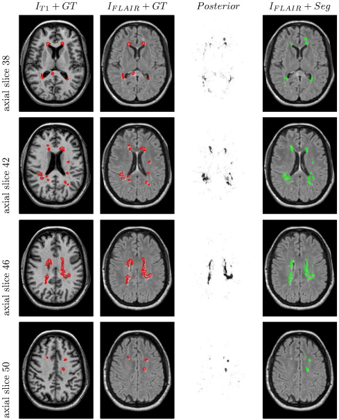
A new approach for MS lesions segmentation was proposed [33]
-
Random forest for automatic segmentation of MS lesions in 3D MR images
-
Features: multi-channel MR intensities, priors, long-range spatial context, symmetry
-
Quantitative evaluation shows significant improvement over the MICCAI Grand Challenge 2008
-
The automatically learned decision sequence mimics the state-of-the-art pipeline
-
Independent validation carried out by the MICCAI Challenge website 2008
-
Exhaustive analysis of the discriminative power of channels and features
-
Analysis of the influence of random forest's meta-parameters on the classification performance
|
Left Ventricle Segmentation from Cardiac 4D Cine MRI Sequences
Participants : Jan Margeta [Correspondant] , Ezequiel Geremia, Nicholas Ayache, Antonio Criminisi [MSR] .
This work was performed in collaboration with Microsoft Research and was partly supported through its PhD Scholarship Programme.
-
We extend the previous work for multiple sclerosis lession segmentation of Geremia et al. [33] for spatio-temporal cardiac images.
-
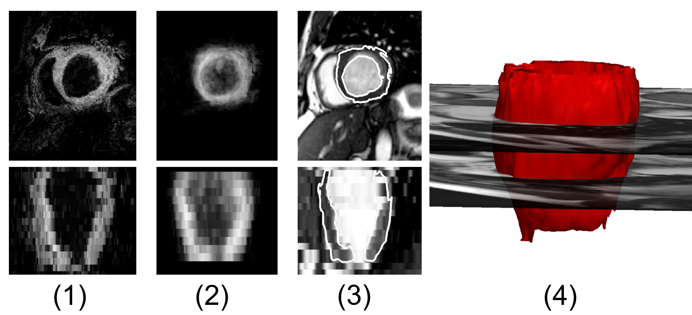
A fully automatic two layer left ventricle segmentation algorithm from 4D cardiac cine MRI sequences (See Fig. 2 ) was proposed for MICCAI STACOM LV segmentation challenge [63] using a random forest classification algorithm.
-
Spatio-temporal features are used in the random forest framework to learn the segmentation task without explicitely defining the segmentation rules.
-
Machine learning based MRI intensity standardization and pose normalization preprocessing pipeline was proposed to deal with diverse cardiac MRI datasets.
|
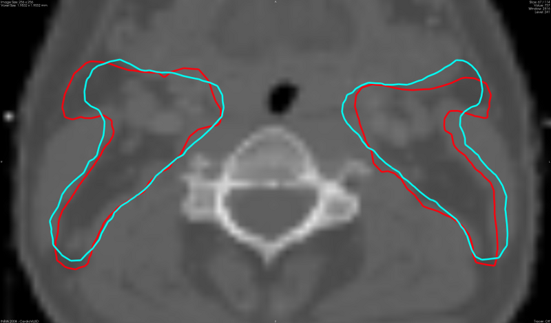
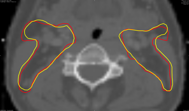
Design and use of anatomical atlases for automatic segmentation: application to radiotherapy of the head and neck region
Participants : Liliane Ramus [Correspondant] , Grégoire Malandain, Vincent Grégoire [UCL] , Juliette Thariat [CAL] .
This work is done in collaboration with DOSIsoft S.A., Centre Antoine Lacassagne (CAL) and Université Catholique de Louvain.
In the context of radiotherapy of the head and neck, we propose different strategies to design anatomical atlases and we compare their performances for automatic segmentation [28] :
-
We investigate average atlas construction, atlas stratification and patient-specific strategies based on the selection and fusion of the most appropriate atlases for each patient. We compared global, regional and local selection and fusion of the atlases.
-
We show that the proposed patient-specific strategies enable to significantly improve the quality of the automatic segmentation in comparison with average atlas strategies. Visual results are presented on figure 3 .
-
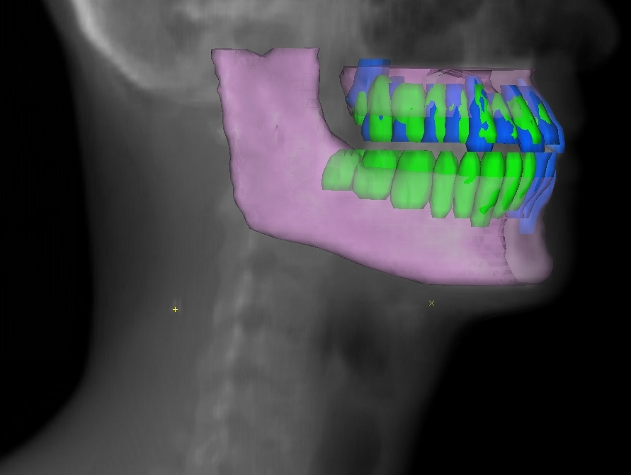
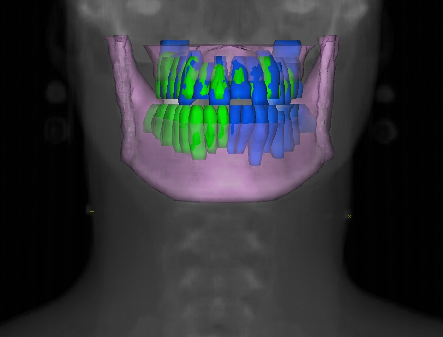
We evaluated the proposed algorithms in two different contexts: segmentation of the lymph node levels and the organs at risk for radiotherapy planning, and segmentation of the teeth for post-irradiation dental care management. Automatic segmentations of the teeth are shown on figure 4 .