Section: New Results
Modeling
Tracking Growing Axons in Fluorescent Microscopy Images
Participants : Huei Fang Yang, Florence Besse, Xavier Descombes.
This work has been done in collaboration with Caroline Medioni from iBV.
Analyzing how growing axons correctly reach their target neurons is essential for biologists to better understand the development of a nervous system. Analysis of the properties of axon growth requires detecting axonal tips and tracking their trajectories within complex and large data sets. When performed manually, the tracking task is arduous and time-consuming. To this end, we proposed a tracking method, based on the particle filtering technique, to follow the traces of axonal tips that appear as small bright spots in the fluorescent two-photon microscopy images exhibiting low signal-to-noise ratios (SNR) and complex background. Our tracking method uses multiple dynamic models in the proposal distribution to predict the positions of the growing axons. Moreover, it incorporates object appearance, motion characteristics of the growing axons, and filament information in the computation of the observation model. The integration of these three sources results in improved accuracy of recovered trajectories. The experimental results obtained from the microscopy images, presented in Figure 15 , showed that the proposed method can successfully estimate trajectories of growing axons, demonstrating its effectiveness even under the presence of noise and complex background.
|
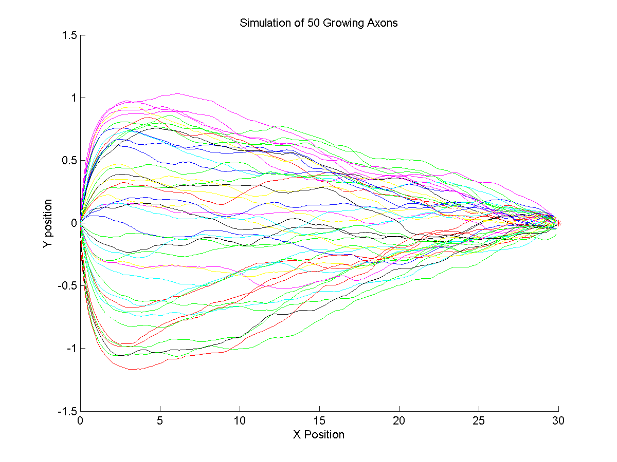
Trajectory Simulation of Growing Axons:
Participants : Huei Fang Yang, Florence Besse, Xavier Descombes.
This work has been done in collaboration with Caroline Medioni from iBV.
It is established in biology that axons reach their target cells in the developing nervous system by the guidance of molecular gradients. To better understand how growing axons react to the molecular cues, either attractant or repellent, we simulated the trajectories of growing axons using a mathematical model that investigates the effect of molecular gradients on the axon's growth angle. Figure 16 shows the simulated trajectories of 50 growing axons. The initial position of axons is , and the red point on the right denotes the target cell that secretes the attractant cue.