

Section: New Results
Interhemispheric Connectivity Characterizes Cortical Reorganization in Motor-Related Networks After Cerebellar Lesions
Participants : Fabrizio de Vico Fallani, Silvia Clausi, Maria Leggio, Mario Chavez, Miguel Valencia, Anton Giulio Maglione, Fabio Babiloni, Febo Cincotti, Donatella Mattia, Marco Molinari [Correspondant] .
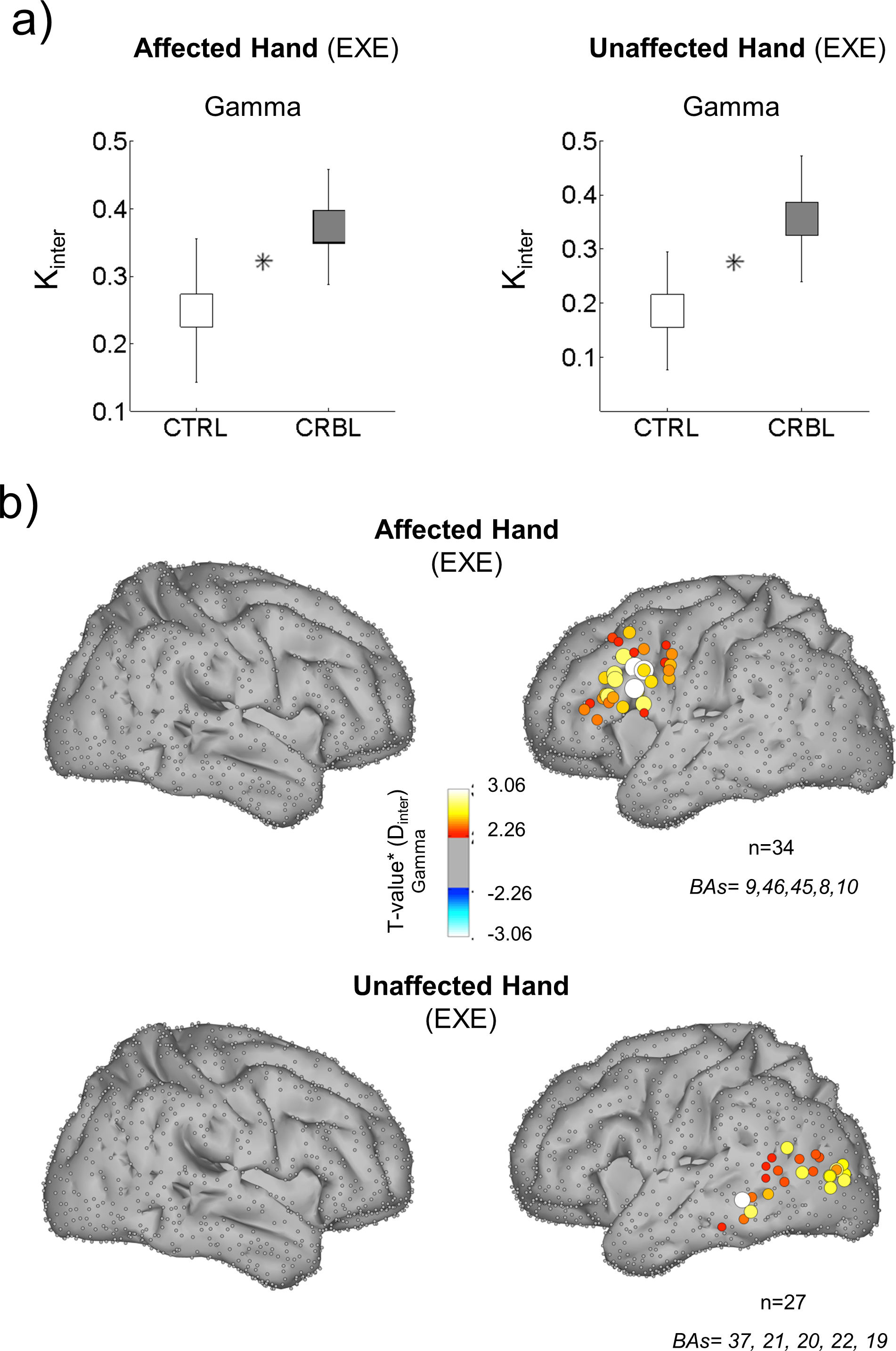
Although cerebellar-cortical interactions have been studied extensively in animal models and humans using modern neuroimaging techniques, the effects of cerebellar stroke and focal lesions on cerebral cortical processing remain unknown. In the present study, we analyzed the large-scale functional connectivity at the cortical level by combining high-density electroencephalography (EEG) and source imaging techniques to evaluate and quantify the compensatory reorganization of brain networks after cerebellar damage. The experimental protocol comprised a repetitive finger extension task by 10 patients with unilateral focal cerebellar lesions and 10 matched healthy controls. A graph theoretical approach was used to investigate the functional reorganization of cortical networks. Our patients, compared with controls, exhibited significant differences at global and local topological level of their brain networks. An abnormal rise in small-world network efficiency was observed in the gamma band (30-40 Hz) during execution of the task, paralleled by increased long-range connectivity between cortical hemispheres (Figure 4). Our findings show that a pervasive reorganization of the brain network is associated with cerebellar focal damage and support the idea that the cerebellum boosts or refines cortical functions. Clinically, these results suggest that cortical changes after cerebellar damage are achieved through an increase in the interactions between remote cortical areas and that rehabilitation should aim to reshape functional activation patterns. Future studies should determine whether these hypotheses are limited to motor tasks or if they also apply to cerebro-cerebellar dysfunction in general.
|
More details in [11].