
Keywords
Computer Science and Digital Science
- A3.1.2. Data management, quering and storage
- A3.1.3. Distributed data
- A3.1.7. Open data
- A3.1.8. Big data (production, storage, transfer)
- A3.2.4. Semantic Web
- A3.3.3. Big data analysis
- A3.4.1. Supervised learning
- A3.4.2. Unsupervised learning
- A3.4.3. Reinforcement learning
- A3.4.4. Optimization and learning
- A3.4.6. Neural networks
- A3.4.8. Deep learning
- A5.1.4. Brain-computer interfaces, physiological computing
- A5.2. Data visualization
- A5.3.2. Sparse modeling and image representation
- A5.3.3. Pattern recognition
- A5.3.4. Registration
- A5.4.1. Object recognition
- A5.4.6. Object localization
- A5.9.2. Estimation, modeling
- A5.9.4. Signal processing over graphs
- A6.2.3. Probabilistic methods
- A6.2.4. Statistical methods
- A6.3.3. Data processing
- A6.3.4. Model reduction
- A9.2. Machine learning
- A9.3. Signal analysis
Other Research Topics and Application Domains
- B1.2. Neuroscience and cognitive science
- B1.2.1. Understanding and simulation of the brain and the nervous system
- B1.2.2. Cognitive science
- B2.1. Well being
- B2.2.2. Nervous system and endocrinology
- B2.2.6. Neurodegenerative diseases
- B2.5.1. Sensorimotor disabilities
- B2.5.2. Cognitive disabilities
- B2.6.1. Brain imaging
1 Team members, visitors, external collaborators
Research Scientists
- Christian Barillot [CNRS, Senior Researcher, until Jun 2020, HDR]
- Emmanuel Caruyer [CNRS, Researcher]
- Julie Coloigner [CNRS, Researcher]
- Olivier Commowick [Inria, Researcher, HDR]
- Claire Cury [Inria, Researcher, from Nov 2020]
- Camille Maumet [Inria, Researcher]
Faculty Members
- Pierre Maurel [Team leader, Univ de Rennes I, Associate Professor]
- Isabelle Bonan [Univ de Rennes I, Professor, HDR]
- Gilles Edan [Univ de Rennes I, Professor, HDR]
- Jean-Christophe Ferré [Univ de Rennes I, Professor, HDR]
- Jean-Yves Gauvrit [Univ de Rennes I, Professor, HDR]
Post-Doctoral Fellows
- Francesca Galassi [Univ de Rennes I]
- Hector Garcia [CNRS, until Sep 2020]
- Lou Scotto Di Covella [Inria, from Feb 2020]
PhD Students
- Thomas Durantel [Univ de Rennes I, from Oct 2020]
- Mathis Fleury [Inria]
- Antoine Legouhy [Inria, until Jan 2020]
- Stephanie Leplaideur [Centre hospitalier régional et universitaire de Rennes]
- Giovanna Orru [Univ de Rennes I]
- Xavier Rolland [CNRS]
- Raphael Truffet [Univ de Rennes I]
- Corentin Vallée [Univ de Rennes I, until Jan 2020]
Technical Staff
- Rémi Adon [Inria, Engineer, 50% Empenn, 50% LACODAM]
- Élise Bannier [Centre hospitalier régional et universitaire de Rennes, Engineer]
- Benoit Combès [Inria, Engineer]
- Aurelien Cornet [INSERM, Engineer, until Apr 2020]
- Isabelle Corouge [Univ de Rennes I, Engineer]
- Claire Cury [Inria, Engineer, from Feb 2020 until Sep 2020]
- Quentin Duché [Univ de Rennes I, Engineer]
- Renaud Hédouin [Inria, Engineer, from Oct 2020]
- Florent Leray [Inria, Engineer]
- Giulia Lioi [Inria, Engineer, until Apr 2020]
- Julien Louis [Inria, Engineer]
- Arthur Masson [Inria, Engineer]
Interns and Apprentices
- Constance Bocquillon [Univ de Rennes I, from May 2020 until Jul 2020]
- Pauline Cloarec [Centre hospitalier régional et universitaire de Rennes, until Oct 2020]
- Thomas Durantel [Univ de Rennes I, from Mar 2020 until Aug 2020]
- Nemo Fournier [École Normale Supérieure de Lyon, until Jun 2020]
- Brandon Le Bon [Univ de Rennes I, from Mar 2020 until Aug 2020]
- Soizic Leguy [Univ de Rennes I, from Jul 2020 until Sep 2020]
- Rosenn Marchand [IRISA, from May 2020 until Jul 2020]
- Caroline Pinte [Centre hospitalier régional et universitaire de Rennes, from Jun 2020 until Nov 2020]
- Pauline Rolland [Centre hospitalier régional et universitaire de Rennes, until Jul 2020]
- Adolfo Veliz [Inria, until Mar 2020]
Administrative Assistant
- Armelle Mozziconacci [CNRS]
Visiting Scientist
- Jonathan Rafael Patino Lopez [École polytechnique fédérale de Lausanne, from Sep 2020 until Oct 2020]
External Collaborators
- Pierre-Yves Jonin [Centre hospitalier régional et universitaire de Rennes, from Oct 2020]
- Jean-Charles Roy [CHGR]
It is with deep sadness that we announce the passing of Christian Barillot on Sunday, June 14. Beyond his international stature in brain imaging research, Christian was for us all in Empenn, a very humane and caring leader. We will miss him terribly!
2 Overall objectives
Empenn (means “Brain” in Breton language) ERL U1228 research team is jointly affiliated with Inria, Inserm (National Institute of Health and Scientific Research), CNRS (INS2I institute), and University of Rennes I. It is a team of IRISA/UMR CNRS 6074. Empenn is based in Rennes, at both the medical and science campuses. The team follows the “VisAGeS” one that was created for 12 years in 2006 by Inria, As for “VisAGeS”, Empenn hosts the accreditation number U1228 renewed by Inserm in 2017, after a competitive evaluation conducted by both HCERES and Inserm.
Through this unique partnership, the ambition of Empenn is to establish a multidisciplinary team bringing together researchers in information sciences and medicine. Our medium- and long-term objective is to introduce our basic research to clinical practice, while maintaining the excellence of our methodological research.
Our goal is to foster research in medical imaging, neuroinformatics and population cohorts. In particular, the Empenn team targets the detection and development of imaging biomarkers for brain diseases and focus its efforts on translating this research to clinics and clinical neurosciences at large.
In particular, the objective of Empenn is to propose new statistical and computing methods, and to measure and model brain morphological, structural and functional states in order to better diagnose, monitor and deliver treatment for mental, neurological and substance use disorders. We propose combining advanced instrumental devices and new computational models to provide advanced diagnosis, therapeutic and neuro-rehabilitation solutions for some of the major disorders of the developing and aging brain.
Generic and challenging research topics in this broad domain include finding new ways to compare models and data, assist decisions and interpretation, and develop feedback from experiments. These activities are performed in close collaboration with the Neurinfo in vivo imaging platform, which is a critical environment for the experimental implementation of our research on challenging clinical research projects and the development of new clinical applications.
3 Research program
3.1 Scientific Foundations
The scientific foundations of our team concern the design and development of new computational solutions for biological images, signals and measurements. Our objective is to develop a better understanding of the normal and pathological brain, at different scales.
This includes imaging brain pathologies in order to better understand pathological behavior from the organ level to the cellular level, and even to the molecular level (using molecule (e.g. through PET-MR imaging), as well as modeling with specific ligands/nanocarriers), and the modelling of normal and pathological large groups of individuals (cohorts) from image descriptors. It also includes the challenge of the discovery of episodic findings (i.e. rare events in large volumes of images and data), data mining and knowledge discovery from image descriptors, the validation and certification of new drugs from imaging features, and, more generally, the integration of neuroimaging into neuroinformatics through the promotion and support of virtual organizations of biomedical actors by means of e-health technologies.
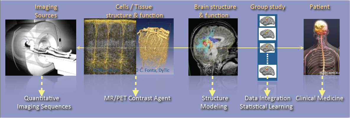
As shown in Figure 2, the research activities of the Empenn team closely link observations and models through the integration of clinical and multiscale data, and phenotypes (cellular, and later molecular, with structural or connectivity patterns in the first stage). Our ambition is to build personalized models of central nervous system organs and pathologies, and to compare these models with clinical research studies in order to establish a quantitative diagnosis, prevent the progression of diseases and provide new digital recovery strategies, while combining all these research areas with clinical validation. This approach is developed within a translational framework, where the data integration process to build the models is informed by specific clinical studies, and where the models are assessed regarding prospective clinical trials for diagnosis and therapy planning. All of these research activities will be conducted in close collaboration with the Neurinfo platform, which benefited in 2018 from a new high-end 3T MRI system dedicated to research (3T Prisma™ system from Siemens), and through the development in the coming years of multimodal hybrid imaging (from the currently available EEG-MRI, to EEG-NIRS and PET-MRI in the future).
In this context, some of our major developments and newly arising issues and challenges will include:
- The generation of new descriptors to study brain structure and function (e.g. the combination of variations in brain perfusion with and without a contrast agent; changes in brain structure in relation to normal, pathological, functional or connectivity patterns; or the modeling of brain state during cognitive stimulation using neurofeedback).
- The integration of additional spatiotemporal and hybrid imaging sequences covering a larger range of observations, from the molecular level to the organ level, via the cellular level (arterial spin labeling, diffusion MRI, MR relaxometry, MR fingerprinting, MR cell labeling imaging, MR-PET molecular imaging, EEG-MRI functional imaging, EEG-NIRS-MRI, etc.).
- The creation of computational models through the data fusion of molecular, cellular (i.e. through dedicated ligands or nanocarriers), structural and functional image descriptors from group studies of normal and/or pathological subjects.
- The evaluation of these models in relation to acute pathologies, especially for the study of degenerative, psychiatric, traumatic or developmental brain diseases (primarily multiple sclerosis, stroke, traumatic brain injury (TBI) and depression, but applicable with a potential additional impact to epilepsy, Parkinson’s disease, dementia, Posttraumatic stress disorder, etc.) within a translational framework.
In terms of new major methodological challenges, we will address the development of models and algorithms to reconstruct, analyze and transform the images, and to manage the mass of data to store, distribute and “semanticize” (i.e. provide a logical division of the model’s components according to their meaning). As such, we expect to make methodological contributions in the fields of model inference; statistical analysis and modeling; the application of sparse representation (compressed sensing and dictionary learning) and machine learning (supervised/unsupervised classification and discrete model learning); data fusion (multimodal integration, registration, patch analysis, etc.); high-dimensional optimization; data integration; and brain-computer interfaces. As a team at the frontier between the digital sciences and clinical research in neuroscience, we do not claim to provide theoretical breakthroughs in these domains but rather to provide significant advances in using these algorithms through to the advanced applications we intend to address. In addition, we believe that by providing these significant advances using this set of algorithms, we will also contribute to exhibiting new theoretical problems that will fuel the domains of theoretical computer sciences and applied mathematics.
In summary, we expect to address the following major challenges:
- Developing new information processing methods able to detect imaging biomarkers in the context of mental, neurological, and substance use disorders.
- Providing new computational solutions for our target applications, allowing a more appropriate representation of data for image analysis and the detection of biomarkers specific to a form or grade of pathology, or specific to a population of subjects.
- Providing, for our target applications, new patient-adapted connectivity atlases for the study and characterization of diseases from quantitative MRI.
- Providing, for our target applications, new analytical models of dynamic regional perfusion, and deriving indices of dynamic brain local perfusion from normal and pathological populations.
- Investigating whether the theragnostics paradigm of rehabilitation from hybrid neurofeedback can be effective in some behavioral and disability pathologies.
These major advances will be primarily developed and validated in the context of several priority applications in which we expect to play a leading role: multiple sclerosis, stroke rehabilitation, and the study and treatment of depression.
4 Application domains
4.1 Basic resarch
4.1.1 Population imaging
One major objective of neuroimaging researchers and clinicians is to be able to stratify brain imaging data in order to derive new and more specific population models. In practice this requires to set up large-scale experiments that, due to the lack of resources and capabilities to recruit locally subjects who meet specific inclusion criteria, motivates the need for sharing the load.
But, building and using multi-site large-scale resources poses specific challenges to deal with the huge quantity of data produced and their diversity. Empenn will focus on two challenges in particular:
- Provide computational environments for the computation and use of imaging biomarkers in the targeted brain diseases, a solution to be used by radiologists and neurologists/psychiatrists for the clinical follow-up of a large patient population.
- Modeling analytic variability of image processing pipelines to better understand and predict the behaviour of imaging biomarker detection solutions and improve reproducibility and productivity in clinical neuroimaging research.
4.1.2 Detection and learning
We intend to make significant contributions with major impacts in learning coupling models between functional recordings during neurofeedback procedures. These advances will provide a breakthrough in brain-computer interfaces for rehabilitation protocols. Our aim is to:
- Provide a computational environment that combines data-driven (machine learning) and Bayesian solutions to improve the detection of abnormal patterns in images through decision or evidence theory data fusion strategies. The major initial application will be for multiple sclerosis. Over the longer term, we also expect to adapt these methods to address a wider range of neurological diseases (epilepsy, stroke, tumors, etc.) in neonate and adult brains.
- Develop solutions for combining brain state measurements from multimodal sensors or sequences (e.g. fMRI, ASL, EEG, NIRS, etc.) with applications in the spatiotemporal reconstruction of brain activity from MRI-EEG or the combined detection of the endogenous hemodynamic and resting state network of the brain from ASL and NIRS. Over the longer term, the advent of new hybrid brain imaging sensors (e.g. PET-MRI) will require these methods to be extended to a larger spectrum of information combining structural, morphological, metabolic, electrophysiological and cellular/molecular information (e.g. through the use of specific ligands/nanocarriers).
4.1.3 Quantitative imaging
The Empenn research group focuses on the development of several quantitative techniques in magnetic resonance imaging of the brain. These methods allow for a characterization of both the function and the structure of the brain with high precision. Arterial spin labelling (ASL) is a contrast agent-free imaging technique which labels arterial blood water as an endogenous tracer for perfusion and can measure resting-state cerebral blood flow. We are interested in estimating multiparametric hemodynamics using ASL, such as combined cerebral blood flow and arterial transit times, and derive statistical descriptors to represent significant differences between groups. In addition to quantitative perfusion parameters, our contributions on tissue compartment imaging aim at delineating neural circuits and characterize their microstructure properties, using both diffusion MRI and relaxometry. In diffusion MRI, arbitrary gradient waveforms were shown to exhibit higher sensitivity to microstructure parameters than standard pulsed gradients. We work on the optimization of sampling protocols in this domain, with the objective to propose sequences compatible with in vivo acquisition. Complementary to diffusion MRI, we develop methods for the reconstruction of myelin-bound, extra-axonal and cerebrospinal fluid water using multi-compartment modelling of the T2-relaxometry signal. We combine these techniques with tractography to identify trajectories of pathologies associated to the evolution of these microstructural parameters along specific fiber bundles in the brain white matter.
4.2 Translational resarch
4.2.1 Behavior
Advances in the field of in vivo imaging offer new opportunities for addressing the management of resistant affective disorders and their consequences (suicide risk and socio-professional impact), and the management of spatial cognition disorders after stroke and their consequences (postural perturbations and the loss of autonomy). Our objective, and the main challenge in this context, will be to introduce medical image computing methods to the multidisciplinary field of behavioral disorders (cognitive disorders, particularly spatial and postural control disorders or anterograde memory impairment, mood disorders, notably resistant depression, schizophrenic disorders, pervasive developmental disorders, attention disorders, etc.) in order to gain a better understanding of the pathology and devise innovative therapeutic approaches.
We also expect to become a major player in the future and make important contributions with significant impacts, primarily in drug-resistant depression in young and old populations. In particular, we expect to provide new image-related metrics combining perfusion, metabolism and microstructural information regarding the brain in order to better characterize pathologies, provide prospective evolution values and potentially provide new brain stimulation targets that could be used in neurofeedback rehabilitation protocols or other types of brain stimulation procedure.
We aim to provide new imaging markers of mental diseases, especially in the context of mood disorders. The new biomarkers will be derived from the metabolic (ASL and later ASL+PET) point of view as well as from the microstructural point of view (multicompartment diffusion MRI and relaxometry). Similarly, we expect to exhibit imaging biomarker regularities combining metabolic and structural information. Over the longer term, we expect these biomarkers to be the target of neurofeedback rehabilitation procedures. Also, over the longer term, we expect to supplement the MRI markers with molecular marker ones coming from new PET tracers, especially those associated with serotonin intake, at one time point or during a rehabilitation protocol under hybrid PET-EEG-MRI neurofeedback procedures.
4.2.2 Neuroinflammation
Some of the major ongoing research issues regarding neuroimaging of neuro-inflammatory diseases concern the definition of new biomarkers to track the development of the pathology using high- dimensional data (e.g. nD+t MRI). This includes the use of white matter-specific imaging, such as magnetization transfer MRI, relaxometry and diffusion-weighted imaging (DW-MRI). Our objective is (1) to develop information-processing tools to tag the spatiotemporal evolutions of MS patterns at the brain parenchyma and spinal cord levels from their different signatures (inflammatory cells visible with USPIO or Gd contrast agents on MRI, persistent black holes, eloquent regional atrophy and microstructure signatures); and (2) to test these new tools on new imaging cohorts. In this respect, we for instance conduct studies on brain and spinal cord imaging, continuing on from the PHRC multicentric EMISEP project (PI: G. Edan), as it is very likely that lesions in the spine will directly affect the ambulatory ability of the patient (and thereby the clinical scores). In order to extend this experiment to a larger MS population, based on our expertise from the OFSEP cohort, we also plan to improve the MS therapeutic decision process through the MUSIC project (Multiple Sclerosis Imaging Check out, a public/private project). Our goal is to develop and assess a standardized monitoring tool that provides a robust, long-term computerized MRI follow-up that will become the gold standard in clinical practice for therapeutic decisions in MS treatment. As part of this project, Empenn will share its expertise in data management systems (Shanoir and FLI-IAM) and automatic processing tools (through the medInria and Anima software repositories) to extract quantitative indices from the images.
4.2.3 Recovery
Mental and neurological disorders are the leading cause of years lived with a disability. Treatment-resistant depression affects approximately 2% of the European population. Meanwhile, in the case of brain disorders, almost 1.5 million Europeans (15 million people worldwide) suffer a stroke event each year. Current recovery methods for brain disorders and traumatic brain injuries remain limited, preventing many from achieving full recuperation. We propose to address the issue of brain recovery by introducing new advances from recent breakthroughs in computational medical imaging, data processing and human-machine interfaces, and demonstrate how these new concepts can be used, in particular for the treatment of stroke and major depressive disorders.
We ambition to combine advanced instrumental devices (hybrid EEG, NIRS and MRI platforms), with new hybrid brain computer interface paradigms and new computational models to provide neurofeedback-based therapeutic and neuro-rehabilitation paradigms in some of the major mental and neurological disorders of the developmental and the aging brain.
Neurofeedback involves using a brain-computer interface that provides an individual with real-time biofeedback about his or her brain activity in the form of sensory feedback. It enables individuals to learn to better control their brain activity, which can be measured in real time using various non-invasive sensors as described above. Although EEG is currently the only modality used by clinical practitioners in that context, it lacks specificity due to its low spatial resolution. Dynamic research into fMRI-neurofeedback has held promise for treating depression, chronic pain and stroke, since it offers the prospect of real-time imagery of the activity in deep brain structures with high spatial resolution. However, the low temporal resolution and high cost of fMRI-Neurofeedback has hampered the development of many applications. We believe that the future belongs to hybrid responses that combine multimodal sensors and intend to demonstrate this in the Empenn project.
5 Social and environmental responsibility
5.1 Footprint of research activities
Elise Bannier took part to meetings with researchers from IRMAR and IRISA to discuss potential actions related to sustainable developement such as the EcoInfo initiative. Also, Inria and the sustainable development commission are working on a national initiative and proposed a survey. Unfortunately, the workshop planned on May 19th dedicated to energy was postponed because of the COVID. We hope 2021 will allow to make more progress.
6 Highlights of the year
6.1 Awards
6.1.1 Prix Harmonie Mutuelle
Pierre-Yves Jonin was awarded the "Prix Harmonie Mutuelle - Maladie d'Alzheimer" in September 2020 (30 k€).
7 New software and platforms
7.1 New software
7.1.1 Anima
- Keywords: Filtering, Medical imaging, Diffusion imaging, Registration, Relaxometry
- Scientific Description: Anima is a set of libraries and tools developed by the team as a common repository of research algorithms. As of now, it contains tools for image registration, statistical analysis (group comparison, patient to group comparison), diffusion imaging (model estimation, tractography, etc.), quantitative MRI processing (quantitative relaxation times estimation, MR simulation), image denoising and filtering, and segmentation tools. All of these tools are based on stable libraries (ITK, VTK), making it simple to maintain.
- Functional Description: Anima is a set of libraries and tools in command line mode for processing and analysing medical images.
-
URL:
https://
anima. irisa. fr - Authors: Olivier Commowick, Sudhanya Chatterjee, Antoine Legouhy, Florent Leray, René-Paul Debroize, Fang Cao, Laurence Catanese, Aymeric Stamm, Christian Barillot, Renaud Hedouin, Sylvain Prima, Aymeric Stamm
- Contact: Olivier Commowick
- Participants: Aymeric Stamm, Fang Cao, Florent Leray, Guillaume Pasquier, Laurence Catanese, Olivier Commowick, Renaud Hedouin, René-Paul Debroize
7.1.2 MedINRIA
- Keywords: Visualization, DWI, Health, Segmentation, Medical imaging
- Scientific Description: MedInria aims at creating an easily extensible platform for the distribution of research algorithms developed at Inria for medical image processing. This project has been funded by the D2T (ADT MedInria-NT) in 2010, renewed in 2012. A fast-track ADT was awarded in 2017 to transition the software core to more recent dependencies and study the possibility of a consortium creation.The Empenn team leads this Inria national project and participates in the development of the common core architecture and features of the software as well as in the development of specific plugins for the team's algorithm.
- Functional Description: MedInria is a free software platform dedicated to medical data visualization and processing.
-
URL:
https://
med. inria. fr - Authors: Michael Buckingham, Nicolas Schnitzler, Florent Leray, Alexandre Abadie, Benoît Bleuzé, Clément Philipot, Fatih Arslan, Florian Vichot, Guillaume Pasquier, Hakim Fadil, Jaime Garcia Guevara, John Stark, Julien Wintz, Loic Cadour, Maxime Sermesant, Michael Knopke, Nicolas Toussaint, Olivier Clatz, Olivier Commowick, Pierre Fillard, René-Paul Debroize, Sergio Medina, Stephan Schmitt, Théodore Papadopoulo
- Contacts: Olivier Commowick, Maxime Sermesant, Théodore Papadopoulo
- Participants: Maxime Sermesant, Olivier Commowick, Théodore Papadopoulo
- Partners: HARVARD Medical School, IHU - LIRYC, NIH
7.1.3 autoMRI
- Keywords: FMRI, MRI, ASL, FASL, SPM, Automation
- Scientific Description: This software is highly configurable in order to fit a wide range of needs. Pre-processing includes segmentation of anatomical data, as well as co-registration, spatial normalization and atlas building of all data types. The analysis pipelines perform either within-group analysis or between-group or one subject-versus-group comparison, and produce statistical maps of regions with significant differences. These pipelines can be applied to structural data to exhibit patterns of atrophy or lesions, to ASL (both pulsed or pseudo-continuous sequences) data to detect perfusion abnormalities, to functional data - either BOLD or ASL - to outline brain activations related to block or event-related paradigms. New functionalities have been implemented to facilitate the management and processing of data coming from complex projects.
- Functional Description: AutoMRI Based on MATLAB and the SPM12 toolbox, autoMRI provides complete pipelines to pre-process and analyze various types of images (anatomical, functional, perfusion).
-
URL:
https://
team. inria. fr/ visages/ software/ - Authors: Camille Maumet, Isabelle Corouge, Pierre Maurel, Quentin Duché, Elise Bannier, Julie Coloigner
- Contacts: Camille Maumet, Isabelle Corouge, Quentin Duché, Pierre Maurel, Elise Bannier
- Participants: Camille Maumet, Elise Bannier, Isabelle Corouge, Pierre Maurel, Quentin Duché, Julie Coloigner
7.1.4 miet
- Name: Medical Imaging Extraction Tools
- Keywords: Brain MRI, Statistics, Data analysis
- Functional Description: In plenty of situations, an MRI study is stored as a hierarchy of folders containing MRI data. Such study may contain a variety of subject/subject-type/center/timeStep/sequences... that structures the corresponding folders naming and hierarchy. Each of the final folder then contains MR image files. These files may consist of a set of raw data as well as post-processed data such as segmentation masks, co-registered volumes, ... that are produced from a variety of image processing tools. Once all this data produced, the next step generally consists in analyzing them. An ubiquitous analysis in medical imaging is the so-called region-of-interest based analysis that consists in analyzing statistics of MR signals over sets of predefined regions. Miet is designed to specify and extract data frames ready for data analysis from a specified folder hierarchy and set of extraction formulas.
-
URL:
https://
gitlab. inria. fr/ miet/ miet - Contact: Benoît Combès
7.1.5 ShanoirUploader
- Keywords: Webservices, PACS, Medical imaging, Neuroimaging, DICOM, Health, Biology, Java, Shanoir
- Scientific Description: ShanoirUploader is a desktop application on base of JavaWebStart (JWS). The application can be downloaded and installed using an internet browser. It interacts with a PACS to query and retrieve the data stored on it. After this ShanoirUploader sends the data to a Shanoir server instance in order to import these data. This application bypasses the situation, that in most of the clinical network infrastructures a server to server connection is complicated to set up between the PACS and a Shanoir server instance.
- Functional Description: ShanoirUploader is a Java desktop application that transfers data securely between a PACS and a Shanoir server instance (e.g., within a hospital). It uses either a DICOM query/retrieve connection or a local CD/DVD access to search and access images from a local PACS or the local CD/DVD. After having retrieved the data, the DICOM files are locally anonymized and then uploaded to the Shanoir server. A possible integration of a hash creation application for patient identifiers is provided as well. The primary goals of that application are to enable mass data transfers between different remote server instances and therefore reduce the waiting time of the users, when importing data into Shanoir. Most of the time during import is spent with data transfers.
-
URL:
http://
shanoir. gforge. inria. fr - Authors: Michael Kain, Christian Barillot
- Contact: Michael Kain
- Participants: Christian Barillot, Inès Fakhfakh, Justine Guillaumont, Michael Kain, Yao Yao
7.1.6 Shanoir-NG
- Keywords: Neuroimaging, DICOM, Nifti
-
Functional Description:
Shanoir-NG is a complete technological remake of the first version of the Shanoir application, but maintaining the key concepts of Shanoir.
Why did we take this big effort to implement Shanoir-NG from scratch? • Over the years of the existence and usage of Shanoir the technological basis of Shanoir has become outdated and most of its original technical frameworks (JBoss 4, Java Server Faces (JSF), Richfaces, JBoss Seam) are not supported and maintained anymore. • Furthermore the architectures and technologies for developing web applications have dynamically progressed in the last 5 years. The arrival of Single-Page-Applications (SPAs), like Gmail and Twitter, the Docker containerization technology and microservices architectures have dramatically changed the way we develop web applications today. • This lead to the consequence, that only migrating Shanoir to newer versions of the existing libraries and code, was far from being sufficient to extend the lifetime and long-time usage of Shanoir. That is why we started to develop Shanoir-NG from scratch with a new architecture (microservices and REST) and new technologies, while keeping most of its functionalities.
Shanoir-NG (SHAring NeurOImaging Resources) is an open-source neuroinformatics platform designed to share, archive, search and visualize neuroimaging data.
It provides a user-friendly secure web access and offers an intuitive workflow to facilitate the collecting and retrieving of neuroimaging data from multiple sources and a wizzard to make the completion of metadata easy. Shanoir-NG comes along many features such as anonymization of data (based on standard profiles), support for multi-centric clinical studies on subjects or group of subjects.
Shanoir-NG offers an ontology-based data organization (OntoNeuroLOG). Among other things, this facilitates the reuse of data and metadata, the integration of processed data and provides traceability trough an evolutionary approach. Shanoir-NG allows researchers, clinicians, PhD students and engineers to undertake quality research projects with an emphasis on remote collaboration. As a secured Jakarta EE web application, it therefore allows you safely storing and archiving, with no more requirements than a computer with an internet connection!
Shanoir-NG has been extended for preclinical data too, it manages your study meta-data and preclinical images: • Pathology models, therapies, anesthetics and physiological data • Imports Bruker file format
Furthermore, Shanoir-NG is not only a web application: it is also a complete neuroinformatics platform in which you can easily integrate your existing processing tools or develop your own ones: see ShanoirTk or ShanoirUploader to import your data directly from the PACS in the hospital.
Using cross-data navigation and advanced search criteria (new Solr search module), the user scan quickly point to a subset of data to be downloaded. Client side applications have as well been developed to illustrate how to locally access and exploit data though the available web services. With regards to security, the system requires authentication and user rights are adjustable for each hosted study. A study responsible can thereby define the users allowed to see, download or import data into his study or simply make it public.
Shanoir-NG serves neuroimaging researchers in organizing efficiently their studies while cooperating with other laboratories. By managing patient privacy, Shanoir allows the exploitation of clinical data in a research context. It is finally a handy solution to publish and share data with a broader community.
It supports the following formats: DICOM (MR, CT, PT, NM), NIfTI, Bruker, EEG(BrainVision/EDF), big zip files
- Release Contributions: Shanoir-NG is a complete technological remake of the first version of the Shanoir application, but maintaining the key concepts of Shanoir.
- News of the Year: Shanoir-NG is a complete technological remake of the first version of the Shanoir application, but maintaining the key concepts of Shanoir.
- Authors: Inès Fakhfakh, Anthony Baire, Christian Barillot, Mathieu Simon, Michael Kain, Yao Yao, Aneta Morawin, Arnaud Touboulic, Julien Louis, Arthur Masson, Julien Lamy, Romain Lahaxe, Farid Ouhmich, Simon Loury, Michel Dojat, Benjamin Lemasson, Olivier Montignon, Marjolaine Bodin, Jean-Côme Douteau, Emmanuel Barbier
- Contacts: Michael Kain, Julien Louis
- Participants: Christian Barillot, Mathieu Simon, Michael Kain, Yao Yao, Aneta Morawin, Arnaud Touboulic, Inès Fakhfakh, Anthony Baire
- Partners: CHU Grenoble, INSERM, CNRS, Université Grenoble Alpes, Université de Strasbourg
7.1.7 Sharpedge_NF
- Keywords: EEG, Neurofeedback, FMRI, Neurorehabilitation
- Scientific Description: Platform for the realization of bimodal EEGIRMf Neurofeedback, including a GUI to set the experimental parameters, a control unit (matlab object) to record the data, synchronised preprocessing and processing of EEG and fMRI data, computation and display of the neurofeedback scores.
- Functional Description: Platform for the realization of bimodal EEGIRMf Neurofeedback, including a GUI to set the experimental parameters, a control unit (matlab object) to record the data, synchronised preprocessing and processing of EEG and fMRI data, computation and display of the neurofeedback scores.
-
URL:
https://
project. inria. fr/ hemisfer/ - Publication: hal-01426072
- Authors: Marsel Mano, Giulia Lioi, Anatole Lécuyer, Christian Barillot, Elise Bannier
- Contacts: Anatole Lécuyer, Elise Bannier
8 New results
8.1 Basic research
8.1.1 Population imaging
In the context of population imaging, we have made progress in three main areas this year. First we proposed an atlas of brain development based on new methodological results. Second, we focused on the topic of analytic variability both by participating to an international project and by proposing our own methodological developments. Finally, we developed guidelines for data sharing in compliance with GDPR as part of an international European collaboration. We also joined two collaborative actions: Modal a regional network for data integration in biological and medical research and GliMR a European action to promote advanced biomarkers of gliomas.
Regional brain development analysis through registration using anisotropic similarity, a constrained affine transformation
Participants: Antoine Legouhy, Olivier Commowick, Christian Barillot.
We propose in 34 a novel method to quantify brain growth in three arbitrary orthogonal directions of the brain or its sub-regions through linear registration. This is achieved by introducing a nine degrees of freedom transformation called anisotropic similarity which is an affine transformation with constrained scaling directions along arbitrarily chosen orthogonal vectors. This gives the opportunity to extract scaling factors describing brain growth along those directions by registering a database of subjects onto a common reference. This information about directional growth brings insights that are not usually available in longitudinal volumetric analysis. The interest of this method is illustrated by studying the anisotropic regional and global brain development of 308 healthy subjects betwen 0 and 19 years old. A gender comparison of those scaling factors is also performed for four age-intervals. We demonstrate through these applications the stability of the method to the chosen reference and its ability to highlight growth differences across regions and gender.Validity of group fMRI studies when combining data from different pipelines
Participants: Camille Maumet, Xavier Rolland, Christian Barillot, Pierre Maurel.
More and more studies in the neuroimaging literature have made their result data publicly accessible, making it possible for other researchers to combine those results quantitatively in order to build robust summaries. But, many factors vary across studies and may impact their compatibility, those include: different scanners, different acquisition protocols, different acquisition sites as well as different post-processing pipelines. It is still unclear whether we can perform group studies with data which have been generated under different conditions. Here, we evaluate the impact of post-processing pipelines in functional MRI (fMRI). Multiple options are typically available to define a pipeline at each step, resulting in a large space of possible processing pipelines, so-called ‘analytic variability’.We used fMRI data from the Human Connectome Project (HCP) and performed between-group analyses comparing two groups of subjects processed using two different pipelines. The two pipelines differed on a predefined set of parameters (smoothing kernel, numbers of motion regressors in the statistical analyses and modeling of the hemodynamic response function). We compared empirical false positive rates to theoretical rates in order to verify the validity of each comparison. We showed that some of the pipelines pairs give false positives rates different from the expected rate, which means that data processed with different pipelines cannot be combined without taking into account the effect of pipelines on the result. A paper is about to be submitted.Variability in the analysis of a single neuroimaging dataset by many teams
Participants: Camille Maumet.
Data analysis workflows in many scientific domains have become increasingly complex and flexible. Here we assess the effect of this flexibility on the results of functional magnetic resonance imaging by asking 70 independent teams to analyse the same dataset, testing the same 9 ex-ante hypotheses. The flexibility of analytical approaches is exemplified by the fact that no two teams chose identical workflows to analyse the data. This flexibility resulted in sizeable variation in the results of hypothesis tests, even for teams whose statistical maps were highly correlated at intermediate stages of the analysis pipeline. Variation in reported results was related to several aspects of analysis methodology. Notably, a meta-analytical approach that aggregated information across teams yielded a significant consensus in activated regions. Furthermore, prediction markets of researchers in the field revealed an overestimation of the likelihood of significant findings, even by researchers with direct knowledge of the dataset. Our findings show that analytical flexibility can have substantial effects on scientific conclusions, and identify factors that may be related to variability in the analysis of functional magnetic resonance imaging. The results emphasize the importance of validating and sharing complex analysis workflows, and demonstrate the need for performing and reporting multiple analyses of the same data. Potential approaches that could be used to mitigate issues related to analytical variability are discussed 13.This work was done as part of an international collaboration with 200 researchers. The project was run by research teams from California Institut of Technology, Stanford University, the Stockholm School of Economics, Tel Aviv University, and the University of Innsbruck. The fMRI data was collected during winter and spring 2018 at Tel Aviv University. The contact person for the analysis teams was Rotem Botvinik-Nezer, from Tel Aviv University.
Every little bit counts: towards data reuse in neuroimaging
Participants: Camille Maumet.
Including open science practices in everyday research is not always straightforward and the wealth of tools available can quickly become overwhelming. This talk proposed approaches to share reserach outputs as part of the scientific process 51The Open Brain Consent: Informing research participants and obtaining consent to share brain imaging data
Participants: Camille Maumet, Elise Bannier.
Having the means to share research data openly is essential to modern science. A key aspect in this endeavour is obtaining consent from participants, not just to take part in a research study, which is a basic bioethical principle, but also to share their data with the scientific community. To ensure that data privacy is respected, national and/or supranational legal rules are in place. It is however not always clear to researchers what the implications are of those regulations, nor how to best comply with them. The Open Brain Consent (https://This work was done as part of a wide international collaboration between members of the The Open Brain Consent working group.
8.1.2 Detection and learning
In this section, we summarised different contributions focusing on information extraction from medical image data. In the field of medical imaging, machine learning methods can be used to detect brain abnormalities, in order to improve the quality of a diagnostic, a pronostic or a disease understanding. Machine learning methods can also be used to predict different scores or brain features. In particular, prediction can be used to transfer knowledge from an imaging modality to another one presenting different specificities. In bimodal EEG-fMRI neurofeedback, it is crucial for a better understanding of brain mechanism, to analyse the relation between EEG and fMRI, two complimentary imaging modalities with different specificities, and to enhance the information extraction from EEG using fMRI signals to reduce the use of the MRI.
A sparse EEG-informed fMRI model for hybrid EEG-fMRI neurofeedback prediction
Participants: Claire Cury, Pierre Maurel, Remi Gribonval, Christian Barillot.
Measures of brain activity through functional magnetic resonance imaging (fMRI) or Electroencephalography (EEG), two complementary modalities, are ground solutions in the context of neurofeedback (NF) mechanisms for brain rehabilitation protocols. Though NF-EEG (real-time neurofeedback scores computed from EEG) have been explored for a very long time, NF-fMRI (real-time neurofeedback scores computed from fMRI) appeared more recently and provides more robust results and more specific brain training. Using simultaneously fMRI and EEG for multimodal neurofeedback sessions (NF-EEG-fMRI, real-time neurofeedback scores computed from fMRI and EEG) is very promising to devise brain rehabilitation protocols. However using fMRI is costly, exhausting and time consuming, and cannot be repeated too many times for the same subject. The original contribution of this work concerns the prediction of multimodal NF scores from EEG recordings only, using a training phase where both EEG and fMRI synchronous signals, and therefore neurofeedback scores, are available. We propose a sparse regression model able to exploit EEG only to predict NF-fMRI or NF-EEG-fMRI in motor imagery tasks. We compare different NF-predictors steming from the proposed model. We show that one of the proposed NF-predictors significanlty improves over what EEG can provide alone (without the learning phase), and correlates at 0.74 in median with the ground-truth 23.This work was done in collaboration with Rémi Gribonval from the PANAMA team.
Impact of 1D and 2D visualisation on EEG-fMRI neurofeedback training during a motor imagery task
Participants: Claire Cury, Giulia Lioi, Lorraine Perronnet, Pierre Maurel, Christian Barillot.
Bi-modal EEG-fMRI neurofeedback (NF) is a new technique of great interest. First, it can improve the quality of NF training by combining different real-time information (hemodynamic and electrophysiological) from the participant’s brain activity; Second, it has potential to better understand the link and the synergy between the two modalities (EEG-fMRI). However there are different ways to show to the participant his NF scores during bi-modal NF sessions. To improve data fusion methodologies, we investigate the impact of a 1D or 2D representation when a visual feedback is given during motor imagery task. Results show a better synergy between EEG and fMRI when a 2D display is used. Subjects have better fMRI scores when 1D is used for bi-modal EEG-fMRI NF sessions; on the other hand, they regulate EEG more specifically when the 2D metaphor is used 44.This work was done in collaboration with Anatole Lécuyer from the Hybrid Inria team, and Lorraine Perronnet following her PhD work between the Empenn and Hybrid teams.
Simultaneous EEG-fMRI during a neurofeedback task, a brain imaging dataset for multimodal data integration
Participants: Giulia Lioi, Claire Cury, Elise Bannier, Christian Barillot.
Combining EEG and fMRI allows for integration of fine spatial and accurate temporal resolution yet presents numerous challenges, noticeably if performed in real-time to implement a Neurofeedback (NF) loop. Here we describe a multimodal dataset of EEG and fMRI acquired simultaneously during a motor imagery NF task, supplemented with MRI structural data. The study involved 30 healthy volunteers undergoing five training sessions. We showed the potential and merit of simultaneous EEG-fMRI NF in previous work. Here we illustrate the type of information that can be extracted from this dataset and show its potential use. This represents one of the first simultaneous recording of EEG and fMRI for NF and we present the first open access bi-modal NF dataset integrating EEG and fMRI. We believe that it will be a valuable tool to (1) advance and test methodologies for multi-modal data integration, (2) improve the quality of NF provided, (3) improve methodologies for de-noising EEG acquired under MRI and (4) investigate the neuromarkers of motor-imagery using multi-modal information 36.This work was done in collaboration with Anatole Lécuyer from the Hybrid Inria team, following the work of Lorraine Perronnet during her PhD between the Empenn and Hybrid teams.
Unsupervised domain adaptation with optimal transport in multi-site segmentation of multiple sclerosis lesions from MRI data
Participants: Antoine Ackaouy, Olivier Commowick, Christian Barillot, Francesca Galassi.
Automatic segmentation of Multiple Sclerosis (MS) lesions from Magnetic Resonance Imaging (MRI) images is essential for clinical assessment and treatment planning of MS. Recent years have seen an increasing use of Convolutional Neural Networks (CNNs) for this task. Although these methods provide accurate segmentation, their applicability in clinical settings remains limited due to a reproducibility issue across different image domains. MS images can have highly variable characteristics across patients, MRI scanners and imaging protocols; retraining a supervised model with data from each new domain is not a feasible solution because it requires manual annotation from expert radiologists. In this work, we explore an unsupervised solution to the problem of domain shift. We present a framework, Seg-JDOT, which adapts a deep model so that samples from a source domain and samples from a target domain sharing similar representations will be similarly segmented. We evaluated the framework on a multi-site dataset, MICCAI 2016, and showed that the adaptation towards a target site can bring remarkable improvements in a model performance over standard training 7.This work was done in collaboration with Nicolas Courty, Obelix team, IRISA laboratory from University of Bretagne Sud.
8.1.3 Quantitative imaging
We develop several quantitative imaging techniques; these methods allow for a characterization of both the function and the structure of the brain with high precision. This year, we contributed to methods in acquisition and processing of diffusion MRI, brain perfusion measured with ASL and BOLD in MRI and we started experimenting fNIRS.
Free water estimation using single-shell diffusion-weighted images
Participants: Emmanuel Caruyer.
Free-water estimation requires the fitting of a bi-compartment model, which is an ill-posed problem when using only single-shell data. Its solution requires optimization, which relies on an initialization step. We propose a novel initialization approach, called "Freewater EstimatoR using iNtErpolated iniTialization" (FERNET), which improves the estimation of free water in edematous and infiltrated peritumoral regions, using single-shell diffusion MRI data. The method has been extensively investigated on simulated data and healthy and brain tumor datasets, demonstrating its applicability on clinically acquired data. Additionally, it has been applied to data from brain tumor patients to demonstrate the improvement in tractography in the peritumoral region 37.This is a collaborative project with the University of Pennsylvania, USA; Synaptive Medical Inc., Toronto, Canada ; Brigham and Women’s Hospital, Boston and Harvard Medical School, USA.
Multi-dimensional diffusion MRI sampling scheme: B-tensor design and accurate signal reconstruction
Participants: Emmanuel Caruyer.
B-tensor encoding enables the separation of isotropic and anisotropic tensors. However, little consideration has been given as to how to design a B-tensor encoding sampling scheme. In this work, we propose the first 4D basis for representing the diffusion signal acquired with B-tensor encoding. We study the properties of the diffusion signal in this basis to give recommendations for optimally sampling the space of axisymmetric b-tensors. We show, using simulations, that the proposed sampling scheme enables accurate reconstruction of the diffusion signal by expansion in this basis using a clinically feasible number of samples 10.This work was done in collaboration with A. Bates, Australian National University and Al. Daducci, University of Verona.
An evolutionary framework for microstructure-sensitive generalized diffusion gradient waveforms
Participants: Raphaël Truffet, Christian Barillot, Emmanuel Caruyer.
In diffusion-weighted MRI, general gradient waveforms became of interest for their sensitivity to microstructure features of the brain white matter. However, the design of such waveforms remains an open problem. In this work, we propose a framework for generalized gradient waveform design with optimized sensitivity to selected microstructure features. In particular, we present a rotation-invariant method based on a genetic algorithm to maximize the sensitivity of the signal to the intra-axonal volume fraction. The sensitivity is evaluated by computing a score based on the Fisher information matrix from Monte-Carlo simulations, which offer greater flexibility and realism than conventional analytical models. As proof of concept, we show that the optimized waveforms have higher scores than the conventional pulsed-field gradients experiments. Finally, the proposed framework can be generalized to optimize the waveforms for any microstructure feature of interest 46.Interpolation and averaging of diffusion MRI multi-compartment models
Participants: Renaud Hedouin, Christian Barillot, Olivier Commowick.
Multi-compartment models (MCM) are increasingly used to characterize the brain white matter microstructure from diffusion-weighted imaging (DWI). Their use in clinical studies is however limited by the inability to resample an MCM image towards a common reference frame, or to construct atlases from such brain microstructure models. We proposed in 26 to solve this problem by first identifying that these two tasks amount to the same problem. We propose to tackle it by viewing it as a simplification problem, solved thanks to spectral clustering and the definition of semi-metrics between several usual compartments encountered in the MCM literature. This generic framework is evaluated for two models: the multi-tensor model where individual fibers are modeled as individual tensors and the diffusion direction imaging (DDI) model that differentiates intra-and extra-axonal components of each fiber. Results on simulated data, simulated transformations and real data showed the ability of our method to well interpolate MCM images of these types. We finally presented as an application an MCM template of normal controls constructed using our approach.Acquisition duration in resting-state arterial spin labeling. How long is enough?
Participants: Corentin Vallée, Pierre Maurel, Isabelle Corouge, Christian Barillot.
Resting-state Arterial Spin Labeling (rs-ASL) is a rather confidential method compared to resting-state BOLD. As ASL allows to quantify the cerebral blood flow, unlike BOLD, rs-ASL can lead to significant clinical subject-scaled applications. Despite directly impacting clinical practicability and functional networks estimation, there is no standard for rs-ASL regarding the acquisition duration. Our work here focuses on assessing the feasibility of ASL as an rs-fMRI method and on studying the effect of the acquisition duration on the estimation of functional networks. To this end, we acquired a long 24 min 30 s rs-ASL sequence and investigated how estimations of six typical functional brain networks evolved with respect to the acquisition duration. Our results show that, after a certain acquisition duration, the estimations of all functional networks reach their best and are stabilized. Since, for clinical application, the acquisition duration should be the shortest possible, we suggest an acquisition duration of 14 min, i.e., 240 volumes with our sequence parameters, as it covers the functional networks estimation stabilization 41, 56.Quantitative perfusion mapping with induced transient hypoxia using BOLD MRI
Participants: Julie Coloigner.
Gadolinium-based dynamic susceptibility contrast (DSC) is commonly used to characterize blood flow in patients with stroke and brain tumors. Unfortunately, gadolinium contrast administration has been associated with adverse reactions and longterm accumulation in tissues. In this work, we propose an alternative deoxygenationbased dynamic susceptibility contrast (dDSC) method that uses a transient hypoxia gas paradigm to deliver a bolus of paramagnetic deoxygenated hemoglobin to the cerebral vasculature for perfusion imaging. Through traditional DSC tracer kinetic modeling, the MR signal change induced by this hypoxic bolus can be used to generate regional perfusion maps of cerebral blood flow, cerebral blood volume and mean transit time. This gas paradigm and BOLD-MR imaging were performed concurrently on a cohort of 66 healthy and chronically anemic subjects (age 23.59.7, female 64%). Our results showed reasonable global and regional agreement between dDSC and other flow techniques like phase contrast and arterial spin labeling. In this proof-of-concept study, we demonstrated the feasibility of using transient hypoxia to generate a contrast bolus that mimics the effect of gadolinium and yields reasonable perfusion estimates. Looking forward, optimization of the hypoxia boluses and measurement of the arterial-input-function is necessary to improve the accuracy of dDSC. Additionally, a cross-validation study of dDSC and DSC in brain tumor and ischemic stroke subjects is warranted to evaluate the clinical diagnostic utility of this approach 42.This work was done in collaboration with Yaqiong Chai, Aart Nederveen, Matthew Borzage, Adam Bush, John Wood from the Children's hospital Los Angeles, University of Southern California.
Functional near-infrared spectroscopy (fNIRS)
Participants: Hector Garcia, Elise Bannier, Julie Coloigner, Isabelle Corouge.
In 2020, we took the opportunity of the new NIRS equipment recently acquired by the Neurinfo platform to initiate new works around the NIRS modality. Functional near-infrared spectroscopy (fNIRS) measures brain activity through the estimation of oxy- and deoxy-hemoglobin concentrations variations over time. Compared to MRI, fNIRS is a light and portable equipment offering a higher temporal resolution suitable for brain function investigation. Limitations of NIRS mainly concern its limited spatial and depth resolution and sensitivity to noise (e.g., motion artifacts, physiological interferences). Our work focused on the experimental setup, the acquisition of concurrent NIRS-MRI data and the design of processing pipelines. We acquired a small database of 12 healthy subjects consisting of NIRS and functional BOLD and ASL MRI data, during task-activation and at resting-state. This dataset constitutes a sandbox for validating our experimental setup and data processing approach. Data analysis is still ongoing.8.2 Translational research
8.2.1 Behavior
Our objective is also to provide new computational solutions for our target clinical applications (e.g., psychiatry,neurology or public health issues), allowing a more appropriate representation of data for image analysis and the detection of biomarkers specific to a form or grade of pathology, or specific to a population of subjects. In this section, we present our contributions in different clinical applications.
Structural abnormalities associated with poor outcome of a major depressive episode: the role of thalamus
Participants: Julie Coloigner, Christian Barillot.
An identification of precise biomarkers contributing to poor outcome of a major depressive episode (MDE) has the potential to improve therapeutic strategies by reducing time to symptomatic relief. In a cross-sectional volumetric study with a 6 month clinical follow-up, we performed baseline brain grey matter volume analysis between 2 groups based on illness improvement: 27 MDD patients in the “responder” (R) group (Clinical Global Impression- Improvement (CGI-I) score 2) and 30 in the “non-responder” (NR) group (CGI-I > 2), using a Voxel Based-Morphometry analysis. NR had significantly smaller Grey Matter (GM) volume in the bilateral thalami, in precentral gyrus, middle temporal gyrus, precuneus and middle cingulum compared to R at baseline. Additionally, they exhibited significant greater GM volume increase in the left anterior lobe of cerebellum and posterior cingulate cortex. The latter result was not significant when participants with bipolar disorder were excluded from the analysis. NR group had higher baseline anxiety scores. Our study has pointed out the role of thalamus in prognosis of MDE. These findings highlight the involvement of emotion regulation in the outcome of MDE. The present study provides a step towards the understanding of neurobiological processes of treatment resistant depression 9.This work was done in collaboration with Jean-Marie Batail, Marine Soulas, Gabriel Robert and Dominique Drapier from the Academic Psychiatry Department / EA 4712 research unit, University of Rennes.
Structural and functional interplay in anxiety related classification: a graph signal processing approach
Participants: Giovanna Orrù, Pierre Maurel, Julie Coloigner.
Anxiety disorders are one of the most common mental health conditions with a high rate of everyday life disability. Connectivity is steadily gaining relevance to increase our knowledge of psychiatric diseases. Graph signal processing (GSP)is a new f ramework to integrate structural connectivity and brain function. We propose here a graph-based analysis using GSP metrics and classification procedure, to identify anxiety biomarkers. Results suggest that the joint consideration of structure-function features improves their discriminatory accuracy, and our understanding of the pathophysiology of anxiety. A conference article has been accepted for publication in 2021.Deviations in early hippocampus development contribute to visual hallucinations in schizophrenia
Participants: Claire Cury.
Auditory hallucinations (AHs) are certainly the most emblematic experiences in schizophrenia, but visual hallucinations (VHs) are also commonly observed in this developmental psychiatric disorder. Notably, several studies have suggested a possible relationship between the clinical variability in hallucinations phenomenology and differences in brain development/maturation. In schizophrenia, impairments of the hippocampus, a medial temporal structure involved in mnesic and neuroplastic processes, have been repeatedly associated with hallucinations, particularly in the visual modality. However, the possible neurodevelopmental origin of hippocampal impairments in VHs has never been directly investigated. A classic marker of early atypical hippocampal development is incomplete hippocampal inversion (IHI). In this study, we compared IHI patterns in healthy volunteers, and two subgroups of carefully selected schizophrenia patients experiencing frequent hallucinations: (a) those with pure AHs and (b) those with audio–visual hallucinations (A+VH). We found that VHs were associated with a specific IHI pattern. Schizophrenia patients with A+VH exhibited flatter left hippocampi than patients with pure AHs or healthy controls. This result first confirms that the greater clinical impairment observed in A+VH patients may relate to an increased neurodevelopmental weight in this subpopulation. More importantly, these findings bring crucial hints to better specify the sensitivity period of A+VH-related IHI during early brain development 16.This work was done in collaboration with Pr. Arnaud Cachia from the Institut de Psychiatrie et Neurosciences de Paris and with the Plateforme CIC - CURE in Lille.
Hippocampal shape is associated with memory deficits in temporal lobe epilepsy
Participants: Claire Cury.
Cognitive problems, especially disturbances in episodic memory, and hippocampal sclerosis are common in temporal lobe epilepsy (TLE) but little is known about the relationship of hippocampal morphology with memory. We aimed to relate hippocampal surface-shape patterns to verbal and visual learning. We analysed hippocampal surface shapes on high-resolution MRI images and the Adult Memory and Information Processing Battery in 145 unilateral refractory TLE patients undergoing epilepsy surgery, a validation set of 55 unilateral refractory TLE patients and 39 age- and sex-matched healthy volunteers. Both left (LTLE) and right (RTLE) TLE patients had lower verbal (LTLE 44 ± 11; RTLE 45 ± 10) and visual learning (LTLE 34 ± 8; RTLE 30 ± 8) scores than healthy controls (verbal 58 ± 8; visual 39 ± 6; p<0.001). Verbal learning was more impaired the greater the atrophy of the left superolateral hippocampal head. In contrast, visual memory was worse with greater bilateral inferiomedial hippocampal atrophy. Postsurgical verbal memory decline was more common in LTLE than in RTLE (reliable change index in LTLE 27% vs. RTLE 7%, p=0.006), whereas there were no differences in postsurgical visual memory decline between those groups. Preoperative atrophy of the left hippocampal tail predicted postsurgical verbal memory decline. Memory deficits in TLE are associated with specific morphological alterations of the hippocampus, which could help stratify TLE patients into those at high vs. low risk of presurgical or postsurgical memory deficits. This knowledge could improve planning and prognosis of selective epilepsy surgery and neuropsychological counselling in TLE 39.This work was done in collaboration with Marian Galovic around the supervision of Tjado Postma's master thesis at the Institut of Neurology, of University College London.
Exposure of pregnant women to organophosphate insecticides and child motor inhibition at the age of 10–12 years evaluated by fMRI
Participants: Elise Bannier, Christian Barillot.
Organophosphate pesticides (OP) are widely used for both agricultural and domestic purposes. Epidemiological studies suggest neurotoxicity in children after exposure to organophosphates pesticides at low levels but possible mechanism is still unclear. We aimed at investigating the effects of prenatal exposure to OPs on inhibitory control of 10-12 year-old children assessed by a motor inhibition task during functional magnetic resonance imaging (fMRI). Ninety-five children from the PELAGIE cohort (Brittany-France, from 2002) underwent a fMRI examination during which inhibition was assessed by a Go/No-Go task. Task performance was assessed by average response latency, commission rate and composite performance score (PS). OP exposure was assessed by measuring six dialkylphosphate (DAP) metabolites in the urine of women in early pregnancy (<19 WG) categorized into levels of exposure: low (reference), moderate or high. The results suggest that prenatal OPs may be associated with altered pattern of brain activity in regions related to inhibition among children 12.This work was done in collaboration with Anne-Claire Binter, Fabienne Pelé, Cécile Chevrier, Christine Montfort and Sylvaine Cordier from the Irset Institute, Dave Saint Amour from the University of Montreal and Grégory Simon from the University of Caen.
BOLD fMRI to assess the impact of alcohol advertisements in young drinkers
Participants: Quentin Duché, Elise Bannier.
The French Evin law (1991) mandates alcohol ads to strictly present the products’ objective qualities. To assess the public health benefits of such a measure, this research aims to measure, using functional magnetic resonance imaging (fMRI), the influence of alcohol ads’ content on the activation of brain structures, notably the reward circuit that is a structure involved in the development of addictive behaviors. During an fMRI experiment (within-subject design), 78 young adult drinkers were exposed to 288 ads for alcohol and water brands. The data collected are still being analyzed but preliminary results have been obtained on 25 participants 47.This work was done in collaboration with Karine Gallopel Morvan (Scientific P.I.) and Arnaud Gatinet from the EHESP, Olivier Droulers, Jacques François Diouf from the IGR in Rennes, Sophie Lacoste-Badie from the University of Lille and Romain Moirand from the CHU Rennes and Numecan (Clinical P.I.) .
Transient Hypoxia Model Revealed Cerebrovascular Impairment in Anemia Using BOLD MRI and Near‐Infrared Spectroscopy
Participants: Julie Coloigner.
Obstructive sleep apnea and nocturnal oxygen desaturations, which are prevalent in sickle cell disease (SCD) and chronic anemia disorders, have been linked to risks of stroke and silent cerebral infarcts (SCI). Cerebrovascular response to intermittent desaturations has not been well-studied and may identify patients at greatest risk. In this study, we investigated cerebral dynamic response to induced desaturation in SCD patients with and without SCI, chronic anemia and healthy subjects. A transient hypoxia challenge of five breaths of 100% nitrogen gas was performed with blood-oxygen-level-dependent (BOLD) MRI and near-infrared spectroscopy (NIRS) acquisitions. Hypoxia responses were characterized by desaturation depth, time-to-peak, return-to-baseline half-life and post-hypoxia recovery in the BOLD and NIRS time courses. SCI were documented by T2-FLAIR. Univariate and multivariate regressions were performed between hypoxic parameters and anemia predictors. Voxel-wise two-sample t-statistic maps were used to assess regional difference in hypoxic responses between anemic and control groups. Compared to controls, SCD and chronically anemic patients demonstrated significantly higher desaturation depth (p<0.01) and shorter return-to-baseline timing response (p<0.01). Patients having SCI had shorter time-to-peak (p<0.01), return-to-baseline (p<0.01) and 5 larger desaturation depth (p<0.01) in both white matter regions at risk and normal appearing white matter than patients without infarcts. On multivariate analysis, desaturation depth and timing varied with age, sex, blood flow, white blood cells and cell-free hemoglobin (r2 =0.25 for desaturation depth; r2 =0.18 for time-to-peak; r2 =0.37 for return-to-baseline). Transient hypoxia revealed global and regional response differences between anemic and healthy subjects. SCI were associated with extensive heterogeneity of desaturation dynamics, consistent with extensive underlying microvascular remodeling 19.This work was done in collaboration with Chau Vu, Matthew Borzage, Adam Bush, Soyoung Choi, Xin Miao, Yaqiong Chai, Cristina Galarza, Natasha Leporé, Benita Tamrazi, Thomas Coates, John Wood from the Children's hospital Los Angeles, University of Southern California.
8.2.2 Neuro-inflammation
This year, we pursued our collaboration with the French observatory of multiple sclerosis (OFSEP) and consolidated our results regarding the relevance of imaging the spinal cord to investigate early biomarkers for MS. Moreover, we developed new spinal cord acquisitions protocols that will drive several of our research projects in the upcoming years.
New OFSEP recommendations for MRI assessment of multiple sclerosis patients: Special consideration for gadolinium deposition and frequent acquisitions
Participants: Elise Bannier, Christian Barillot, Olivier Commowick, Jean-Christophe Ferré, Gilles Edan.
New multiple sclerosis (MS) disease-modifying therapies (DMTs), which exert beneficial effects through prevention of relapse, limitation of disability progression, and improvement of patients’ quality of life, have recently emerged. Nonetheless, these DMTs are not without associated complications (severe adverse events like. progressive multifocal leukoencephalopathy). Patient follow-up requires regular clinical evaluations and close monitoring with magnetic resonance imaging (MRI). Detection of new T2 lesions and potential brain atrophy measurements contribute to the evaluation of treatment effectiveness. Current MRI protocols for MS recommend the acquisition of an annual gadolinium (Gd) enhanced MRI, resulting in administration of high volume of contrast agents over time and Gd accumulation in the brain. A consensus report was established by neuroradiologists and neurologists from the French Observatory of MS, which aimed at reducing the number of Gd injections required during MS patient follow-up. The French Observatory of MS recommends the use of macrocyclic Gd enhancement at time of diagnosis, when a new DMT is introduced, at 6-month re-baseline, and when previous scans are unavailable for comparison. Gd administration can be performed as an option in case of relapse or suspicion of intercurrent disease such as progressive multifocal leukoencephalopathy. Other follow-up MRIs do not require contrast enhancement, provided current and previous MRI acquisitions follow the same standardized protocol including 3D FLAIR sequences.This article 14 results from the collaboration between Empenn and OFSEP.
Prognostic value of spinal cord MRI in multiple sclerosis patients
Participants: Soizic Leguy, Benoît Combès, Elise Bannier.
Multiple sclerosis (MS) is a common inflammatory, demyelinating and neurodegenerative disease of the central nervous system that affects both the brain and the spinal cord. In clinical practice, spinal cord MRI is performed far less frequently than brain MRI, mainly owing to technical limitations and time constraints. However, improvements of acquisition techniques, combined with a strong diagnosis and prognostic value, suggest an increasing use of spinal cord MRI in the near future. We provided a review of the current data from the literature on the prognostic value of spinal cord MRI in MS patients in the early and later stages of their disease. Both conventional and quantitative MRI techniques are discussed. The prognostic value of spinal cord lesions is clearly established at the onset of disease, underlining the interest of spinal cord conventional MRI at this stage. However, studies are currently lacking to affirm the prognostic role of spinal cord lesions later in the disease, and therefore the added value of regular follow-up with spinal cord MRI in addition to brain MRI. Besides, spinal cord atrophy, as measured by the loss of cervical spinal cord area, is also associated with disability progression, independently of other clinical and MRI factors including spinal cord lesions. Although potentially interesting, this measurement is not currently performed as a routine clinical procedure. Finally, other measures extracted from quantitative MRI have been established as valuable for a better understanding of the physiopathology of MS, but still remain a field of research 43.This work was done in collaboration with Anne Kerbrat from the Neurology Department, University Hospital of Rennes.
Multiple sclerosis lesions in motor tracts from brain to cervical cord: spatial distribution and correlation with disability
Participants: Benoît Combès, Francesca Galassi, Elise Bannier, Gilles Edan.
Despite important efforts to solve the clinico-radiological paradox, correlation between lesion load and physical disability in patients with multiple sclerosis remains modest. One hypothesis could be that lesion location in corticospinal tracts plays a key role in explaining motor impairment. In this work, we describe the distribution of lesions along the corticospinal tracts from the cortex to the cervical spinal cord in patients with various disease phenotypes and disability status. We also assess the link between lesion load and location within corticospinal tracts, and disability at baseline and 2-year follow-up. We retrospectively included 290 patients (22 clinically isolated syndrome, 198 relapsing remitting, 39 secondary progressive, 31 primary progressive multiple sclerosis) from eight sites. Lesions were segmented on both brain (T2-FLAIR or T2-weighted) and cervical (axial T2- or T2*-weighted) MRI scans. Data were processed using an automated and publicly available pipeline. Brain, brainstem and spinal cord portions of the corticospinal tracts were identified using probabilistic atlases to measure the lesion volume fraction. Lesion frequency maps were produced for each phenotype and disability scores assessed with Expanded Disability Status Scale score and pyramidal functional system score. Results show that lesions were not homogeneously distributed along the corticospinal tracts, with the highest lesion frequency in the corona radiata and between C2 and C4 vertebral levels. The lesion volume fraction in the corticospinal tracts was higher in secondary and primary progressive patients (mean = 3.6 2.7% and 2.9 2.4%), compared to relapsing-remitting patients (1.6 2.1%, both P < 0.0001). Voxel-wise analyses confirmed that lesion frequency was higher in progressive compared to relapsing-remitting patients, with significant bilateral clusters in the spinal cord corticospinal tracts (P < 0.01). The baseline Expanded Disability Status Scale score was associated with lesion volume fraction within the brain (r = 0.31, P < 0.0001), brainstem (r = 0.45, P < 0.0001) and spinal cord (r = 0.57, P < 0.0001) corticospinal tracts. The spinal cord corticospinal tracts lesion volume fraction remained the strongest factor in the multiple linear regression model, independently from cord atrophy. Baseline spinal cord corticospinal tracts lesion volume fraction was also associated with disability progression at 2-year follow-up (P = 0.003). Our results suggest a cumulative effect of lesions within the corticospinal tracts along the brain, brainstem and spinal cord portions to explain physical disability in multiple sclerosis patients, with a predominant impact of intramedullary lesions 32.Anne Kerbrat from the Neurology Department, University Hospital of Rennes, is the leading author of this publication involving a large number of collaborators. Indeed, this work results from a collaboration at a national (Rennes, Montpellier, Paris, Marseille, Strasbourg) and international (France, Canada, Sweden, USA, Japan, Italy) levels.
8.2.3 Recovery
This axis aims at developing and evaluating new rehabilitation protocols involving imaging. In particular, hybrid EEG-IRM bimodal neurofeedback protocols were carried out as well as protocols with sensory stimulation and haptic feedback. These protocols were evaluated in healthy controls and applied to chronic stroke.
The first work reported in this section is a proof of concept study evaluating the bimodal feedback computation platform in four patients who underwent EEG-fMRI neurofeedback twice. The second work is the full clinical study which is still ongoing, aiming at including 36 patients, in two arms (with and without EEG-fMRI neurofeedback) with the computation method validated in the first work.
A multi-target motor imagery training using bimodal EEG-fMRI neurofeedback: A pilot study in chronic stroke patients
Participants: Giulia Lioi, Mathis Fleury, Elise Bannier, Isabelle Bonan, Christian Barillot.
Traditional rehabilitation techniques present limitations and the majority of patients show poor 1-year post-stroke recovery. Thus, Neurofeedback (NF) or Brain-Computer-Interface applications for stroke rehabilitation purposes are gaining increased attention. Indeed, NF has the potential to enhance volitional control of targeted cortical areas and thus impact on motor function recovery. However, current implementations are limited by temporal, spatial or practical constraints of the specific imaging modality used. In this pilot work and for the first time in literature, we applied bimodal EEG-fMRI NF for upper limb stroke recovery on four stroke-patients with different stroke characteristics and motor impairment severity as ilustrated in Figure 3. We also propose a novel, multi-target training approach that guides the training towards the activation of the ipsilesional primary motor cortex. In addition to fMRI and EEG outcomes, we assess the integrity of the corticospinal tract (CST) with tractography. Preliminary results suggest the feasibility of our approach and show its potential to induce an augmented activation of ipsilesional motor areas, depending on the severity of the stroke deficit. Only the two patients with a preserved CST and subcortical lesions succeeded in upregulating the ipsilesional primary motor cortex and exhibited a functional improvement of upper limb motricity. These findings highlight the importance of taking into account the variability of the stroke patients’ population and enabled to identify inclusion criteria for the design of future clinical studies 35, 15.This work was done in collaboration with Simon Butet, Physical and Rehabilitation Medicine Department, University Hospital of Rennes and with Anatole Lécuyer, Hybrid Inria team.
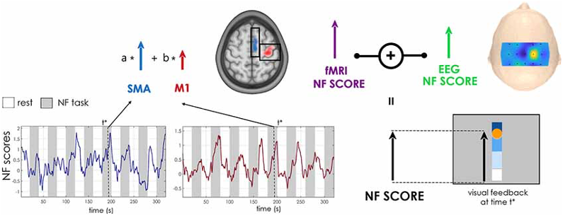
Efficacy of bimodal EEG-fMRI Neurofeedback for stroke rehabilitation
Participants: Giulia Lioi, Mathis Fleury, Quentin Duché, Lou Scotto di Covella, Elise Bannier, Pierre Maurel, Isabelle Bonan, Christian Barillot.
Neurofeedback (NF) is a brain rehabilitation technique that have shown potential for motor rehabilitation after a stroke. It has mostly been using either EEG or fMRI, both methods having their advantages and drawbacks. Therefore, there has been a growing interest in using simultaneous NF tasks, using both EEG and fMRI signals, which could powerfully enhance a more specific regulation. In this project, we investigate the effect of bi-modal EEG-fMRI NF (interventional group (IG)) versus motor imagery (non-interventional group (NIG)) in patients. The feasibility of the NF training was investigated with respect to the integrity of the corticospinal tract (CST), a well-established predictor of the potential for clinical improvement. Publications of this work are forthcoming since the study is still going on, so far we have included 14 patients in the IG (out of 16) and 11 patients in the NIG (out of 16), and the team is currently working on several analysis: fMRI data, EEG data and NF scores.This work was done in collaboration with Simon Butet, Physical and Rehabilitation Medicine Department, University Hospital of Rennes and with Anatole Lécuyer, Hybrid Inria team.
Implementation of an original visual metaphor in a bimodal EEG-fMRI Neurofeedback protocol
Participants: Giulia Lioi, Pauline Rolland, Lou Scotto di Covella, Pierre Maurel, Christian Barillot.
Neurofeedback (NF) is a technique based on operant conditioning which helps a subject to learn how to modulate his own cerebral activity. A brain activity of interest is acquired in real-time, treated and provided to the subject thanks to a feedback (also called “metaphor”). It is an innovating non-pharmacological treatment for motor rehabilitation in psychiatric diseases such as major depressive disorder. The frontal alpha asymmetry in EEG is a well-known marker of depression underlying deficiencies in the reward system and continuous attention. The left amygdala is responsible for encoding emotional memories and is excessively triggered by negative stimuli in depressed patients. The alpha band and the amygdala are also respectively involved in visual attention and color perception. We proposed to associate the alpha asymmetry with visual blur and the BOLD signal of the amygdala with color to create an innovative metaphor. The subjects were asked to realize a positive memory recall task during the NF sessions and received feedback from a personal photograph with varying blur and colorization depending on the prefrontal alpha asymmetry and BOLD signal of the amygdala. Our main objective was to implement an original visual metaphor in a bimodal NF protocol through a proof-of-concept study. The acquisitions on healthy volunteers are still going on. So far we have acquired data for 17 participants out of 30 expected. Then, we plan to analyze the fMRI data, the EEG data and the NF scores to assess if our new metaphor is indeed more efficient to guide participants towards a better control of the two brain targets.This work was done in collaboration with Jean-Marie Batail, Psychiatry Hospital, Rennes and with Anatole Lécuyer, Hybrid Inria team.
A Survey on the Use of Haptic Feedback for Brain-Computer Interfaces and Neurofeedback
Participants: Mathis Fleury, Giulia Lioi, Christian Barillot.
Neurofeedback (NF) and brain-computer interface (BCI) applications rely on the registration and real-time feedback of individual patterns of brain activity with the aim of achieving self-regulation of specific neural substrates or control of external devices. These approaches have historically employed visual stimuli. However, in some cases vision is unsuitable or inadequately engaging. Other sensory modalities, such as auditory or haptic feedback have been explored, and multisensory stimulation is expected to improve the quality of the interaction loop. Moreover, for motor imagery tasks, closing the sensorimotor loop through haptic feedback may be relevant for motor rehabilitation applications, as it can promote plasticity mechanisms. We conducted a survey reviewing the various haptic technologies and describing their application to BCIs and NF as illustrated in Figure 4. We identified major trends in the use of haptic interfaces for BCI and NF systems and discussed crucial aspects that could motivate further studies 24.This work was done in collaboration with Anatole Lécuyer from the Hybrid Inria team.
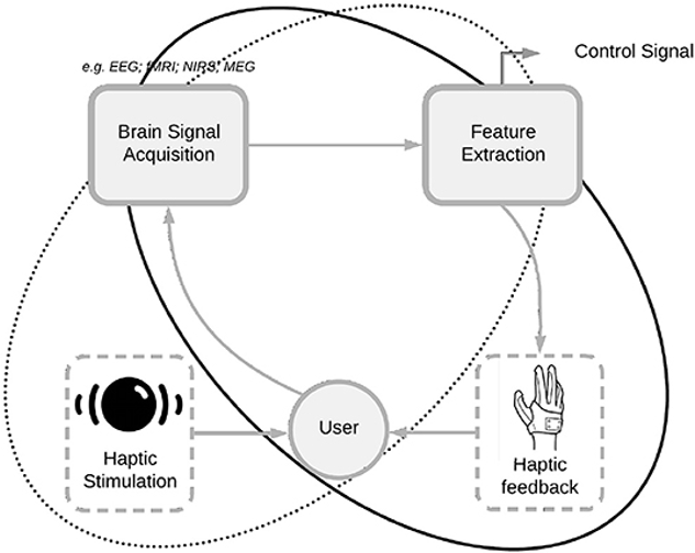
The effects of neck muscle vibration on postural orientation and spatial perception: A systematic review
Participants: Stéphanie Leplaideur, Isabelle Bonan.
Neck muscle vibration (NMV) is increasingly used for its modulation of body orientation and spatial perception, but its mechanisms of action are still not well known. The objectives of this work were to describe the effects of NMV on postural orientation and spatial perception, in both healthy people and patients with disturbed balance potentially related to distorted body orientation perception. Following the PRISMA guidelines, a systematic search was performed using the databases MEDLINE, EMBASE, Cochrane library and PEDrO with the key words ((Postural balance) OR (Spatial reference)) AND (Neck muscle vibration) for articles published through to July 2016. A total of 67 articles were assessed; these exhibited wide heterogeneity and generally poor quality methodology. In healthy subjects, under bilateral NMV, the body tilts in the anterior direction (Level of Evidence LoE II). Under unilateral NMV,the visual environment moves towards the side opposite the vibration (LoE II) and the subject’s experience of ‘‘straight ahead’’ is shifted towards the side of the vibration (LoE II). NMV also modulates both spatial and postural bias between stroke and vestibular patients. NMV modulates both spatial and postural bias and could thus be proposed as a tool in rehabilitative therapy. However, due to the heterogeneity of published data and the various significant shortfalls highlighted, current research does not allow clear guidelines to be proposed 30.This work was done in collaboration with Karim Jamal, Physical and Rehabilitation Medicine Department, University Hospital of Rennes/M2S laboratory–EA 1274, University of Rennes 2; Frédérique Leblanche, Physical and Rehabilitation Medicine Department, University Hospital of Rennes; Annelise Moulinet Raillon, Physical and rehabilitation medicine department, Hospital of Saint-Vallier; Thibaud Honoré, Physical and Rehabilitation Medicine Department, University Hospital of Rennes.
The effects of repetitive neck-muscle vibration on postural disturbances after a chronic stroke
Participants: Stéphanie Leplaideur, Isabelle Bonan.
We aimed to test a repeated program of vibration sessions of the neck muscles on postural disturbances and spatial perception in patients with right versus left vascular brain damage. Thirty-two chronic stroke patients underwent a program of 10 sessions over two weeks. Posturography parameters,balance rating, Timed Up and Go, space representation (subjective straight ahead (SSA), longitudinal body axis (LBA), subjective visual vertical (SVV)), and post-stroke deficiencies (motricity index, sensitivity, and spasticity) were tested and the data analyzed by ANOVA or a linear rank-based model, depending on whether the data were normally distributed, with lesion side and time factor. The ANOVA revealed a significant interaction between lesion side and time for WBA (P < 0.0001) with a significant shift towards the paretic lower limb in the RBD patients only (P = 0.0001), whereas there was no effect in the LBD patients (P = 0.98). Neither group showed a significant modification of spatial representation. Nonetheless, there was a significant improvement in motricity (P = 0.02), TUG (P = 0.0005), and BBS (P < 0.0001) in both groups at the end of treatment and afterwards. rNMV appeared to correct WBA in RBD patients only. This suggests that rNMV could be effective in treating sustainable imbalance due to spatial cognition disorders 29.This work was done in collaboration with Karim Jamal, Physical and Rehabilitation Medicine Department, University Hospital of Rennes/M2S laboratory–EA 1274, University of Rennes 2; Chloé Rousseau, Department of Clinical Pharmacology, Clinical Investigation, Center INSERM 1414, University Hospital of Rennes; Sébastien Cordillet, Physical and Rehabilitation Medicine Department, University Hospital of Rennes; Annelise Moulinet Raillon, Physical and rehabilitation medicine department, Hospital of Saint-Vallier; Simon Butet, Physical and Rehabilitation Medicine Department, University Hospital of Rennes; Armel Crétual, M2S laboratory–EA 1274, University of Rennes 2.
Effect of prism adaptation and neck muscle vibration on body weight asymmetry after recent right hemisphere stroke: a multicentre randomized controlled study
Participants: Stéphanie Leplaideur, Isabelle Bonan.
Spatial cognition disorders may contribute to body weight asymmetry after stroke. The main objective of this study was to test an intervention with prism adaptation and/or neck muscle vibration on postural asymmetry after recent right hemisphere stroke. We found a Vibration effect exclusively with eyes open without an additive effect of sessions. In contrast, the prism adaptation effect occured with eyes closed and tended to be cumulative. Surprisingly, the combination of these two interventions had a negative effect, revealing a conflicting effect in the sensory stimulation (such as egocentric vs allocentric stimulation) or temporo-parietal overstimulation 48.This work was done in collaboration with Karim Jamal, Physical and Rehabilitation Medicine Department, University Hospital of Rennes/M2S laboratory–EA 1274, University of Rennes 2; Etienne Allart, Neuro-rehabilitation Department, University Hospital of Lille; Lucie Chochina, centre Mutualiste de Rééducation et de Réadaptation Fonctionnelles de KERPAPE, Ploemeur; Dominique Perennou, Physical and Rehabilitation Medicine Department, University Hospital of Grenoble; Gilles Rode, Physical and Rehabilitation Medicine Department, Hospices Civils de Lyon; François Boyer, University Hospital of Reims; Jean Paysant, Institut Régional de Médecine Physique et de Réadaptation, Nancy; Alain Yelnik, Hôpital Lariboisière, APHP, Paris.
Influence of virtual reality visual feedback on the illusion of movement induced by tendon vibration of wrist in healthy participants
Participants: Mathis Fleury, Christian Barillot, Isabelle Bonan.
Illusion of movement induced by tendon vibration is an effective approach for motor and sensory rehabilitation in case of neurological impairments. The aim of our study was to investigate which modality of visual feedback in Virtual Reality (VR) associated with tendon vibration of the wrist could induce the best illusion of movement. We included 30 healthy participants in the experiment (see Figure 5). Tendon vibration inducing illusion of movement (wrist extension, 100Hz) was applied on their wrist during 3 VR visual conditions (10 times each): a moving virtual hand corresponding to the movement that the participants could feel during the tendon vibration (Moving condition), a static virtual hand (Static condition), or no virtual hand at all (Hidden condition). After each trial, the participants had to quantify the intensity of the illusory movement on a Likert scale, the subjective degree of extension of their wrist and afterwards they answered a questionnaire. There was a significant difference between the 3 visual feedback conditions concerning the Likert scale ranking and the degree of wrist’s extension (p<0.001). The Moving condition induced a higher intensity of illusion of movement and a higher sensation of wrist’s extension than the Hidden condition (p<0.001 and p<0.001 respectively) than that of the Static condition (p<0.001 and p<0.001 respectively). The Hidden condition also induced a higher intensity of illusion of movement and a higher sensation of wrist’s extension than the Static condition (p<0.01 and p<0.01 respectively). The preferred condition to facilitate movement illusion was the Moving condition (63.3%). This study demonstrated the importance of carefully selecting a visual feedback to improve the illusion of movement induced by tendon vibration, and the increase of illusion by adding VR visual cues congruent to the illusion of movement. Further work will consist in testing the same hypothesis with stroke patients 33.This work was done in collaboration with Salomé Le Franc, Mélanie Cogné, Simon Butet from the Rehabilitation Medicine Unit, University Hospital of Rennes; and with Anatole Lécuyer from the Hybrid Inria team.
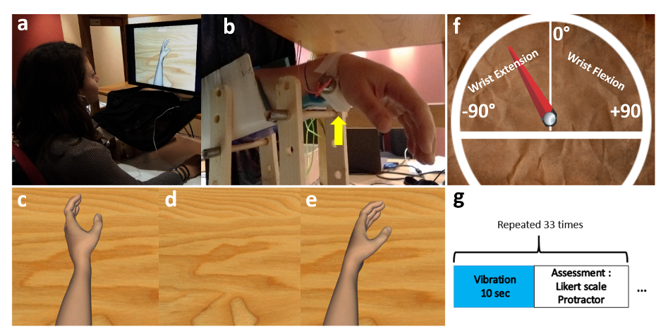
9 Bilateral contracts and grants with industry
9.1 Bilateral contracts with industry
9.1.1 Siemens
Participants: Elise Bannier, Christian Barillot, Emmanuel Caruyer, Olivier Commowick, Isabelle Corouge, Jean-Christophe Ferré, Jean-Yves Gauvrit.
In the context of the Neurinfo imaging platform, a master research agreement between Siemens SAS - Healthcare and University of Rennes 1 defines the terms of the collaboration between Siemens, Empenn and the Neurinfo platform. Relying on this research agreement contract, Neurinfo has received work in progress (WIP) sequences from Siemens in the form of object code for evaluation in the context of clinical research. The Neurinfo platform has also received source code of selected MRI sequences. As an example, the diffusion sequence code was modified to load arbitrary diffusion gradient waveforms for the FastMicroDiff project led by E. Caruyer. This is crucial in the collaboration since it enables the development of MRI sequences on site. The MR Diffusion pulse sequence source code was modified in collaboration with our Siemens clinical scientist as part of our Master Research Agreement, Marc Lapert, in order to play arbitrary gradient waveforms. This was done on the Syngo VB17 software version and again VE11C. Acquisitions on healthy controls have started to evaluate several sets of waveforms.10 Partnerships and cooperations
10.1 International initiatives
10.1.1 Inria International Labs
MMINCARAV
- Title: Multimodal Microstructure-Informed Connectivity: Acquisition, Reconstruction, Analysis and Validation
- Duration: 2019 - 2021
- Coordinator: Emmanuel Caruyer
-
Partners:
- Laboratoire de Traitement du Signal 5, Ecole Polytechnique Fédérale de Lausanne (Switzerland)
- Inria contact: Emmanuel Caruyer
- Summary: The MMINCARAV associate team is part of the Inria-EPFL International lab program. The objectives of this associate team will be to address new scientific challenges related to the use of multimodal magnetic resonance imaging (MRI) to derive microstructure indices and apply them to the measure of brain connectivity. We will focus on 4 aspects of this: first we will develop novel sampling techniques, with the objective to reduce acquisition time for the accurate reconstruction of microstructure indices using diffusion MRI; next we will propose joint T2 relaxometry and diffusion models for the description of microstructure, to take advantage of the complementarity of both modalities in the estimation of microstructure indices; in continuation, we will propose new statistical and network analysis methods using the microstructure-informed connectome, and evaluate its potential to reduce bias and false positives; last we will develop a realistic simulation tool combining a fine macroscopic description of fiber bundles, with a fast and realistic simulator at the mesoscopic scale developed by LTS5.
- Élise Bannier, Emmanuel Caruyer, Julie Coloigner and Raphaël Truffet visited the signal processing lab 5 (LTS5), EPFL (Lausanne, Switzerland), for a 2 days workshop organized in the context of this MMINCARAV associate team.
10.1.2 Inria international partners
Informal international partners
- Pierre-Yves Jonin collaborates with
- Dr Gabriel Besson, University of Coimbra, Portugual: Behavioral and neuroimaging studies of ultra-fast familiarity for visual objects
- Dr Ann-Kathrin Zaiser, University of Heidelberg, Germany: Neuropsychological and neuroimaging studies of the implicit learning of new concepts
- Camille Maumet collaborates with
- Prof. Thomas Nichols and his group, NISOx at the Oxford Big Data Institute on neuroimaging statistics,
- Prof. Jean-Baptiste Poline and his group at McGill University on neuroimaging data sharing,
- Prof. Satrajit Ghosh and his group at MIT,
- Dr David Keator at UCI Irvine, Dr. Karl Helmer at MGH, on neuroinformatics and neuroimaging standards,
- international members of the INCF on neuroimaging data sharing.
- Olivier Commowick collaborates with Prof. Simon K. Warfield and his group, Computational Radiology Laboratory at Childrens Hospital Boston, on topics ranging from diffusion image processing to acquisition and processing of relaxometry MRI data.
- Claire Cury collaborates with Pr, Arnaud Cachia from the Institut de Psychiatrie et Neurosciences de Paris on the contribution of deviations in early hippocampus development to visual hallucinations in schizophrenia.
- Elise Bannier collaborates with
- the CEMEREM lab in Marseille, in particular Virginie Callot, Bertrand Audoin, Jean Pelletier and Adil Maarouf on multiple sclerosis multicenter MRI studies (PHRC EMISEP and MS-TRACTS projects)
- members of the REMI network, in particular members from the bureau, Gabriel Hossu from the CHU Nancy, Irène Troprès from the Irmage Grenobles Alpes MRI facility, Emmanuelle le Bars form the CHU Montpellier, Marie Chupin from the CATI in Paris and Jean-Luc Anton from the CERIMED in Marseille, on recommendations for multicenter MR imaging, expertise sharing on data acquisition, analysis and quality control.
- international members of the COST action on neuroimaging data sharing (e.g, the Open Brain Consent initiative).
10.2 International research visitors
10.2.1 Visits of international scientists
- Jonathan Rafael-Patiño, PhD candidate from EPFL, visited Empenn for 4 weeks in september 2020 in the context of the MMINCARAV associate team.
10.3 European initiatives
10.3.1 European COST Action GLIMR
Participants: Camille Maumet.
The GLIMR COST Action (PI: Esther Warnert, Erasmus MC, Netherlands) aims to build a pan-European and multidisciplinary network of international experts in glioma research, patient organisations, data scientists, and MR imaging scientists by uniting the glioma imaging community within Europe and progressing the development and application of advanced MR imaging for improved decision making in diagnosis, patient monitoring, and assessment of treatment response in clinical trials and clinical practice. Camille Maumet leads the work package "WG2 - Multi-site data integration" with Cyril Pernet (University of Edinburgh, UK).
GliMR’s first grant period ran from September 2019 to April 2020, during which several meetings were held and projects were initiated, such as reviewing the current knowledge on advanced MRI; developing a General Data Protection Regulation (GDPR) compliant consent form; and setting up the website. A publication led by Patricia Clement (Ghent University, Belgium) was published in 2020 18 describing the results of this GliMR’s first grant period. The Action overcomes the pre-existing limitations of glioma research and is funded until September 2023. New members will be accepted during its entire duration.
10.4 National initiatives
10.4.1 Rapid Neocortical Declarative Learning in normal aging and memory disorders
Participants: Pierre-Yves Jonin, Julie Coloigner.
Funding: Fondation de l'Avenir - Duration: 2020-2022 - Budget: 40k€
Our project aims at making the case for the existence of a rapid declarative learning system largely independent from the extended hippocampal system and characterizing its neural bases, by use of experimental psychology, cognitive neuropsychology and neuroimaging methods. This project is led in collaboration with Dr Audrey Noël, Assistant Prof., University of Rennes 2, France, with Dr Gabriel Besson, Associate Researcher, University of Coimbra, Portugal, with Dr Ann-Kathrin Zaiser, Associate Researcher, University of Heidelberg, Germany, with Dr Serge Belliard, PhD, MD, Rennes University Hospital, Neurology Dept., France, and with Dr Anca Pasnicu, MD, Rennes University Hospital, Neurology Dept., France.
10.4.2 Effect of prenatal exposures to neurotoxicants on the developing brain: an MRI study (PERINE)
Participants: Élise Bannier, Isabelle Corouge, Julie Coloigner, Jean-Christophe Ferré, Christian Barillot.
Funding: Fondation de France - Duration: 2015-2021 - Budget: 100k€
The PELAGIE cohort evaluates the effect of prenatal exposure to neurotoxicants on child development. Following previous studies, the PERINE study focuses on the assessment of brain developement at 10-12 years old using MRI (ASL, Diffusion imaging, working memory as well as motor inhibition BOLD fMRI together with neuropsychological tests). A total of 101 children were included. A PhD of Anne-Claire Binter was defended in December 2019 linking epidemiology with functional imaging during a GoNoGo task and neuropsychological scores and two publications were co-authored. This work is done in collaboration with Fabienne Pelé and Cécile Chevrier (IRSET).
10.4.3 Connectivity of the amygdala in depression
Participants: Christian Barillot, Olivier Commowick, Emmanuel Caruyer, Julie Coloigner, Claire Cury.
Funding: Fondation de France - Duration: 2019-2021 - Budget: 200k€
The onset of depression in teenagers and young adults increases the risk to develop a drug-resistant depression in the adulthood. This project aims at evaluating the role of early changes in the microstructure and connectivity of the amygdala. Using a cohort of drug-resistant patients (N=30), non drug-resistant patients (N=30) and controls (N=30), the aim is to identify imaging biomarkers of the pathology and to compare these with emotional and cognitive phenotypes in this population, searching for early differences in the development of the amygdala connectivity. Inclusions are ongoing.
This is a colloborative project with M.-L. Paillère Martinot from Paris-Descartes University, as Principal Investigator.
10.4.4 Multimodal Imaging of the Limbic Amygdala for the Prognosis of Depression (IMpAirED)
Participants: Julie Coloigner, Olivier Commowick, Élise Bannier, Emmanuel Caruyer, Christian Barillot.
Funding: CNRS-Inserm Défi Santé numérique AAP 2019 - Start: 2019 - Budget: 19k€
This grant is an extension of the Projet Fondation de France: Connectivity of the amygdala in depression.
In order to identify early features of this depression disease, the aim of this project is to develop multimodal modeling of the limb amygdala and its network from MR imaging combining activation and rest functional imaging and MR brain microstructure quantitative imaging (diffusion and relaxometry). The development of this model will allow us to define three imaging biotypes corresponding to depressed adult patients responding to antidepressant treatments, depressed resistant patients and controls. These multimodal imaging biomarkers will be used to stratify a large longitudinal cohort of young adults into three sub-groups, in order to retrospectively identify early differences in development trajectories of amygdala.
10.4.5 Brain modeling from multi-scale, multimodal and dynamic graphs and development of statistical prediction models
Participants: Julie Coloigner.
Funding: Rennes Métropole, Allocation d'installation scientifique - Duration: 2020-2023 - Budget: 10k€
10.4.6 ANR "MAIA" Multiphysics image-based AnalysIs for premature brAin development understanding
Participants: Pierre Maurel, Antoine Legouhy, Olivier Commowick, Isabelle Corouge, Jean-Christophe Ferré, Christian Barillot.
Funding: ANR, generic projects program - Duration: 2016-2021 - Budget: 150k€ - PI: F. Rousseau, IMT Atlantique, Brest
Each year in France, 55 000 children are born prematurely, i.e., before the 37th week of gestation. Long-term studies of the outcome of prematurely born infants have clearly documented that the majority of such infants may have significant motor, cognitive, and behavioral deficits. However, there is a limited understanding of the nature of the cerebral abnormality underlying these adverse neurologic outcomes. In this context, the emergence of new modalities of 3D functional MRI, e.g., Arterial Spin Labeling(ASL) or optical imaging technologies, e.g., Near InfraRed Spectroscopy (NIRS), brings new perspectives for extracting cognitive information, via metabolic activity measures. Other classical techniques devoted to cerebral signal measurement, such as ElectroEncephaloGraphy (EEG), provide cognitive information at the cortical level. Each of these various non-invasive imaging technologies brings substantial and specific information for the understanding of newborn brain development.
This project is developing innovative approaches for multi-image / multi-signal analysis, in order to improve neurodevelopment understanding methods. From a fundamental point of view, mathematics and computer science have to be considered in association with imaging physics and medicine, to deal with open issues of signal and image analysis from heterogeneous data (image, signal), considered in the multiphysics contexts related to data acquisition (magnetic, optic, electric signals) and biophysics modeling of the newborn brain. A sustained synergy between all these scientific domains is then necessary. Finally, the sine qua non condition to reach a better understanding of the coupled morphological cognitive development of premature newborns, is the development of effective software tools, and their distribution to the whole medical community. The very target of this project is the design of such software tools for medical image / signal analysis, actually operational in clinical routine, and freely available. Academic researchers and industrial partners are working in close collaboration to reach that ambitious goal.
In 2020, Empenn produced two publications in the scope of the ANR MAIA project (34, 55).
10.4.7 Hybrid EEG/IRM Neurofeedback for rehabilitation of brain pathologies
Participants: Élise Bannier, Isabelle Bonan, Isabelle Corouge, Jean-Christophe Ferré, Jean-Yves Gauvrit, Pierre Maurel, Mathis Fleury, Giulia Lioi, Christian Barillot.
Funding: Fondation pour la recherche médicale (FRM) - Duration: 2017-2021 - Budget: 370k€
This project is a continuation of the HEMISFER project ("Hybrid Eeg-MrI and Simultaneous neuro-FEedback for brain Rehabilitation") conducted at Inria Rennes with the support of the Labex "CominLabs" 1.
The goal of this project is to make full use of neurofeedback (NF) paradigm in the context of brain rehabilitation. The major breakthrough will come from the coupling associating functional and metabolic information from Magnetic Resonance Imaging (fMRI) to Electro-encephalography (EEG) to “optimize” the neurofeedback protocol. We propose to combine advanced instrumental devices (Hybrid EEG and MRI platforms), with new hybrid Brain computer interface (BCI) paradigms and new computational models to provide novel therapeutic and neuro-rehabilitation paradigms in some of the major mental and neurological disorders of the developmental and the aging brain (stroke, language disorders, Mood Depressive Disorder (MDD), …). Though the concept of using neurofeedback paradigms for brain therapy has somehow been experimented recently (mostly through case studies), performing neurofeedback through simultaneous fMRI and EEG has almost never been done before so far (two teams in the world including us within the HEMISFER CominLabs project). This project will be conducted through a very complementary set of competences over the different involved teams: Empenn U1228, HYBRID and PANAMA Teams from Inria/Irisa Rennes and EA 4712 team from University of Rennes I.
10.4.8 PHRC EMISEP: Evaluation of early spinal cord injury and late physical disability in Relapsing Remitting Multiple Sclerosis
Participants: Élise Bannier, Christian Barillot, Emmanuel Caruyer, Benoit Combès, Olivier Commowick, Gilles Edan, Jean-Christophe Ferré, Haykel Snoussi.
Funding: PHRC - Duration: 2016-2021 - Budget: 200k€
Multiple Sclerosis (MS) is the most frequent acquired neurological disease affecting young adults (1 over 1000 inhabitants in France) and leading to impairment. Early and well adapted treatment is essential for patients presenting aggressive forms of MS. This PHRC (Programme hospitalier de recherche clinique) project focuses on physical impairment and especially on the ability to walk. Several studies, whether epidemiologic or based on brain MRI, have shown that several factors are likely to announce aggressive development of the disease, such as age, number of focal lesions on baseline MRI, clinical activity. However, these factors only partially explain physical impairment progression, preventing their use at the individual level. Spinal cord is often affected in MS, as demonstrated in postmortem or imaging studies. Yet, early radiological depiction of spinal cord lesions is not always correlated with clinical symptoms. Preliminary data, on reduced number of patients, and only investigating the cervical spinal cord, have shown that diffuse spinal cord injury, observed via diffusion or magnetisation transfer imaging, would be correlated with physical impairment as evaluated by the (EDSS) Expanded Disability Status Scale score. Besides, the role of early spinal cord affection (first two years) in the evolution of physical impairment remains unknown.
In this project, we propose to address these different issues and perform a longitudinal study on Relapsing Remitting Multiple Sclerosis (RRMS) patients, recruited in the first year of the disease. Our goal is to show that diffuse and focal lesions detected spinal cord MRI in the first two years can be used to predict disease evolution and physical impairment at 5 years. Twelve centers are involved in the study to include 80 patients.
To date, all subjects have been included. The EMISEP data consists of brain and spinal cord structural and quantitative MR images of early MS patients followed over 5 years. From November 2016 to August 2020, B. Combès processed and analyzed the data corresponding to the two first years of follow-up. Four papers have been published so far and three additional papers are under submission or in preparation. Haykel Snoussi defended his PhD Thesis on diffusion imaging in the spinal cord starting with distortion correction.
10.4.9 Estimating the impact of multiple sclerosis lesions in motor and proprioceptive tracts, from the brain to the thoracic spinal cord, on their functions, assessed from clinical tests and electrophysiological measurements (MS-TRACTS)
Participants: Élise Bannier, Benoit Combès.
Funding: ARSEP and COREC - Duration: 2019-2021 - Budget: 100k€
Previous studies, whether epidemiologic or based on brain MRI, have shown that several factors were likely to announce aggressive development of the disease, such as age, clinical relapses, number of focal lesions on baseline MRI. However, these factors only partially explain physical disability progression, preventing their use at the individual level. We hypothesize that a fine assessment of damage on specific networks, from the brain to the thoracic cord, offers a relevant biomarker of disability progression in MS. Such damage assessments must take into account both lesion location, assessed on structural brain and cord MR images and lesion severity, assessed using advanced brain and cord imaging through quantitative MRI. We propose to test this hypothesis by combining assessments of lesion location and severity on corticospinal and proprioceptive tracts from the brain to the thoracic cord with clinical and electrophysiological measurements.
This study includes two French centers (Rennes, Marseille) and includes a total of 60 patients. The expected outcome is to obtain early biomarkers of physical impairment evolution in RRMS patients, first treated with immunomodulatory treatment. The long-term goal is to provide the clinician with biomarkers able to anticipate therapeutic decisions and support the switch to alternative more aggressive treatment. Inclusions are ongoing.
10.4.10 PIA projects
France Life Imaging (FLI)
Participants: Christian Barillot, Olivier Commowick.
Funding: FLI - Duration: 2012-2023 - Total budget: 2000k€ (phase 1) + 1200k€ (phase 2) + 800k€ (phase 3)
France Life Imaging (FLI) is a large-scale research infrastructure project to establish a coordinated and harmonized network of biomedical imaging in France. This project was selected by the call “Investissements d’Avenir - Infrastructure en Biologie et Santé”. One node of this project is the node Information Analysis and Management (IAM), a transversal node built by a consortium of teams that contribute to the construction of a network for data storage and information processing. Instead of building yet other dedicated facilities, the IAM node use already existing data storage and information processing facilities (LaTIM Brest; CREATIS Lyon; CIC-IT Nancy; Empenn U1228 Inria Rennes; CATI CEA Saclay; ICube Strasbourg) that increase their capacities for the FLI infrastructure. Inter-connections and access to services are achieved through a dedicated software platform that is developed based on the expertise gained through successful existing developments. The IAM node has several goals. It is building a versatile facility for data management that inter-connects the data production sites and data processing for which state-of-the-art solutions, hardware and software, are available to infrastructure users. Modular solutions are preferred to accommodate the large variety of modalities acquisitions, scientific problems, data size, and to be adapted for future challenges. Second, it offers the latest development that are made available to image processing research teams. The team Empenn fulfills multiple roles in this nation-wide project. Christian Barillot is the chair of the node IAM, Olivier Commowick is participating in the working group workflow and image processing and Michael Kain is the technical manager. Apart from the team members, software solutions like MedInria and Shanoir are part of the software platform.
OFSEP: French Multiple Sclerosis Observatory
Participants: Élise Bannier, Christian Barillot, Olivier Commowick, Gilles Edan, Jean-Christophe Ferré, Francesca Galassi, Arthur Masson, Benoît Combès, Brandon Le Bon.
Funding: ANR-PIA - Duration: 2017-2021 - Budget: 175k€
The French Observatory of Multiple Sclerosis (OFSEP) is one of ten projects selected in January 2011 in response to the call for proposal in the “Investissements d’Avenir - Cohorts 2010” program launched by the French Government. It allows support from the National Agency for Research (ANR) of approximately 10 million € for 10 years. It is coordinated by the Department of Neurology at the Neurological Hospital Pierre Wertheimer in Lyon (Professor Christian Confavreux), and it is supported by the EDMUS Foundation against multiple sclerosis, the University Claude Bernard Lyon 1 and the Hospices Civils de Lyon. OFSEP is based on a network of neurologists and radiologists distributed throughout the French territory and linked to 61 centers. OFSEP national cohort includes more than 50,000 people with Multiple Sclerosis, approximately half of the patients residing in France. The generalization of longitudinal monitoring and systematic association of clinical data and neuroimaging data is one of the objectives of OFSEP in order to improve the quality, efficiency and safety of care and promote clinical, basic and translational research in MS. For the concern of data management, the Shanoir platform of Inria has been retained to manage the imaging data of the National OFSEP cohort in multiple sclerosis.
One long term objective of the OFSEP project is to identify prognostic factors of the evolution of Multiple Sclerosis. The HD Cohort is an enhanced cohort specifically designed for this purpose in which some patients are followed-up on a yearly basis. Additional clinical, quality of life and other patient-reported data is also collected. This study aims at developing personalized predictive tools to improve patient care management, and help in making decision to start, maintain or adapt medical care. Collected data will be processed to extract valuable information enabling to determine specific biomarkers of the evolution of the disease. Multiple Sclerosis brain lesions are of particular interest, hence the need for a careful comparison of lesion segmentation methods. A litterature review enabled to gather most promising cross-sectionnal methods, designed to identify and localize lesions with precise measurement of the lesion load at one particular point in time ; and longitudinal methods which gives more insight on the evolution of those lesions over the different time points. Those later methods are particularly interesting for clinicians for whom the type of lesion evolution is of foremost importance. A ground truth has been carefully designed with images taken from a group of 100 patients selected from the HD Cohort. Those images were meticulously segmented by four experts and the obtained semgentations where reviewed by another experienced radiologist. Four cross-sectionnal methods and one longitudinal methods where trained and evaluated to select the ones which will be used to analyze the entire HD Cohort dataset.
10.5 Regional initiatives
Region Bretagne: project VARANASI
Participants: Christian Barillot, Camille Maumet, Xavier Rolland, Pierre Maurel.
Budget: 0.5 PhD thesis
Thanks to the development of open science practices, more and more public datasets are available to the research community. In the field of brain imaging, these data, combined, bring a critical increase in sample size, necessary to build robust models of the typical and atypical brain. However, in order to build valid inferences on these data, we need to take into account their heterogeneity. Variability can arise due to multiple factors such as: differences in imaging instruments, in acquisitions protocols and even, in post-processing pipelines. In particular, the expansion of open source machine learning workflows creates a multitude of possible outputs out of the same dataset. The variations induced by this methodological plurality can be referred to as ‘analytic variability’ which will be the focus of the thesis funded in half by region Bretagne. The thesis of Xavier Rolland (2018-2021) will address two challenges: 1) How to combine neuroimaging data generated by different analysis pipelines? 2) How to publish neuroimages with an adequate level of metadata to enable their reuse? Methodological developments will combine machine learning techniques with methods from knowledge representation.
Grand Ouest: project MoDaL
Participants: Aurélien Cornet, Christian Barillot, Camille Maumet.
Biological research exploring is increasingly confronted with the need to link heterogeneous multi-scale data. In spite of existing national initiatives of mutalization and standardization through large research infrastructures such as FLI (France Life Imaging), FBI (France BioImaging), or IFB (Institut Français de Bioinformatics), it is very challenging to perform a joint analysis of these various data sources. MoDaL (Multiscale Data Links), is a federating project bringing together the Brittany and Pays de la Loire regions, funded by Biogenouest, led by Christian Barillot of IRISA Rennes and Richard Redon of the Thorax Institute in Nantes with Olivier Dameron, Camille Maumet and Anne Siegeld of IRISA Rennes and Alban Gaignard of the Thorax Institute in Nantes. In 2020, we organized a workshop 58 and proposed an overview of the Grand Ouest landscape in biomedical, vegetal biology and marine biology 57.11 Dissemination
11.1 Promoting scientific activities
11.1.1 Scientific events: organisation
General chair, scientific chair
- Camille Maumet, Chair of OHBM Open Science Special Interest Group, overseeing the Open Science Room and OHBM Hackathon, 1000+ attendees, Montreal, Canada (Online).
- Camille Maumet was chair of the symposium "Open Science in Action: Doing research as a community!" at the annual congree of Organisation of Human Brain Mapping, 2020 (Online).
- Emmanuel Caruyer was chair of the session "fMRI Analysis I" at the IEEE International Symposium on Biomedical Imaging, organized virtually from April 3rd to April 7th.
Member of the organizing committees
- Emmanuel Caruyer co-organized the "Journées nouvelles imageries en hommage à Christian Barillot", an initiative by INS2I, CNRS, organized on December 16th and 17th.
- Elise Bannier co-organized the "Journée REMI", an initiative from the REMI network, which took place on January 21st (Paris) and on December 3rd (Online).
- Elise Bannier co-organized the "Workshop IRM ARSEP - Imaging GLymphatic system in Multiple Sclerosis", an initiative from the ARSEP Foundation, which took place on February 7th in Paris.
11.1.2 Scientific events: selection
Member of the conference program committees
Reviewer
- Information Processing in Medical Imaging - IPMI (Olivier Commowick, Claire Cury)
- Annual congress of the Organisation of Human Brain Mapping (Camille Maumet)
- IEEE International Symposium on Biomedical Imaging (Olivier Commowick)
- Medical image computing and Computer assisted intervention - MICCAI (Olivier Commowick)
- Workshop on Biomedical Image Registration - WBIR (Olivier Commowick)
- Congress from the European Society for Magnetic Resonance in Medicine and Biology - ESMRMB (Elise Bannier)
- Congress from the French Society for Magnetic Resonance in Medicine and Biology - SFRMBM (Elise Bannier)
- SIPAIM 2020 International Symposium on Medical Information Processing and Analysis (Julie Coloigner)
11.1.3 Journal
Member of the editorial boards
- Camille Maumet is member of Editorial Boards of Neuroinformatics
Reviewer - reviewing activities
- Brain Science (4 papers, Claire Cury)
- Brain Connectivity (Julie Coloigner)
- Molecular Psychiatry (1 paper, Claire Cury)
- Neuroimage (1 paper, Claire Cury; 2 papers, Emmanuel Caruyer; 1 paper, Olivier Commowick)
- Neuroimage: Clinical (Julie Coloigner)
- Nature Communications (1 paper, Camille Maumet)
- Magnetic Resonance in Medicine (1 paper, Emmanuel Caruyer)
- Medical Image Analysis (1 paper, Emmanuel Caruyer; 2 papers, Olivier Commowick)
- Memory (Pierre-Yves Jonin)
- Neurocase (Pierre-Yves Jonin)
- SN Applied Sciences (1 paper, Emmanuel Caruyer)
11.1.4 Invited talks
- Isabelle Corouge, "Functional NIRS: Principles and applications in neuro-imaging", Gen2Bio; November 2020 (remote)
- Pierre-Yves Jonin, "Les modèles de la mémoire humaine en neurosciences cognitives"; Journées de Neurologie de Bretagne; October 2020 (remote)
- Camille Maumet, "Building a more collaborative neuroimaging science" Think Open Rovereto Workshop 2020, July 2020 (online) 49.
- Camille Maumet, "Collaboratively Building Large Neuroimaging Datasets for Glioma Research", ESRMBM 2020, September 2020 (Online) 50.
- Camille Maumet, "Sharing more than research papers for transparent and reusable research", Workshop Open and reproducible neuroimaging, University of Oldenburg, November 2020 (Online) 53.
- Camille Maumet, "Towards large-scale brain imaging studies". Workshop Inria Chile, Inteligencia Articicial en imagenes medicas, Santiago, Chile, December 2020 (Online) 54.
11.1.5 Leadership within the scientific community
- Camille Maumet. Chair of the Open Science special interest group of the international Organization of Human Brain Mapping. This group is known for the organization of the Open Science Room and the OHBM Brainhack, two international events for the open neuroscience community.
- Camille Maumet. Member (by selection) of the national committee on Open Science, Working group "open software" led by Roberto Di Cosmo and François Pellegrini.
- Elise Bannier. Member (by election) of the national SFRMBM society's internal office.
- Elise Bannier. Member (Founding member) of the national REMI network for mutual aid in MRI clinical research.
11.1.6 Scientific expertise
-
Benoît Combès is the scientific leader of the metric4MS project. Metric4MS is a software for the computation of metrics from MRIs for the monitoring of multiple sclerosis. This prototype is developed by InriaTech in collaboration with the clinicians of the Rennes University Hospital and is funded by the Institute of Neurosciences of Rennes. It is intended to serve as a basis for possible industrial developments.
Duration: February 2020-June 2021.
11.2 Teaching - Supervision - Juries
11.2.1 Teaching
- L2 Informatique: Raphaël Truffet, Génie Logiciel, (TP: 20h), L2, ISTIC
- L2 Mathématiques: Raphaël Truffet, Calculabilité (TD/TP: 24h), University of Rennes 1
- L3 bioinformatique: Corentin Vallée, Statistics, 32h, University of Rennes 1, France.
- L3 SIF (ENS Rennes/ISTIC): Raphaël Truffet, Algorithmic (TD: 20h)
- L3 SIF (ENS Rennes/ISTIC): Emmanuel Caruyer, Digital image processing (20h)
- M2 SIF (ENS Rennes/ISTIC): Julie Coloigner, Computer vision (Plenary: 10h)
- Master SIBM, M2, University of Angers-Brest-Rennes:
- Jean-Christophe Ferré is head of the master.
- Benoît Combès is co-head of the UE “Méthodes avancées de traitement des données spatio-temporelles”
- Élise Bannier, “IRM fonctionnelle BOLD” (Plenary: 1h).
- Emmanuel Caruyer, “Introduction to diffusion MRI” (Plenary: 3h).
- Julie Coloigner, "Méthodes d'analyse de la connectivité cérébrale" (Plenary: 3h).
- Benoit Combès, “Méthodes de segmentation pr l'imagerie médicale” (Plenary: 3h).
- Benoit Combès, “Méthodes de recalage linéaire et non-linéaires des images médicales” (Plenary: 6h)
- Benoit Combès, “Applications des méthodes de traitement des images médicales” (Plenary: 3h)
- Benoit Combès, “Soutenance de présentation critiques d'articles scientifiques” (TD: 3h)
- Isabelle Corouge, “Bio-marqueurs d’imagerie et IRM métabolique et fonctionnelle” (Plenary: 3h).
- Quentin Duché, “Traitement des données d’IRM fonctionnelle” (Plenary: 1h).
- Claire Cury, “Evaluation des performances en imagerie médicale”(Plenary: 3h).
- Camille Maumet, “Imaging processing pipelines” (Plenary: 3h).
- L3 Psychologie: Pierre-Yves Jonin, Travaux Dirigés UES Handicap et neurosciences cliniques, 6h, L3, Université de Rennes 2, France.
- L3 Psychologie: Pierre-Yves Jonin, Travaux Dirigés "Les syndromes neuropsychologiques", 18h, L3, Université de Rennes 2, France.
- M2 « Psychologie et Neuropsychologie de l’Enfant et de l’Adulte »: Pierre-Yves Jonin, Méthodologie psychométrique chez le sujet âgé, 4h, M2, Université de Poitiers, France.
- M2 « Psychologie, parcours Neuropsychologie »: Pierre-Yves Jonin, Limites et apports du bilan neuropsychologique dans le diagnostic des maladies neurodégénératives, 4h, M2, Université de Savoie, France.
- M2 « Neurosciences cliniques » Pierre-Yves Jonin, Neurosciences cognitives et cliniques de la mémoire humaine, 4h, M2, Faculté de Médecine de Rennes, France.
- M2 «Psychologie clinique, Psychopathologie et psychologie de la santé, parcours Handicap et troubles neurodéveloppementaux »: Pierre-Yves Jonin, Examen neuropsychologique du sujet âgé, 3h, M2, Université de Rennes 2, France.
- DCEM 4, UE Psychologie et Neurobiologie: Pierre-Yves Jonin, Exploration neuropsychologique des maladies neurologiques et psychiatriques, 4h, DCEM 4, Faculté de médecine de Brest, France.
- DIU « Sémiologie et diagnostic des démences »: Pierre-Yves Jonin, Diagnostic neuropsychologique de la maladie d'Alzheimer au stade pré-démentiel, 4h, DIU, Université de Caen, France.
- ENS Rennes: Pierre Maurel, Introduction to image processing (24h)
- ESIR, École Supérieure d’Ingénieur de Rennes:
- Pierre Maurel, General image processing (60h), Algorithmics and complexity (60h), Medical imaging (60h).
- Francesca Galassi, Databases (TP, 32h), Graph algorithms (TD/TP, 24h), Algorithms and complexity (TP 28h), Artificial Intelligence (15h), Data mining (TP 24), Medical Imaging (TP 20h).
- Licences 2 & 3 Biologie: Xavier Rolland, "Introduction aux Biostatistiques" 1 (TP sur R: 64h), Université de Rennes 1
- L2 Biologie: Xavier Rolland, TP biostatistiques sur R, 40h, University of Rennes 1
- L1 Mathématiques: Xavier Rolland, Préparation aux Concours 1, 24h, University of Rennes 1
- L1: Xavier Rolland, TD/TP Statistiques, 20h, ESIR
- Rémi Adon, git, DASK and docker, Intermediate level, 5 days, Yotta Academy
11.2.2 Supervision
PhD & HdR
- PhD: Antoine Legouhy, “Longitudinal brain altas creation, application to development studies”, CNRS, defended on June 23, 2020, Christian Barillot, François Rousseau, Olivier Commowick 55.
- PhD: Corentin Vallée, "Joint estimation of neuronal activation, resting-state and basal metabolism from Arterial Spin Labeling", defended on June 26, 2020, Univ. Rennes, Christian Barillot, Isabelle Corouge, Pierre Maurel 56.
- PhD in progress: Xavier Rolland, “Modeling analytic variability in brain imaging”, CNRS, from Oct 2018, Camille Maumet, Christian Barillot and Pierre Maurel.
- PhD in progress: Raphaël Truffet, “Compressed sensing for microstructure-enabled diffusion MRI”, Univ. Rennes / ENS Rennes, from Oct 2018, Emmanuel Caruyer.
- PhD in progress: Giovanna Orrù, “Modeling brain structural and functional connectivity ”, Univ. Rennes, from Oct 2019, Julie Coloigner and Pierre Maurel.
- PhD in progress: Stéphanie Leplaideur, "Equilibre de la rééducation par vibrations cervicales, adaptation prismatique et association aux deux techniques, chez des patients cérébro-lésés droits– Protocole AVC POSTIM " , from Oct 2017, Isabelle Bonan and Élise Bannier.
- PhD in progress; Mathis Fleury, "Multimodal Neurofeedback EEG/fMRI for brain rehabilitation", from Nov 2017, Christian Barillot and Anatole Lecuyer
- PhD in progress; Thomas Durantel, "Anatomy and microstructure informed tractography for connectivity evaluation in neurological pathologies", from Nov 2020, Olivier Commowick and Julie coloigner
Other supervisions
- L3 SIF (ENS Rennes/ISTIC): Constance Bocquillon, "Application du traitement du signal sur les graphes au problème de l'échantillonnage du signal en IRM de diffusion", supervised by Emmanuel Caruyer.
- Master Student (Ecole Superieure d'Ingenieurs de Rennes, second year) : Caroline Pinte, "Detection of EEG electrodes on fMRI acquisitions with U-Net neural", Mar-Jul 2020, co-supervised by Mathis Fleury and Pierre Maurel
- Master Student (master 2 EEA from Toulouse) : Thomas Durantel, "Informed tractography", Mar-Jul 2020, supervised by Olivier Commowick
- Co-supervised project : Alexander Bowring, Exploring what part of the fMRI pipeline has the strongest impact on Task fMRI Results, 2020, Camille Maumet in collaboration with Thomas Nichols (University of Oxford).
- Co-supervised project : Freya Acar, Best practices reporting for fMRI meta-analysis 2020, Camille Maumet in collaboration with Beatrijs Moerkerke (University of Ghent).
- M1 Physique Médicale: Aurélien Hervouin, "Evaluation of QC procedures at Neurinfo and optimisation", supervised by Elise Bannier.
- M2 SIBM: Soizic Leguy, "MTR asymmetry in the spinal cord as a biomarker for MS", Mar-Aug 2020, co-supervised by Benoit Combès.
- ESIR 3: Brandon Le Bon, "Deep learning for detection of new MS lesions from serial brain MR scans", Feb-Jul 2020, supervised by Benoit Combès.
- ESIR 3, tutor project 'BIPOSCHIZ'Appli', Centre Hospitalier Guillaume Régnier, tutored by Francesca Galassi.
11.2.3 Juries
- Emmanuel Caruyer. PhD committee: Yann Bihan-Poudec, Université Claude Bernard Lyon 1; December 17, 2019.
- Olivier Commowick. MD thesis committee: Marine Dubois "Analyse multiparamétrique du développement cérébral des enfants prématurés par IRM", Université de Rennes 1; October 21, 2020.
- Camille Maumet. PhD committee: Hannes Almgren, Ghent University, Belgium, August 27, 2020.
- Camille Maumet. PhD committee: Kamalaker Reddy Dadi, Université Paris-Saclay, September 14, 2020.
- Camille Maumet. PhD committee: Céline Delettre, Université de Paris, December 16, 2020.
11.3 Popularization
11.3.1 My thesis in 180 seconds
- Raphaël Truffet participated in the 2020 edition of the French edition of the Three Minutes Thesis competition".
11.3.2 Sciences en Cour[t]s
- Raphaël Truffet participated in the organisation and animation of the 2020 edition of "Sciences en Cour[t]s". This event is a festival of short films, which offers doctoral students the opportunity to make short films about their thesis work.
11.3.3 Articles and contents
- Camille Maumet. Interview for Le Monde "Les données de santé, un trésor mondialement convoité" par Laure Belot, mars 2020.
- Camille Maumet. Interview for Inria.fr "Projet NARPS : la robustesse de la méthode scientifique à l’épreuve des faits", septembre 2020.
- Camille Maumet. Interview "5 Outils pour Ouvrir les Sciences", Entretien Science Ouverte par Rudy Patard, octobre 2020.
11.3.4 Education
- Camille Maumet. L codent L créent - An outreach program to send PhD students to teach Python to middle school students in 8 sessions of 45 minutes. Camille Maumet is a co-organizer of the local version of this program, initiated in Lille, with Anne-Cecile Orgerie and Tassadit Bouadi. The program is currently supported by: Alstom, Fondation Blaise Pascal, ED MathSTIC, Inria and Fondation Rennes 1.
- Camille Maumet. Participation to the mini-forum with other profesionnal during the "D'Connectées !" days, Collège de Janzé.
- Brain awareness week - March 2020 - Canceled due to Covid19 lockdown - Participation of Empenn / Neurinfo supervised by Mathis Fleury and Raphaël Truffet.
11.3.5 Interventions
- Camille Maumet. Journée Santé et IA, CNRS INS2I. Talk "Quand les données s'ouvrent : Opportunités et nouveaux défis pour mieux comprendre notre cerveau", Paris, janvier 2020 52.
- Claire Cury. Journée Nouvelles Imageries - en hommage à, Christian Barillot, CNRS INS2I. Talk "Projet Hemisfer : l’imagerie cérébrale au service de la rééducation grâce au Neurofeedback", Online, December the 16-17th 2020.
12 Scientific production
12.1 Major publications
- 1 articleExploring the Impact of Analysis Software on Task fMRI ResultsHuman Brain Mapping40112019, 3362-3384
- 2 articleFocal and diffuse cervical spinal cord damage in patients with early relapsing--remitting MS: A multicentre magnetisation transfer ratio studyMultiple Sclerosis Journal258February 2019, 1113-1123
- 3 article A sparse EEG-informed fMRI model for hybrid EEG-fMRI neurofeedback prediction Frontiers in Neuroscience 13 January 2020
- 4 articleMultiple sclerosis lesions in motor tracts from brain to cervical cord: spatial distribution and correlation with disabilityBrain - A Journal of Neurology 1437July 2020, 2089-2105
- 5 articleA Multi-Target Motor Imagery Training Using Bimodal EEG-fMRI Neurofeedback: A Pilot Study in Chronic Stroke PatientsFrontiers in Human Neuroscience14February 2020, 1-13
- 6 articlePatch-Based Super-Resolution of Arterial Spin Labeling Magnetic Resonance ImagesNeuroImage189January 2019, 85-94
12.2 Publications of the year
International journals
National journals
International peer-reviewed conferences
Conferences without proceedings
Doctoral dissertations and habilitation theses
Reports & preprints