Section: New Results
Biological Image Analysis
Pre-clinical molecular imaging: breath-hold reconstruction in micro-SPECT and segmentation of IHC stomach slices
Participants : Marine Breuilly [Correspondant] , Grégoire Malandain, Nicholas Ayache, Jacques Darcourt [CAL] , Philippe Franken [CAL] , Thierry Pourcher [CEA] .
This work is jointly conducted with the Transporter in Imagery and Oncologic Radiotherapy team (TIRO, CEA-CAL-UNSA) located in Nice.
SPECT/CT, small animal, respiratory motion, respiratory gating, 4D images, stomach, segmentation, immunohistochemistry
Using the coupled CT and SPECT device, both the anatomy (with the CT) and physiology information targeted by a dedicated radio-pharmaceutical tracer (here the tumors, with the SPECT) can be imaged. However, tumor quantification is impaired by the respiratory motion that induces an artifical enlargement of the moving structures. Thus, the characterization of respiratory motion in dynamic images was studied.
An ad hoc method for motion detection in dynamic image was developped and tested on two different modalities (4D-SPECT and 4D-CT).
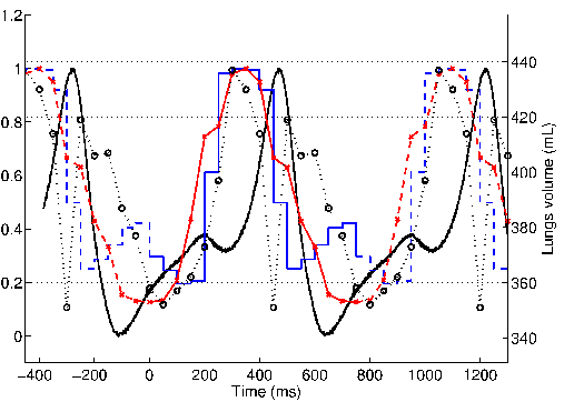
Image-based motion detection results were compared to the pressure signal and to lung volume variation. A temporal shift between the peak of motion in images and the ones in the pressure signal was observed (see Figure 3 ).
The temporal shift suggested to carefully select data from the non moving phase for a motionless 3D-SPECT image reconstruction. This step was incorporated in a breath-hold like reconstruction method [66] , [68] , [67] .
|


