Section: New Results
Other detection approaches
Multiple-instance object detection using a higher-order active shape prior
Participants : Ikhlef Bechar, Josiane Zerubia [contact] .
This work is done in collaboration with Dr. Ian Jermyn of Durham University (United Kingdom, https://www.dur.ac.uk/mathematical.sciences/ ) and was funded by a contract with the EADS foundation (http://www.fondation.eads.com/ ).
object detection, shape prior, transformation invariance, higher-order active contours, energy minimization, non-convex energy, exact convex relaxation.
The problem under consideration is the multiple-instance object detection from imagery using prior shape knowledge. As mathematical and algorithmic framework, we have used the higher-order active contour (HOAC) model framework in order to incoporate prior shape knowledge about a class of objects of interest. On top of its robustness and its computational attractiveness (due to its parameter-estimation free method), the HOAC object-detection framework allows to incorporate shape knowledge about multiple occurrences of an object of interest in an image and to carry out object detection in a single algorithmic framework via the minimization of energy of the form:
where stands for the contour an image object, stands for its image-based energy and stands for a prior energy which is only a function of an objet's shape (and not of image data). The goal of this project is thus to model using the HOAC methodology.
In this work, we have developed a fourth-order active contour (FOAC) framework for incorporating prior shape knowledge about target shapes. Typically, we express a FOAC energy model as
where and stand respectively for the length and the area of a contour and models fourth-order interactions between quadruples of contour points, and , and stand for some tradeoff parameters that control the contribution of each term of the FOAC energy. Note that the parameters of the method include both the real coefficients , and and the bivariate kernel . These parameters need to be tuned optimally for a given target shape . Thus we have developed a direct method for the optimal estimation of the FOAC parameters.
We have then shown that shapes with arbitrary geometric complexity can be modeled using such the FOAC framework 2 , and we have developed a direct method for the estimation of the parameters for a given class of shapes. In order to be able to detect multiple occurrences of a target object in an image, one needs to re-express such an originally contour-based energy 2 by replacing appropriately in formula 2 the one-dimensional contour quantity with an equivalent two-dimensional quantity (ie. with respect to the image domain) such as the characteristic function of and to minimize with respect to it the resulting energy functional. This allows topological changes of an evolving contour and hence the detection of possible multiple instances of a target object in an image. We have shown that such a new formalism is a third-order Markov Random Field (MRF) which practical optimization was a challenging question. Therefore, we have also developed a method for the exact minimization of the energy of the resulting MRF model (using a equivalent convex-relaxation approach, see Fig. 12 ).
|
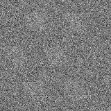
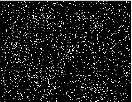
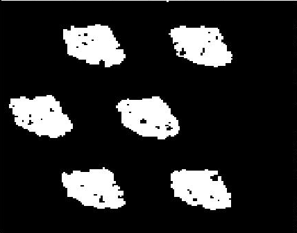
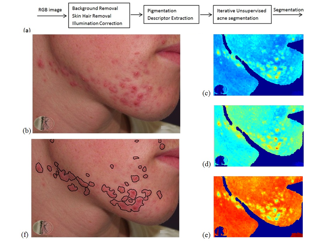
Image analysis for automatic facial acne detection and evaluation
Participants : Zhao Liu, Josiane Zerubia [contact] .
This work is part of LIRA Skin Care Project, which includes four key partners: Philips R&D (Netherlands, http://www.philips.nl ), CWI (Netherlands, http://www.cwi.nl/ ), Fraunhofer Institutes (Germany, http://www.fraunhofer.de/en.html ) and Inria (France).
image processing, feature extraction, pigmentation distributions, acne, cosmetology
Acne vulgaris is a highly prevalent skin disease, which has a significant life quality impact on sufferers. Although acne severity is readily observed by the human eye, it is an extremely challenging task to relate this visual inspection to measurable quantities of various skin tones and types. So far there is no golden standard for acne diagnosis in clinics, and it entirely depends on dermatologists' experience for evaluation of acne severity. But significant inter-rater variability among individual assessment may lead to less trustworthy diagnosis when several clinicians get involved in the study. In addition, less reproducibility of human evaluation makes comparison of acne changes over time difficult. Therefore, the long-term objective of this study is to construct an automatic acne grading system through applying spectroscopy imaging techniques and image processing methods, to objectively evaluate severity of skin disorder. Such a computer-based tool would also significantly benefit the development of better skin care products, if it can reliably characterize treatment effects of products in individual skin layers in agreement with physiological understanding.
Acne segmentation is normally considered as the first significant step in an automatic acne grading system, because segmentation accuracy directly influences the definition of acne pigmentation level, what has an impact on the goodness of acne severity evaluation. An initial unsupervised segmentation method is proposed for conventional RGB images, whose process is demonstrated in Fig. 13 (a). After several pre-processing steps (background and skin hair removal, illumination corrections), nine pigmentation descriptors were extracted from three RGB channels based on colorimetric transformations and absorption spectroscopy of major chromophores. It has been proved that the derived hemoglobin, normalized red, and normalized green descriptors can properly characterize pigmentation distributions of acne, and they are used as segmentation features. Finally, an iterative unsupervised segmentation was performed to maximize pigmentation distributions between acne and normal skin. Fig. 13 (b) shows an example of acne image on human face captured by a conventional RGB camera, while experimental result in Fig. 13 (f) illustrates that suspicious acne areas and healthy human skin can be automatically discriminated by applying the proposed method. Moreover, it only takes 90.8 seconds to segment the example image with the size of pixels, which demonstrates the computation efficiency of the algorithm.
It should be noted that the segmentation method stated above is an initial approach. Shadows around non-flatten areas on human face (e.g. areas around nose) have a large influence on accuracy of automatic acne detection. However, based on the initial experimental results, it is difficult to entirely get rid of these effects using RGB channels only. Our finding is actually consistent with the existing studies, where researchers divided human face into several sub-regions and worked on these sub-regions individually to avoid shadow influence. Therefore, the next step study will compare acne segmentation results derived from RGB images and multi- or hyperspectral images, to investigate the most effective bands for describing acne pigmentation, as well as whether the introduction of multi- or hyperspectral analysis to the automatic acne detection and evaluation is necessary.
|