Section: New Results
Vesicle segmentation method with automatic scale selection in TIRF microscopy
Participants : Antoine Basset, Charles Kervrann, Patrick Bouthemy.
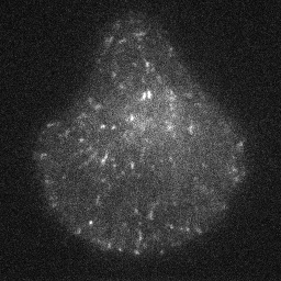
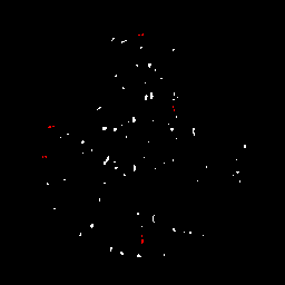
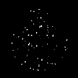
Accurately detecting subcellular particles in fluorescence microscopy is of primary interest for further quantitative analyses such as counting, tracking or classification. Our primary goal was to segment vesicles in fluorescence microscopy images. In [15] we proposed a first spot detection method with automatic scale selection. We have now dramatically improved the precision of the scale selection step, yielding to a more reliable detection of the spots [23] . The method relies on a Laplacian of Gaussian (LoG) filter to first enhance the spots while reducing noise. To obtain good detection results, the scale of the Gaussian filter must be precisely set, according to the spots size [23] . In order to cope with very small spots, we rely on the discrete analog of the Gaussian filter [45] , instead of the previously used sampled Gaussian filter. With this filter, we can find the optimal Gaussian scale with an arbitrary precision by minimizing a statistical criterion. We have introduced two criteria for this purpose and compared them. Once the optimal scale is selected, we threshold the lowest values of the LoG-filtered image, which correspond to spots. To cope with inhomogeneous background, thresholding must be adapted to local statistics so that a single probability of false alarm (PFA) setting can be defined for the whole image or even the collection of images to be processed. In short, we automatically infer from image data the optimal parameters usually left to the user guidance in other methods, that is, spot scale and detection threshold. We have carried out an extensive comparative evaluation, which demonstrates that our new scale selection approach improves detection performances, and that our spot detection method outperforms state-of-the-art detectors [23] .
Collaborators: Jean Salamero (UMR 144 CNRS-Institut Curie, STED team and PICT-IBiSA)
Jérôme Boulanger (UMR 144 CNRS-Institut Curie, STED team)
|