Section: New Results
Spot localization and segmentation for Tissue MicroArray (TMA) de-arrying
Participants : Hoai Nam Nguyen, Charles Kervrann.
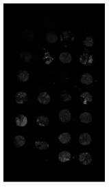
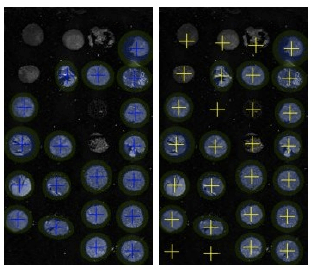
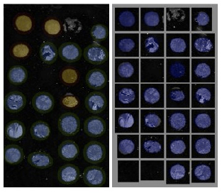
Tissue core de-arraying is one of the most important steps in tissue microarray (TMA) image analysis. A very first task of TMA (Tissue MicroArray) image analysis is to accurately localize spots (separate tissue core) representing arrays of pixels each, in very large images of several thousands of pixels. However, few solutions and frameworks are available and none of them covers images provided by fluorescent scanners. We developed a robust TMA de-arraying method adapted for digital images from classical optical and new fluorescent devices. The proposed algorithm is composed of three modules: i) detection, ii) segmentation, and iii) array indexing. The detection of TMA cores is performed by local adaptive thresholding of isotropic wavelet transform coefficients. We demonstrated how a wavelet decomposition at any desired scale can be performed faster than usual techniques by exploiting explicit formula of the analysis wavelet. Our core detection strategy enables to deal with images having significant noise level, inhomogeneous background, and high dynamic range such as fluorescence images, without any assumption on image noise and intensity value range. The detected cores are furhter segmented by using parametric ellipse model to improve detection accuracy. Combining these two modules, we can handle complex background and artifacts, particularly in fluorescence imaging, and thus reduce false detections. After the segmentation step, the position of detected cores is determined by the centroid of relevant segments. Finally, to compute array indices of cores, we estimate the deformation of a theoretical grid under a thin-plate model by using an iterative scheme. After each iteration, the initial regular grid is progressively transformed for fitting computed core positions. Our main contribution is the reformulation of the array indexing problem as an estimation of the deformation function, which is solved with a iterative algorithm. Moreover, when design layout of TMA slide is known, our estimator of deformation yields quantitative information about grid deformation such as average translation, rotation angle, shearing coefficients, bending energies along axis, etc. They can be used as quality indicators of the manufactured TMA slide.
Collaborator: Vincent Paveau (Innopys company)
|