Section: New Results
Modelling and identification of the sensory-motor system
Implementation and Validation of a Stride Length Estimation Algorithm, Using a Single Basic Inertial Sensor on Healthy Subjects and Patients Suffering from Parkinson's Disease
Participants : Christine Azevedo Coste, Benoît Sijobert, Mourad Benoussaad [ENIT, Tarbes, France] , Christian Geny [CHU Montpellier, Neurology, France] , Jennifer Denys [stagiaire M2 STIC SANTE - DEMAR] .
Providing a clinical oriented solution, our study presented a gyrometer and accelerometer based algorithm for stride length estimation. Compared to most of the numerous existing works where only an averaged stride length is computed from several IMU, or where the use of the magnetometer is incompatible with everyday use, our challenge here has been to extract each individual stride length in an easy-to-use algorithm requiring only one inertial sensor attached to the subject shank. Our results were validated on healthy subjects and patients suffering from Parkinson's disease (PD). Estimated stride lengths were compared to GAITRite walkway system data: the mean error over all the strides was less than 6 percents for healthy group and 10.3 percents for PD group. This method provides a reliable portable solution for monitoring the instantaneous stride length and opens the way to promising applications ([27] ).
Dynamic mapping of upper limb tremor by muscle ultrasonography
Participants : Olivier Tassaert [stagiaire M1 - DEMAR / ICAR] , Benjamin Gilles, Olivier Strauss [LIRMM] , Christian Geny [CHU Montpellier, Neurology, France] , Christine Azevedo Coste.
Focal treatment of action tremor by botulinum toxin injections has been inadequately investigated and at best provides modest relief with significant muscle weakness. Complexity of multi-joint tremulous movements results in non-individualized dosing regimens. Tremor is complex, especially in the upper extremity, and its manifestation can change depending on posture, task, and bodypart. Proper characterization of the tremor based on visual inspection alone is a daunting task for the clinician. Identification of the main trembling muscles task disturbing is challenging because many upper limb muscles are bi-functional. The performance of electromyographic( EMG) pattern-recognition based method in classifying movements strongly depends on arm positions and needs multiple measurements. High density-surface EMG (HD-sEMG) is a non-invasive promising technique to measure electrical muscle activity but has not been used for tremor research because deep muscles could not be investigated Quantification of tremor dynamics by kinematics may be a feasible assessment and guidance tool which can be used to optimize injection conditions for focal tremor therapy. This approach is limited by the redundancy of the upper limb muscle organization Contribution of synergistic muscles toward specific movements over multi joint systems may change with varying position of distal or proximal joints. The choice of injected muscles remains highly subjective and variable. In the study of Rahami, ten different arm or forearm muscles have been injected and improvement was mild and delayed and associated with muscle weakness. In recent years, muscle ultrasonography has become a promising tool for diagnosing neuromuscular disorders . This technique is a non-invasive, low-cost, imaging modality that may be used to characterize normal and pathological muscle tissue but also subtle muscular activity (fasciculatios) in amyotrophic lateral sclerosis. The frequency of tremor remains stable during movement (3 to 8 Hz). We have initiated the investigation of the use of standard ultrasound as a technique to identify muscle groups responsible of upper limb tremor in patient with essential tremor or Parkinson's disease. The feasibility of the overall procedure has been validated: the acquisition procedure on patients, the possibility to track and segment the apparent motion in images using optical flow, and the ability to segment muscle groups by registering a 3D anatomical template.
Understanding electrophysiological effects of direct electrical stimulation of the brain during wide awake surgery
Participants : Marion Vincent, Olivier Rossel, Mitsuhiro Hayashibe, Hugues Duffau [CHU Montpellier] , David Guiraud, François Bonnetblanc.
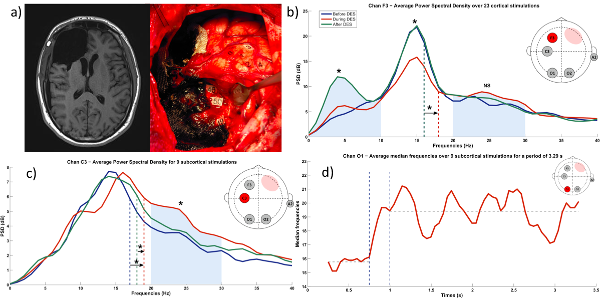
Direct electrical stimulation (DES) have been recently introduced in the neurosurgery of slow-growing and infiltrative brain tumors to guide the resection. By generating transient perturbations, this method allows the real-time identification of both cortical areas and subcortical networks that are essential for the function. Thus, as much as possible, non-functional tissue can be removed while minimizing the sequelae. However, there is much controversy as to whether the use of DES during wideawake surgery is the gold standard for studying the brain function. It is sometimes wrongly assumed that electrical microstimulation (EMS) and DES induce similar effects in the nervous tissues and have comparable behavioural consequences. Both of them are used to perform functional brain mapping: EMS for animal fundamental neuroscience experiments, and DES for neurosurgery patients. We tried to shed new light on electrical stimulation (ES) techniques in brain mapping by comparing EMS and DES [1]. In fact, their effects cannot be directly compared - especially in the electrophysiological domain. There is a gap between theory and practice for ES of the brain. We do not know exactly how ES and especially DES influence the electrophysiological state of networks in the brain; a strong biophysical rationale is lacking. In contrast, the gap between EMS and DES highlights the potential for new experimental paradigms in electrical stimulation for functional brain mapping. In view of this gap and recent technical developments in stimulator design, it may now be time to move towards alternative, innovative protocols. Moreover, the understanding of the electrophysiological effects of DES remains an open and key question. Intra-operative EEG (iEEG) recordings were studied to analyze if and how stimulation currents spread at distant sites. Data were collected during an awake brain surgery for one patient. We observed significant changes in the frequency content at different iEEG sites during DES [2]. Subcortical DES led to neuromodulation at more sites than cortical DES (Figure 4 ). This may be due to (i) a better conduction and propagation following the direct stimulation of large, myelinated axons and (ii) the greater current intensity in subcortical DES. Further research will have to characterize these aspects more carefully and apply cortical and subcortical DES with identical current intensities [30] , [31] .
|
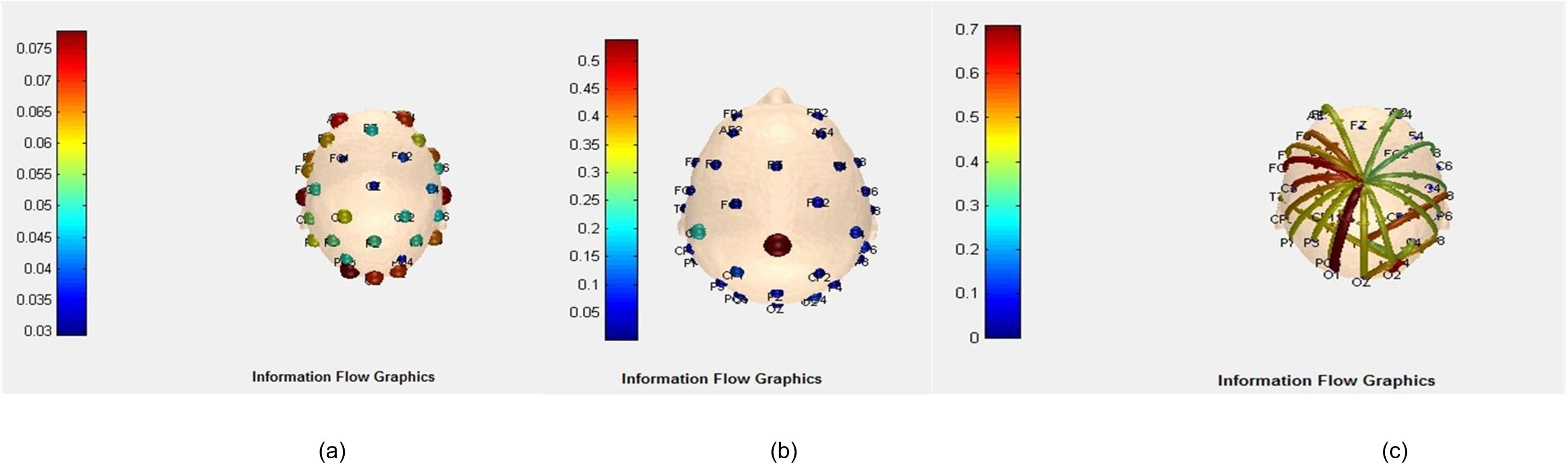
Functional Connectivity Analysis of Motor Imagery EEG signal for Brain-computer Interfacing Application: A preliminary study
Participants : Saugat Bhattacharyya, Poulami Ghosh [Jadavpur University, India] , Ankita Mazumder [Jadavpur University, India] , D.n. Tibarewala [Jadavpur University, India] , Mitsuhiro Hayashibe.
The human brain can be considered as a graphical network having different regions with specific functionality and it can be said that a virtual functional connectivity are present between these regions. These regions are regarded as nodes and the functional links are regarded as the edges between them. The intensity of these functional links depend on the activation of the lobes while performing a specific task(e.g. motor imagery tasks, cognitive tasks and likewise). The analysis of these networks are performed by using a very useful mathematical tool called graph theory. Graph theory basically represents the entire functional network with a number of nodes and edges between them and the amount of connectivity existing between two nodes is depicted by assigning weights to the edges between them. In this study we have tried to utilize functional connectivity between different parts of the human brain for classifying a motor imagery task.
Brain connectivity patterns can be determined by using two types of measures, namely, Bivariate and Multivariate. Here we have considered a multivariate measure known as multivariate autoregressive (MVAR) model. One of the most widely investigated connectivity measure is the Directed Transfer Function (DTF). This function basically computes the directional influences between any two given nodes. There are a number of theoretical indices for defining a graph. In this preliminary work, two indices, namely node strength and network density are measured from the DTF values. In the current study, the BCI competition Dataset III is used for computing different multivariate measures.
The inflow-outflow graph of subject 1 while imagining right hand movement in the first training set are given in Fig.5 . Fig. 5 (a) describes the amount of inflow of functional connectivity going out of all the 32 electrodes and these are color coded to indicate the intensity of these inflows. From Fig. 5 (a) it is quite evident that the inflows are maximum in the frontal, temporal and occipital lobes. Figure 5 (b) depicts the functional outflow from the nodes and in contrast to Fig.5 (a) it shows that the outflows are maximum from the Central lobe(Cz). In Fig 5 (c), the direction of the flow between different nodes are shown and it can be seen clearly that majority of the paths are going from Cz to different nodes of the frontal, parietal and temporal lobes.
|
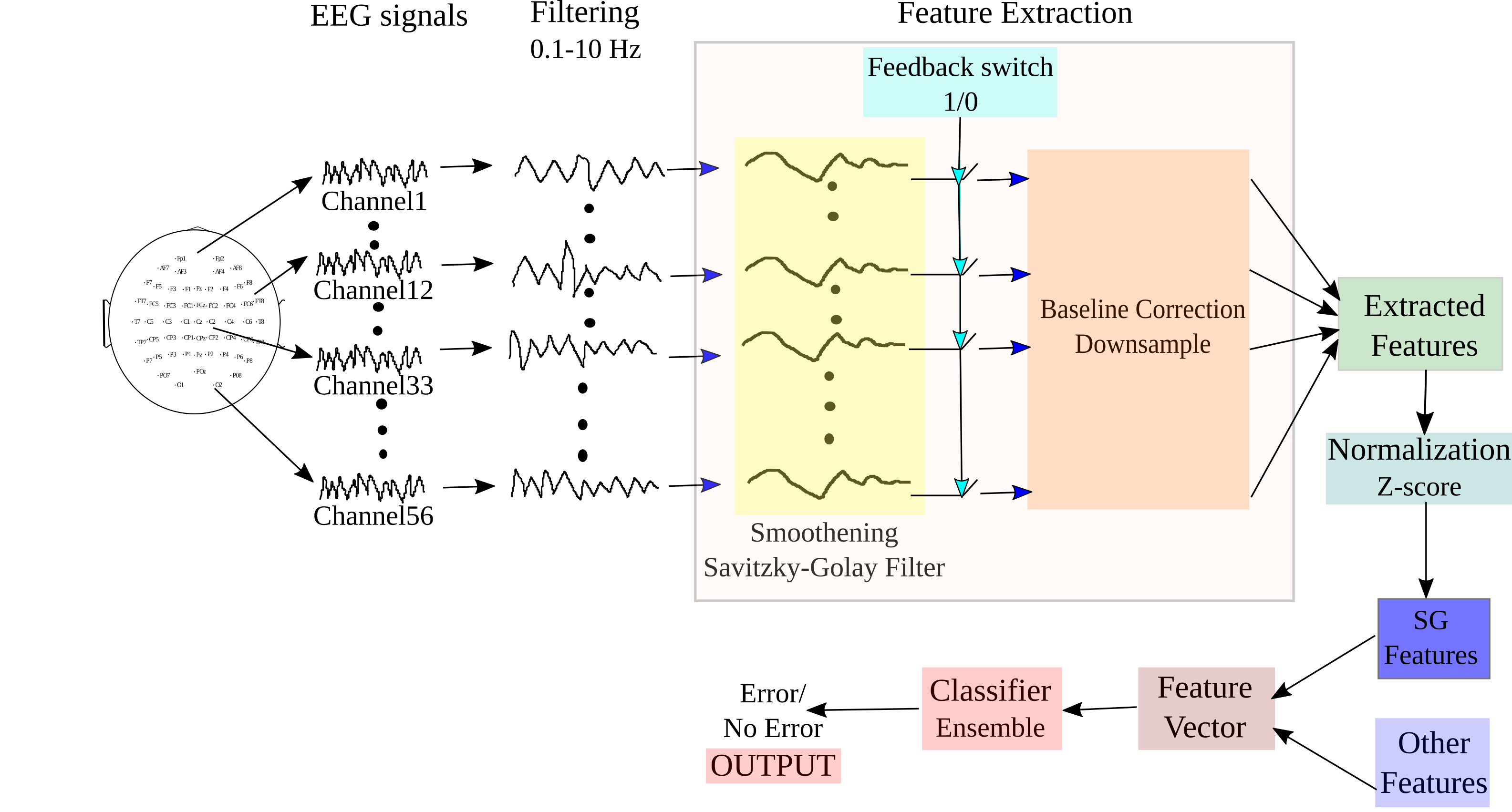
A Generic Transferable EEG Decoder for Online Detection of Error Potential in Target Selection
Participants : Saugat Bhattacharyya, Amit Konar [Jadavpur University, India] , D.n. Tibarewala [Jadavpur University, India] , Mitsuhiro Hayashibe.
Detection of error from electroencephalography (EEG) signals as feedback while performing a discrete target selection task is beneficial for general Brain-computer Interfacing (BCI) systems including rehabilitative application. Error Related Potentials (ErrP) are EEG signals which occur when the participant observes an erroneous feedback from the system.
In this study, we have designed a novel scheme for detection of error feedback directly from the EEG signal. For this purpose, we have used a P300-speller dataset from the `BCI Challenge @ NER 2015' competition hosted at Kaggle. The task involves the subject to select a letter of a word which is followed by a feedback period. The feedback period displays the letter selected and if the selection is wrong, the subject perceives it by the generation of ErrP signal. Our proposed system is designed to detect whether the feedback is erroneous or not. The decoder designed for this task is an ensemble of linear discriminant analysis, quadratic discriminant analysis and logistic regression classifier. The decoder is also transferable in nature as it is should work with single-trial on new subject without any prior subject-specific training.
The block diagram of the BCI system adopted for online ErrP detection from input EEG signals is shown in Fig.6 . The system implements three main processes: i) Pre-processing of the signal, i.e., temporal filtering in the bandwidth [0.1, 10]Hz, ii) Extraction of relevant features corresponding to the mental state from the signal using savitzsky-golay filter and meta-data of the features, and iii) Classification of the features, using our proposed decoder, to detect the intention of the participant from two given states: Error and No-Error. A switch is incorporated in the design to detect the beginning of feedback period in the trials, which is marked in the datasets. We have tested the online functionality of the BCI system on the test dataset provided in the website. To simulate a real-time condition, the EEG is continuously streamed until an onset of the feedback period is detected. On detection of the feedback period, the system extracts a pre-defined length of signal for further processing and the rest are rejected.