Section: New Results
Attenuation and delay of remote potentials evoked by direct electrical stimulation during brain surgery.
Participants : Anthony Boyer, Sofiane Ramdani [LIRMM] , Hugues Duffau [CHU Montpellier] , David Guiraud, François Bonnetblanc.
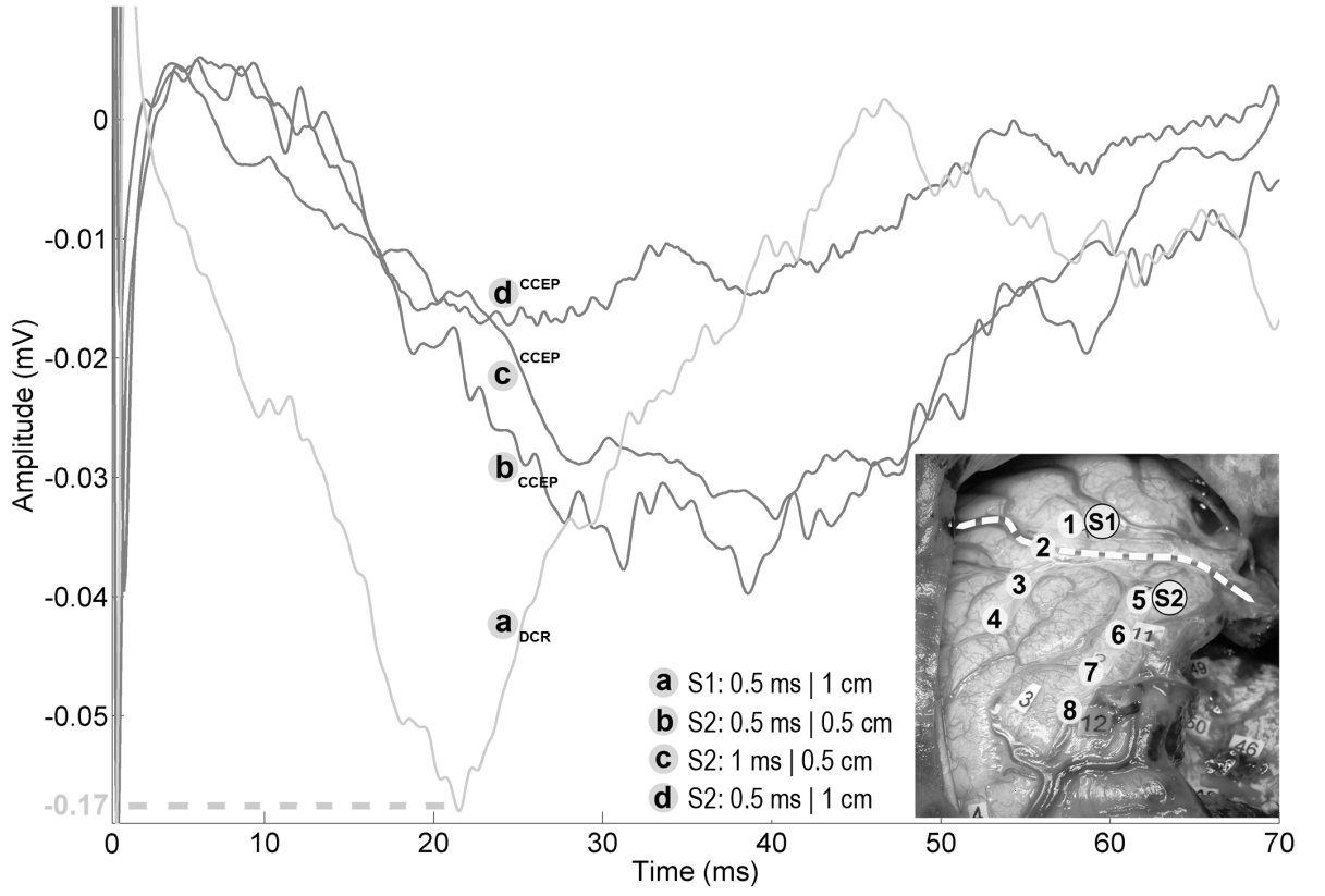
Direct electrical stimulation (DES) is used during awake brain surgery for functional mapping as it generates transient behavioural disturbances, allowing the identification of both cortical areas and subcortical white matter pathways which are essential to the function. However, the electrophysiological effects of DES remain by far unknown. DES may be coupled with the measurement of Evoked Potentials (EPs) to study the conductive and integrative properties of activated neural ensembles and probe the spatiotemporal dynamics of short- and long- range networks. We recorded ECoG signals on two patients undergoing awake brain surgery and measured EPs on functional sites after cortical stimulations, using combinations of stimulation parameters (Figure 11). We were more particularly interested in the generation of evoked potentials (EPs) triggered by both close and remote stimulations. Obtained EPs were very similar in shape, suggesting a stereotyped electrophysiological response, but delayed in time and attenuated in amplitude when elicited from a different gyrus or remotely from the recording site. We were also able to observe the bidirectional nature of the arcuate fasciculus triggering EPs on 2 anatomically connected sites. We propose different activation and electrophysiological propagation mechanisms following DES based on recruited neural elements. The variations in amplitude and delay of EPs are most likely due to different propagation mechanisms, which can be intra- or sub- cortical, and correspond to commonly described DCRs and CCEPs.
|