Section: Research Program
Cardiac Electrophysiology at the Microscopic Scale
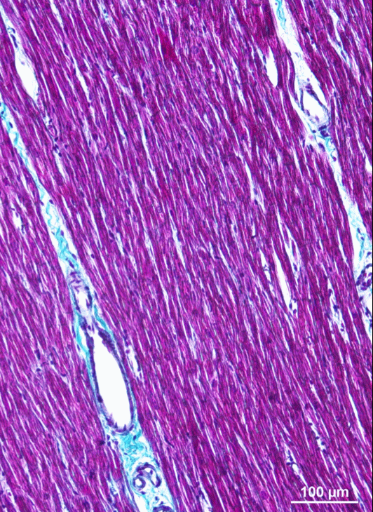
Numerical models of whole-heart physiology are based on the approximation of a perfect muscle using homogenisation methods. However, due to aging and cardiomyopathies, the cellular structure of the tissue changes. These modifications can give rise to life-threatening arrhythmias. For our research on this subject and with cardiologists of the IHU LIRYC Bordeaux, we aim to design and implement models that describe the strong heterogeneity of the tissue at the cellular level and to numerically explore the mechanisms of these diseases.
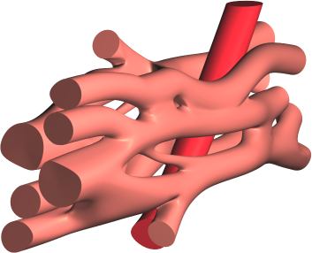
The literature on this type of model is still very limited [67]. Existing models are two-dimensional [60] or limited to idealized geometries, and use a linear (purely resistive) behaviour of the gap-juction channels that connect the cells. We propose a three-dimensional approach using realistic cellular geometry (figure 1), nonlinear gap-junction behaviour, and a numerical approach that can scale to hundreds of cells while maintaining a sub-micrometer spatial resolution (10 to 100 times smaller than the size of a cardiomyocyte) [52], [51], [50]. P-E. Bécue defended his PhD thesis on this topic in December 2018.
|