Section: New Results
Automatic and robust 2D/3D registration on fluoroscopic images
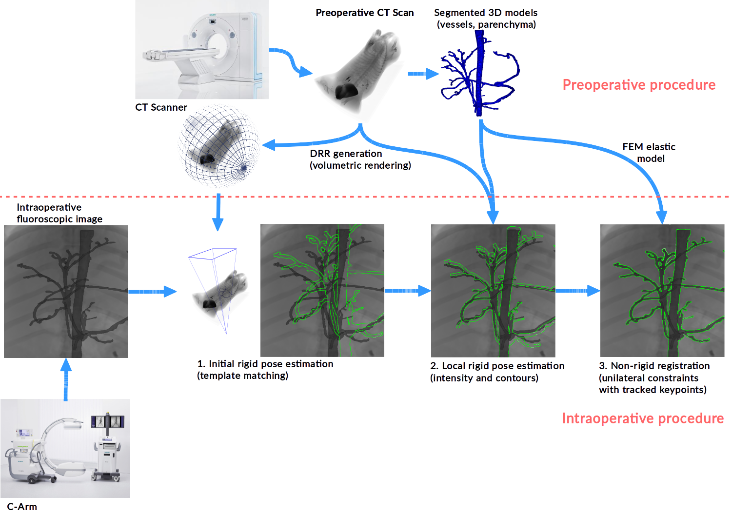
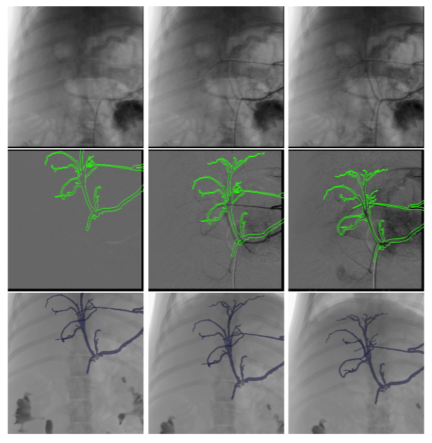
We introduce a unified solution to detect, register and track the liver in 2D live fluoroscopic X-ray images, in order to provide augmented reality and guidance during surgery (see Fig. 9). The solution can be decomposed into two phases, with an initial phase to globally estimate the rigid pose through template matching, and a second local rigid refinement step. A main contribution lies in the combination, for the pose refinement step, of intensity and contour based features over the contrasted vessels of the liver and surrounding organs, by integrating corresponding visual cues in a local optimization framework with respect to the pose. The method does not need any 2D segmentation of the contrasted vessels but relies on a synthetic X-ray rendering algorithm, and requires very few assumptions or priors. Our solution has been tested on synthetic and porcine data, showing its efficiency on realistic scenarios.
A non-rigid registration technique to account for local deformations of the target is also investigated. Once the model is rigidly aligned, local estimation of the deformations undergone by the vasculature and the parenchyma, given a linear or volumetric elastic deformation model of the vessels and the parenchyma, driven by local optical flow features.
The method has been implemented through software developed within the team; for more details see sec. 6.4-6.3. Validation has been performed on porcine in-vivo data, acquired in accordance with UE norms, in collaboration with Pr. Mario GIMENEZ and Dr. Alain GARCIA, Pr. Federico Davrieux, and Pim Hendriks, Daan Kuppens and from IHU-Strasbourg.