

Section: New Results
Breathing detection via tracheal sounds
Participants : Xinyue Lu, David Guiraud, Christine Azevedo, Serge Renaux [Neuroresp] , Thomas Similowski [Hosp. LA Salpêtrière, Paris] .
Individuals with a respiratory paralysis are essentially supplied by mechanical ventilation. However, severe drawbacks of mechanical ventilation were reported: low autonomy, high health costs, infection risk, etc. If patients' phrenic nerves and diaphragms are still functional, implanted diaphragm pacing can provide them a more natural respiration. Compared to classic mechanical ventilation, implanted diaphragm pacing can cancel some of the disadvantages mentioned above, and can also help to significantly improve speech and recover some olfactory sensation.
But existing implanted diaphragm pacing systems can not monitor patient’s induced respiration and they stimulate at constant intensity and frequency - they work in open-loop. It means that stimulation intensity, pulse width and frequency are fixed at the installation of the implant, updated at each control visit, but do not adapt to patient's continuous situation evolution because of the absence of respiratory monitoring. To close the loop, an ambulatory respiratory monitoring solution needs to be developed. Adding adaptive abilities to existing systems would improve the efficiency of the delivered stimulation.
The gold standard for apnea/hypoventilation evaluation is the polygraph, which includes an pulse oximeter and at least one respiratory flow sensor. In a clinical use, flow sensors could be nasal cannula, pneumotachograph, thermistor or plethysmograph. But these sensors need to be placed over the face or are sensitive to patient's movements. They are therefore not compatible with an implanted diaphragm pacing system which is portable and for a daily living use. With this in mind, this study investigated an acoustic method. The proposed tracheal sounds recording requires only one tiny microphone fixed on the neck with a support, which is the only physical contact with the patient.
Many previous studies have shown some positive results on respiration analysis from tracheal sounds in sleep apnea, especially for obstructive sleep apnea. But only few methods are developed for real-time applications (processing delay within seconds) with robustness requirements, indeed, all these studies have been carried out in quiet and controlled acoustic environments with stable sources of noises, and with limited movements of the subjects (during sleep).
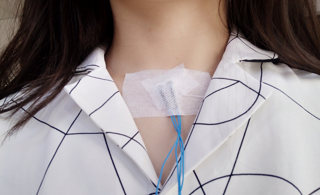
In collaboration with NEURORESP company and La Salpêtrière Gospital (Paris) we are investigating the possibility to perform a real-time and continuous breathing detection (day and night), even during wakefulness in noisy environments. We proposed thea method with tracheal sounds recorded on the neck at suprasternal notch (Fig.19). This method is noninvasive and easy to apply. And the recorded tracheal sounds contain not only respiratory sounds, but also heart beats sounds (as phonocardiogram: PCG) so that some basic cardiac information, as cardiac rhythm, could be calculated. Furthermore, inspired by ECG-derived respiration, the similar method could also be applied on obtained PCG to get respiratory information.
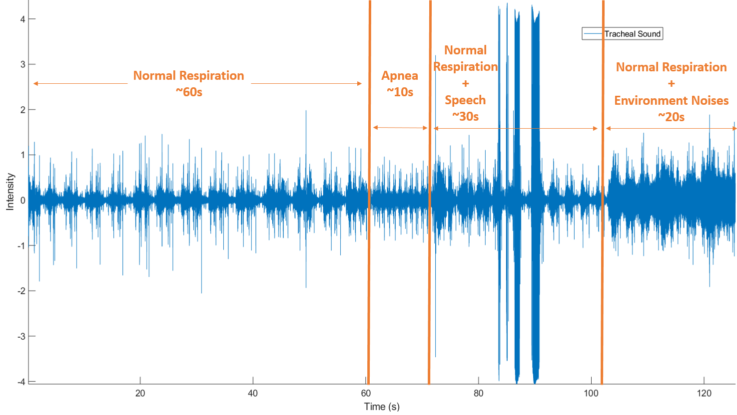
The proposed method has been tested on 30 recordings from 15 healthy subjects with different respiratory condition, one example is shown in Fig.20. We proposed a new algorithm to detect respiration phases, by combining the signal processing both in the temporal (envelope and PCG-derived respiration) and the frequency domains. We assessed the performances of the algorithm in emulated noisy environments. The accuracy, sensibility and specificity of system are all superior to 90%. The result is good enough to show a proof of such a conception. Furthermore, a tracheal sounds recording from a patient under implanted phrenic nerve stimulation has shown that the recording system has the possibility to capt an image of stimulation impulse from the wireless transmission. Getting the synchronization with respiratory sounds and stimulation signals can help to verify and even to adjust patient's stimulation parameters.