Section: New Results
Elastic registration based on biomechanical graph matching
Participants : Jaime Garcia Guevara, Igor Peterlik, Marie-Odile Berger, Stéphane Cotin.
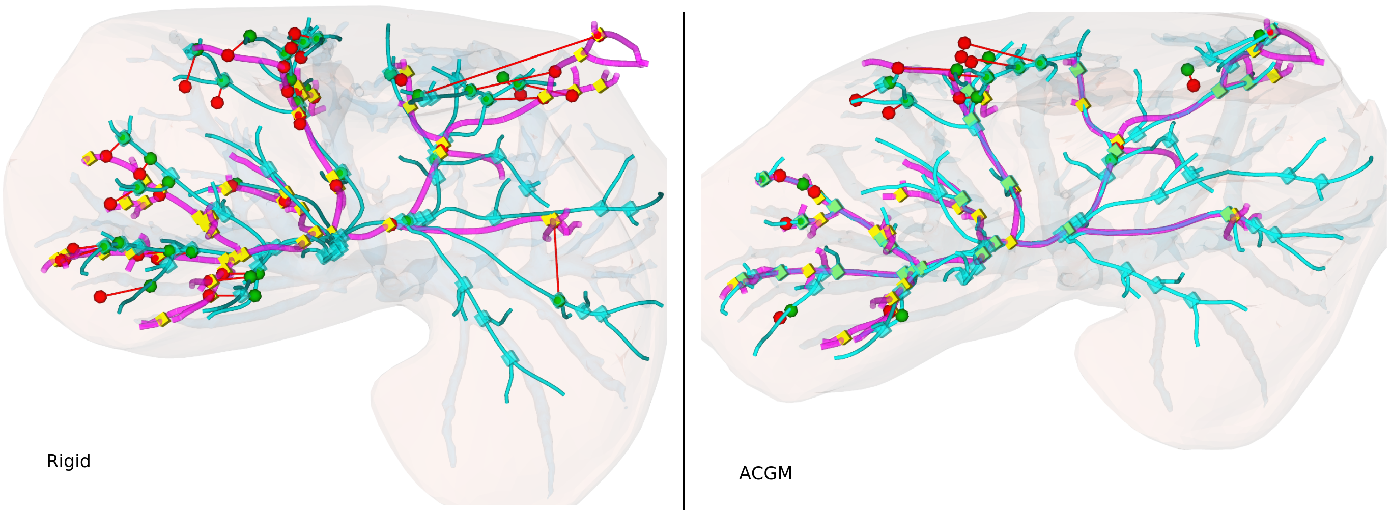
An automatic elastic registration method suited for vascularized organs is proposed. The vasculature in both the preoperative and intra-operative images is represented as a graph. A typical application of this method is the fusion of pre-operative information onto the organ during surgery, to compensate for the limited details provided by the intra-operative imaging modality (e.g. CBCT) and to cope with changes in the shape of the organ. Due differences in image modalities and organ deformation, each graph has a different topology and shape. The Adaptive Compliance Graph Matching (ACGM) method presented does not require any manual initialization, handles intra-operative nonrigid deformations of up to 65 mm and computes a complete displacement field over the organ from only the matched vasculature. ACGM is better than the previous Biomechanical Graph Matching method [3] (BGM) because it uses an efficient biomechanical vascularized liver model to compute the organ's transformation and compliance of vessel bifurcations. It allows to efficiently find the best graph matches with a novel compliance-based adaptive search. These contributions are evaluated on ten realistic synthetic and two real porcine automatically segmented datasets. ACGM obtains better target registration error (TRE) than BGM, with an average TRE in the real datasets of 4.2 mm compared to 6.5 mm, respectively. It also is up to one order of magnitude faster, less dependent on the parameters used and more robust to noise. Figure 8 depicts the large deformation and the registered porcine CBCT and CTA data. Results were published in Annals of Biomedical Engineering (2019) [4].
|