
2024Activity reportProject-TeamEMPENN
RNSR: 200518339S- Research center Inria Centre at Rennes University
- In partnership with:CNRS, INSERM, Université de Rennes
- Team name: Neuroimaging: methods and applications
- In collaboration with:Institut de recherche en informatique et systèmes aléatoires (IRISA)
- Domain:Digital Health, Biology and Earth
- Theme:Computational Neuroscience and Medicine
Keywords
Computer Science and Digital Science
- A3.1.2. Data management, quering and storage
- A3.1.3. Distributed data
- A3.1.7. Open data
- A3.1.8. Big data (production, storage, transfer)
- A3.2.4. Semantic Web
- A3.3.3. Big data analysis
- A3.4.1. Supervised learning
- A3.4.2. Unsupervised learning
- A3.4.3. Reinforcement learning
- A3.4.4. Optimization and learning
- A3.4.6. Neural networks
- A3.4.8. Deep learning
- A5.1.4. Brain-computer interfaces, physiological computing
- A5.2. Data visualization
- A5.3.2. Sparse modeling and image representation
- A5.3.3. Pattern recognition
- A5.3.4. Registration
- A5.4.1. Object recognition
- A5.4.6. Object localization
- A5.9.2. Estimation, modeling
- A5.9.4. Signal processing over graphs
- A6.2.3. Probabilistic methods
- A6.2.4. Statistical methods
- A6.3.3. Data processing
- A6.3.4. Model reduction
- A9.2. Machine learning
- A9.3. Signal analysis
Other Research Topics and Application Domains
- B1.2. Neuroscience and cognitive science
- B1.2.1. Understanding and simulation of the brain and the nervous system
- B1.2.2. Cognitive science
- B2.1. Well being
- B2.2.2. Nervous system and endocrinology
- B2.2.6. Neurodegenerative diseases
- B2.5.1. Sensorimotor disabilities
- B2.5.2. Cognitive disabilities
- B2.6.1. Brain imaging
1 Team members, visitors, external collaborators
Research Scientists
- Emmanuel Caruyer [CNRS, Researcher]
- Julie Coloigner [CNRS, Researcher]
- Benoit Combes [INRIA, Researcher]
- Claire Cury [INRIA, Researcher]
- Fanny Degeilh [INSERM, Researcher]
- Camille Maumet [INRIA, Researcher]
Faculty Members
- Pierre Maurel [Team leader, Université de Rennes , Professor]
- Isabelle Bonan [Université de Rennes , Professor]
- Gilles Edan [Université de Rennes , Professor]
- Jean-Christophe Ferré [Université de Rennes , Professor]
- Francesca Galassi [Université de Rennes , Associate Professor]
- Jean-Yves Gauvrit [Université de Rennes , Professor]
- Anne Kerbrat [Université de Rennes , Associate Professor]
- Gabriel Robert [Université de Rennes , Professor]
Post-Doctoral Fellows
- Julie Fournier [INCR, Post-Doctoral Fellow, from Oct 2024]
- Agustina Fragueiro [INRIA, Post-Doctoral Fellow, until Apr 2024]
- Burhan Rashid Hussein [INRIA, Post-Doctoral Fellow, until Oct 2024]
- Jeremy Lefort-Besnard [INRIA, until Oct 2024]
- Anne Lise Marais [INRIA, Post-Doctoral Fellow, from Sep 2024]
- Camille Muller [Université de Rennes , Post-Doctoral Fellow]
PhD Students
- Melvin Selim Atay [INRIA, from Sep 2024]
- Constance Bocquillon [Université de Rennes ]
- Valentine Chouquet [INRIA, from Sep 2024]
- Sebastien Dam [INRIA]
- Carlo Ferritto [Université de Rennes ]
- Mathys Georgeais [Université de Rennes , from Oct 2024]
- Elodie Germani [Université de Rennes , until Sep 2024]
- Malo Gicquel [INRIA, from Nov 2024]
- Maud Guillen [Université de Rennes ]
- Nolwenn Jegou [INRIA]
- Carla Joud [Université de Rennes ]
- Mathilde Liffran [Université de Rennes , from Sep 2024]
- Youwan Mahe [CIFRE Siemens, from Nov 2024]
- Youenn Merel [INRIA, from Oct 2024]
- Caroline Pinte [Université de Rennes , until Nov 2024]
- Marie Poirier [Université de Rennes ]
- Benjamin Prigent [INRIA, from Dec 2024]
- Adele Savalle [INRIA, from Oct 2024]
- Gregoire Ville [INRIA, from Oct 2024]
- Ricky Walsh [Université de Rennes ]
Technical Staff
- Gwenael Ambrosino-Ielpo [INRIA, Engineer, from Dec 2024]
- Elise Bannier [CHRU RENNES]
- Boris Clenet [INRIA, Engineer]
- Isabelle Corouge [Université de Rennes ]
- Pierre-Henri Dauvergne [INRIA, Engineer]
- Rene-Paul Debroize [INRIA, Engineer, until Mar 2024]
- Jean-Côme Douteau [INRIA, Engineer, until Nov 2024]
- Quentin Duché [Université de Rennes ]
- Malo Gaubert [CHRU RENNES]
- Guewen Hubert [INRIA, Engineer, from Nov 2024]
- Michael Kain [INRIA (SED), Engineer]
- Florent Leray [INRIA (SED), Engineer]
- Julien Louis [INRIA (SED), Engineer]
- Youenn Merel [INRIA, Engineer, until Aug 2024]
- Cédric Meurée [INRIA, Engineer]
- Sandesh Patil [INRIA, Engineer]
- Mathis Piquet [INRIA, Engineer, from Dec 2024]
- Alexandre Pron [INRIA, Engineer]
- Gwendal Soisnard [INRIA, Engineer, from Apr 2024]
- Benjamin Streichenberger [INRIA, Engineer]
Interns and Apprentices
- Jeanne Beraud-Morin [Université de Rennes , from Sep 2024 until Sep 2024]
- Valentine Chouquet [INRIA, Intern, until Jul 2024]
- Andjela Dimitrijevic [UNIV MONTREAL, Intern, from Apr 2024 until Jul 2024]
- Mathilde Liffran [INRIA, Intern, until Jul 2024]
- Baptiste Lode [INRIA, Intern, until Jul 2024]
- Youwan Mahe [INRIA, Intern, from Feb 2024 until Jun 2024]
- Solene Painchaud [INRIA, Intern, until Jul 2024]
- Emma Redor [INRIA, until May 2024]
- Mathis Relion [INRIA, Intern, from Feb 2024 until Jul 2024]
- Sebastien Resche-Rigon [INRIA, Intern, from Apr 2024 until Oct 2024]
- Gregoire Ville [INRIA, Intern, from Feb 2024 until Jul 2024]
Administrative Assistant
- Armelle Mozziconacci [CNRS]
2 Overall objectives
The research team Empenn ("Brain" in Breton language) ERL U1228 is co-affiliated with Inria, Inserm (National Institute for Health and Scientific Research), CNRS (INS2I institute), and the University of Rennes. It is a team of IRISA/UMR CNRS 6074. Empenn is located in Rennes, on the medical and scientific campus. It succeeded in 2019 to the "VisAGeS" team, created in 2006 by Inria. As for "VisAGeS", Empenn holds the accreditation number U1228, renewed by Inserm in 2022 and for a period of 6 years, after an evaluation conducted by the HCERES and Inserm.
Thanks to this unique partnership, Empenn's ambition is to establish a multidisciplinary team of researchers in information sciences and medicine. Our medium and long term objective is to introduce our fundamental research into clinical practice, while maintaining the excellence of our methodological research.
Our goal is to foster research in medical imaging, neuroinformatics and population cohorts. In particular, the Empenn team aims at the detection and development of imaging biomarkers for brain diseases and focuses its efforts on transferring this research to the clinic and clinical neuroscience in general. More specifically, the objective of Empenn is to propose new statistical and computational methods, and to measure and model morphological, structural and functional states of the brain to better diagnose, monitor and treat mental, neurological and substance use disorders. We propose to combine advanced instrumental devices and novel computational models to provide advanced diagnostic, therapeutic, and neurorehabilitation solutions for some of the major developing and aging brain disorders.
Generic and challenging research topics in this broad area include finding new ways to compare models and data, aid in decision making and interpretation, and develop feedback. These activities are carried out in close collaboration with the Neurinfo imaging platform in vivo, which is an essential environment for the experimental implementation of our research on ambitious clinical research projects and the development of new clinical applications.
3 Research program
3.1 Glossary
-
Magnetic Resonance Imaging
- MR - Magnetic Resonance
- MRI - Magnetic Resonance Imaging
- fMRI - Functional Magnetic Resonance Imaging
- DWI - Diffusion-Weighted Imaging
- ASL - Arterial Spin Labeling
-
Other modalities
- PET - Positron Emission Tomography
- EEG - Eletroencephalograpy
- NIRS - Near InfraRed Spectroscopy
-
Medical terminology
- MS - Multiple Sclerosis
- TBI - Traumatic Brain Injury
-
Methodological terminology
- GLM - General Linear Model
- MCM - Multi-compartment models
- NF - Neurofeedback
3.2 Scientific Foundations
The scientific foundations of our team concern the design and development of new computational solutions for biological images, signals and measurements. Our goal is to develop a better understanding of the normal and pathological brain, at different scales.
This includes imaging brain pathologies in order to better understand pathological behavior from the organ level to the cellular level, and even to the molecular level (PET-MR imaging), and modeling of large groups of normal and pathological individuals (cohorts) from image descriptors. It also addresses the challenge of the discovery of episodic findings (i.e., rare events in large volumes of images and data), data mining and knowledge discovery from image descriptors, validation and certification of new drugs from imaging features, and, more generally, the integration of neuroimaging into neuroinformatics by promoting and supporting virtual organizations of biomedical actors using e-health technologies.
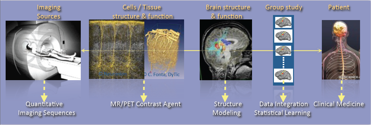
Illustration of the major scientific objectives of the Empenn team, including Imaging sources, Cells and tissue structure and function, Brain structure and function, Group study and Patient.
As shown in Figure 1, the research activities of the Empenn team closely link observations and models through the integration of clinical and multiscale data, and phenotypes (cellular, and later molecular, with structural or connectivity patterns in the first stage). Our ambition is to build personalized models of central nervous system organs and pathologies, and to compare these models with clinical research studies in order to establish a quantitative diagnosis, prevent the progression of diseases and provide new digital recovery strategies, while combining all these research areas with clinical validation. This approach is developed within a translational framework, where the data integration process to build the models is informed by specific clinical studies, and where the models are assessed regarding prospective clinical trials for diagnosis and therapy planning. All of these research activities are conducted in close collaboration with the Neurinfo platform, which benefited in 2018 from a new high-end 3T MRI system dedicated to research (3T Prisma™ system from Siemens), and through the development in the coming years of multimodal hybrid imaging (from the currently available EEG-MRI, to EEG-NIRS and PET-MRI in the future).
In this context, some of our major developments and newly arising issues and challenges include:
- The generation of new descriptors to study brain structure and function (e.g. the combination of variations in brain perfusion with and without a contrast agent; changes in brain structure in relation to normal, pathological, functional or connectivity patterns; or the modeling of brain state during cognitive stimulation using neurofeedback).
- The integration of additional spatiotemporal and hybrid imaging sequences covering a larger range of observations, from the molecular level to the organ level, via the cellular level (arterial spin labeling, diffusion MRI, MR relaxometry, MR cell labeling imaging, EEG-MRI functional imaging, EEG-NIRS-MRI).
- The creation of computational models through the data fusion of multimodal MR images, structural and functional image descriptors from group studies of normal and/or pathological subjects.
- The evaluation of these models in relation to acute pathologies, especially for the study of degenerative, psychiatric, traumatic or developmental brain diseases (primarily multiple sclerosis, stroke, traumatic brain injury (TBI) and depression, but applicable with a potential additional impact to epilepsy, Parkinson’s disease, dementia, post-traumatic stress disorder, etc.) within a translational framework.
In terms of new major methodological challenges, we address the development of models and algorithms to reconstruct, analyze and transform the images, and to manage the mass of data to store, distribute and “semanticize” (i.e. provide a logical division of the model’s components according to their meaning). As such, we expect to make methodological contributions in the fields of model inference; statistical analysis and modeling; the application of sparse representation (compressed sensing and dictionary learning) and machine learning (supervised/unsupervised classification and discrete model learning); data fusion (multimodal integration, registration, patch analysis, etc.); high-dimensional optimization; data integration; and brain-computer interfaces. As a team at the frontier between the digital sciences and clinical research in neuroscience, we do not claim to provide theoretical breakthroughs in these domains but rather to provide significant advances in using these algorithms through to the advanced applications we intend to address. In addition, we believe that by providing these significant advances using this set of algorithms, we will also contribute to exhibiting new theoretical problems that will fuel the domains of theoretical computer sciences and applied mathematics.
In summary, we expect to address the following major challenges:
- Developing new information processing methods able to detect imaging biomarkers in the context of mental, neurological, and substance use disorders.
- Providing new computational solutions for our target applications, allowing a more appropriate representation of data for image analysis and the detection of biomarkers specific to a form or grade of pathology, or specific to a population of subjects.
- Providing, for our target applications, new patient-adapted connectivity atlases for the study and characterization of diseases from quantitative MRI.
- Providing, for our target applications, new analytical models of dynamic regional perfusion, and deriving indices of dynamic brain local perfusion from normal and pathological populations.
- Investigating whether the theragnostics paradigm of rehabilitation from hybrid neurofeedback can be effective in some behavioral and disability pathologies.
These major advances are primarily developed and validated in the context of several priority applications in which we expect to play a leading role: multiple sclerosis, stroke rehabilitation, and the study and treatment of depression.
3.3 Research axes
Figure 2 summarizes the scientific organization of the research team through three basic research topics in information sciences: Population Imaging (see 3.3.1), Detection and Learning (see 3.3.2), and Quantitative Imaging (see 3.3.3) and three translational axes on central nervous system diseases: Behavior, Neuro-inflammation and Recovery (see section 4).
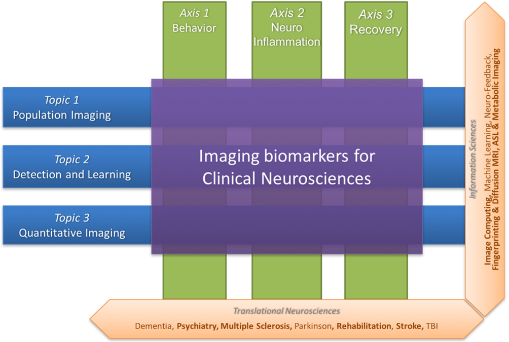
Illustration of the research topics and research axes of the Empenn team. The former (Population imaging, detection and learning and quantitative imaging) concern information sciences, and these topics intersect with the latter (Behavior, neuro-inflammation and recovery), which are translational neurosciences axes.
3.3.1 Population imaging
One major objective of neuroimaging researchers and clinicians is to be able to stratify brain imaging data in order to derive new and more specific population models. In practice, this requires to set up large-scale experiments that, due to the lack of resources and capabilities to recruit locally subjects who meet specific inclusion criteria, motivates the need for sharing the load.
However, building and using multi-site large-scale resources pose specific challenges to deal with the huge quantity of data produced and their diversity. Empenn focuses on two challenges in particular:
- Providing computational environments for the computation and use of imaging biomarkers in the targeted brain diseases, a solution to be used by radiologists and neurologists/psychiatrists for the clinical follow-up of a large patient population.
- Modeling analytic variability of image processing pipelines to better understand and predict the behaviour of imaging biomarker detection solutions and improve reproducibility and productivity in clinical neuroimaging research.
3.3.2 Detection and learning
We intend to make significant contributions with major impacts in learning coupling models between functional recordings during neurofeedback procedures. These advances will provide a breakthrough in brain-computer interfaces for rehabilitation protocols. Our aim is to:
- Our research employs data-driven approaches, encompassing machine learning and deep learning, to enhance the detection and segmentation of abnormal patterns in medical images. Our primary focus is on multiple sclerosis (MS) and, more recently, on stroke. The findings from our studies indicate promising outcomes in automated tools for accurate disease activity assessment and lesion segmentation within large MRI databases. Special attention is given to the integration of multimodal information and the utilization of labeled and unlabeled data. As we progress, our aim is to adapt these methods to address a broader range of neurological diseases, including epilepsy, tumors, etc., in both neonate and adult brains. This research contributes to advancing diagnostic tools and methodologies in the field of medical imaging.
- Develop solutions for combining brain state measurements from multimodal sensors or sequences (e.g. fMRI, ASL, EEG, NIRS, etc.) with applications in the spatiotemporal reconstruction of brain activity from MRI-EEG or the combined detection of the endogenous hemodynamic and resting state network of the brain from ASL and NIRS. Over the longer term, the advent of new hybrid brain imaging sensors (e.g. PET-MRI) will require these methods to be extended to a larger spectrum of information combining structural, morphological, metabolic, electrophysiological and cellular/molecular information (e.g. through the use of specific ligands/nanocarriers).
3.3.3 Quantitative imaging
The Empenn research group focuses on the development of several quantitative techniques in magnetic resonance imaging of the brain. These methods allow for characterization of both the function and the structure of the brain with high precision. Arterial spin labelling (ASL) is a contrast agent-free imaging technique which labels arterial blood water as an endogenous tracer for perfusion and can measure resting-state cerebral blood flow. We are interested in estimating multiparametric hemodynamics using ASL, such as combined cerebral blood flow and arterial transit times, and derive statistical descriptors to represent significant differences between groups. In addition to quantitative perfusion parameters, our contributions on tissue compartment imaging aim at delineating neural circuits and characterize their microstructure properties, using both diffusion MRI and relaxometry. In diffusion MRI, arbitrary gradient waveforms were shown to exhibit higher sensitivity to microstructure parameters than standard pulsed gradients. We work on the optimization of sampling protocols in this domain, with the objective to propose sequences compatible with in vivo acquisition. Complementary to diffusion MRI, we develop methods for the reconstruction of myelin-bound, extra-axonal and cerebrospinal fluid water using multi-compartment modelling of the T2-relaxometry signal. We combine these techniques with tractography to identify trajectories of pathologies associated to the evolution of these microstructural parameters along specific fiber bundles in the brain white matter. Finally, we are also focusing on assessing the characteristics (repeatability, reproducibility and sensitivity) of several quantitative metrics variability (e.g. MTR, T1 relaxometry) in the spinal cord of patients with MS.
3.3.4 Translational research
The three translational axes focus on the central nervous system and are presented in details in the following section.
4 Application domains
The team develops three translational research axes focused on the central nervous system: Behavior (see 4.1) Neuro-inflammation (see 4.2) and Recovery (see 4.3).
4.1 Behavior
Advances in the field of in vivo imaging offer new opportunities for addressing the management of resistant affective disorders and their consequences (suicide risk and socio-professional impact), and the management of spatial cognition disorders after stroke and their consequences (postural perturbations and the loss of autonomy). Our objective, and the main challenge in this context, is to introduce medical image computing methods to the multidisciplinary field of behavioral disorders (cognitive disorders, particularly spatial and postural control disorders or anterograde memory impairment, mood disorders, notably resistant depression, schizophrenic disorders, pervasive developmental disorders, attention disorders, etc.) in order to gain a better understanding of the pathology and devise innovative therapeutic approaches.
We also expect to become a major player in the future and make important contributions with significant impacts, primarily in drug-resistant depression in young and old populations. In particular, we expect to provide new image-related metrics combining perfusion, metabolism and microstructural information regarding the brain in order to better characterize pathologies, provide prospective evolution values and potentially provide new brain stimulation targets that could be used in neurofeedback rehabilitation protocols or other types of brain stimulation procedures.
We aim to provide new imaging markers of mental diseases, especially in the context of mood disorders. The new biomarkers are derived from the metabolic (ASL and later ASL+PET) point of view as well as from the microstructural point of view (multicompartment diffusion MRI and relaxometry). Similarly, we expect to exhibit imaging biomarker regularities combining metabolic and structural information. Over the longer term, we expect these biomarkers to be the target of neurofeedback rehabilitation procedures. Also, over the longer term, we expect to supplement the MRI markers with molecular markers coming from new PET tracers, especially those associated with serotonin intake, at one time point or during a rehabilitation protocol under hybrid PET-EEG-MRI neurofeedback procedures.
4.2 Neuroinflammation
Some of the major ongoing research issues regarding neuroimaging of neuro-inflammatory diseases concern the definition of new biomarkers to track the development of the pathology using high- dimensional data (e.g. nD+t MRI). This includes the use of white matter-specific imaging, such as magnetization transfer MRI, relaxometry and diffusion-weighted imaging (DW-MRI). Our objective is (1) to develop information-processing tools to tag the spatiotemporal evolutions of Multiple Sclerosis patterns at the brain parenchyma and spinal cord levels from their different signatures (inflammatory cells visible with USPIO or Gd contrast agents on MRI, persistent black holes, eloquent regional atrophy and microstructure signatures); and (2) to test these new tools on new imaging cohorts. In this respect, we for instance conduct studies on brain and spinal cord imaging, continuing on from the PHRC multicentric EMISEP project (PI: G. Edan), as it is very likely that lesions in the spine will directly affect the ambulatory ability of the patient (and thereby the clinical scores). In order to extend this experiment to a larger MS population, based on our expertise from the OFSEP cohort, we also plan to improve the MS therapeutic decision process notably through the RHU PRIMUS (PRojection In MUltiple Sclerosis) project (PI: G. Edan). Our goal is to develop and assess a standardized monitoring tool that provides a robust, long-term computerized MRI follow-up that will become the gold standard in clinical practice for therapeutic decisions in MS treatment. As part of this project, Empenn will share its expertise in data management systems (Shanoir and FLI-IAM), automatic processing tools (through the medInria and Anima software repositories) to extract quantitative indices from the images and the assessment of the added-value of promising quantitative sequences.
4.3 Recovery
Mental and neurological disorders are the leading cause of years lived with a disability. Treatment-resistant depression affects approximately 2% of the European population. Meanwhile, in the case of brain disorders, almost 1.5 million Europeans (15 million people worldwide) suffer a stroke event each year. Current recovery methods for brain disorders and traumatic brain injuries remain limited, preventing many from achieving full recuperation. We propose to address the issue of brain recovery by introducing new advances from recent breakthroughs in computational medical imaging, data processing and human-machine interfaces, and demonstrate how these new concepts can be used, in particular for the treatment of stroke and major depressive disorders.
We ambition to combine advanced instrumental devices (hybrid EEG, NIRS and MRI platforms), with new hybrid brain computer interface paradigms and new computational models to provide neurofeedback-based therapeutic and neuro-rehabilitation paradigms in some of the major mental and neurological disorders of the developmental and aging brain.
Neurofeedback involves using a brain-computer interface that provides an individual with real-time biofeedback about his or her brain activity in the form of sensory feedback. It enables individuals to learn to better control their brain activity, which can be measured in real time using various non-invasive sensors as described above. Although EEG is currently the only modality used by clinical practitioners in that context, it lacks specificity due to its low spatial resolution. Dynamic research into fMRI-neurofeedback has held promise for treating depression, chronic pain and stroke, since it offers the prospect of real-time imagery of the activity in deep brain structures with high spatial resolution. However, the low temporal resolution and high cost of fMRI-neurofeedback has hampered the development of many applications. We believe that the future belongs to hybrid responses that combine multimodal sensors and intend to demonstrate this in the Empenn project.
5 Social and environmental responsibility
- Elise Bannier and Francesca Galassi: members of the Groupe Développement Durable de l'IRISA et du Centre Inria - for the assessment and mitigation of the impact of our research activities on the environment. A report was published setting out the work carried out by the working group on the environmental impact of our research activities, and in particular greenhouse gas (GHG) emissions due to air travel. The working group, made up of around fifteen members from the various supervisory bodies, addressed these issues between September 2022 and December 2023. After a preliminary reflection phase aimed at mapping travel practices and similar actions carried out outside the research center, the group set up a participative action phase in three acts : I) realization of the center's GHG balance sheet for the year 2022; II) organization of a participative workshop aimed at questioning our travel practices and building reduction measures; III) democratic consultation via a general assembly and an online survey, making it possible to estimate the acceptability of these various reduction measures and validate them 55.
-
Participation to gender-equality
- Camille Maumet: co-chair of the women-men equality group at Inria Rennes / IRISA until August 2024.
- Jérémy Lefort-Besnard, Nolwenn Jégou, Camille Maumet: members of the women-men equality group at Inria Rennes / IRISA
- Elise Bannier: member of the Pairing Committee for the mentoring program of Inria Rennes / IRISA.
6 Highlights of the year
6.1 New Member
Jean-Marie Batail, MD-PhD in Psychiatry joined the Empenn team.
6.2 Habilitation degrees
- Fanny Dégeilh defended her HDR entitled "Pediatric traumatic brain injury and brain development" in January 2024
- Camille Maumet defended her HDR entitled "Towards reproducible neuroimaging: Solutions for sharing and re-using brain imaging data" 54 in February 2024.
- Julie Coloigner defended her HDR entitled "Cerebral functional and structural connectivity analysis" in June 2024
- Jean-Marie Batail defended his HDR entitled "Vers une médecine de précision en psychiatrie : phénotypes, psychophysiologie et neuromodulation dans le traitement de la dépression" in December 2024.
6.3 PhD degrees
- Elodie Germani defended her PhD thesis entitled "Explore and mitigate analytical variability in fMRI with representation learning" in September 2024
- Caroline Pinte defended her PhD thesis entitled "Apprentissage automatique pour le neurofeedback bi-modal EEG-IRMf : localisation des électrodes EEG et prédiction des scores NF IRMf" in November 2024.
6.4 Scientific events
Claire Cury and Pierre Maurel organised the symposium "Multi-modal neurofeedback methods for post-stroke rehabilitation" at the rt-FIN 2024 conference, Heidelberg, Germany.
6.5 Awards
Elodie Germani was winner of the L'Oréal-Unesco 2024 Young Talent Award for Women in Science: News on the IRISA website and on the Inria website.
6.6 New grants
- ANR-JCJC-CoYoKi: "Concussion in Young Kids : From acute brain alteration to neurodevelopment" led by Fanny Dégeilh with Claire Cury and Pierre Maurel. Funding: Appel à projets générique 2024.
- ANR-JCJC-NIRVAVA: "Unravelling bimodal neurofeedback efficiency for dynamic non-invasive brain rehabilitation" led by Claire Cury with Elise Bannier. Funding: Appel à projets générique 2024.
- Inria Exploratory Action INCLUDE "Integrating fuNctional MRI and EEG with Carbon-wire Loops : towards the characterization of mUltimoDal functional biomarkErs" led by Julie Coloigner with Elise Bannier, Claire Cury, Mathis Piquet.
7 New software, platforms, open data
7.1 New software
7.1.1 Anima
-
Keywords:
Medical imaging, Neuroimaging, Image processing
-
Scientific Description:
Anima is a set of libraries and tools developed by the team as a common repository of research algorithms. As of now, it contains tools for image registration, statistical analysis (group comparison, patient to group comparison), diffusion imaging (model estimation, tractography, etc.), quantitative MRI processing (quantitative relaxation times estimation, MR simulation), image denoising and filtering, and segmentation tools. All of these tools are based on stable libraries (ITK, VTK), making it simple to maintain.
-
Functional Description:
Anima is a set of libraries and tools in command line mode for processing and analysing medical images.
- URL:
-
Contact:
Julie Coloigner
-
Participants:
Aymeric Stamm, Fang Cao, Florent Leray, Guillaume Pasquier, Laurence Catanese, Olivier Commowick, Renaud Hedouin, Rene-Paul Debroize
7.1.2 MedINRIA
-
Keywords:
Visualization, DWI, Health, Segmentation, Medical imaging
-
Scientific Description:
MedInria aims at creating an easily extensible platform for the distribution of research algorithms developed at Inria for medical image processing. This project has been funded by the D2T (ADT MedInria-NT) in 2010, renewed in 2012. A fast-track ADT was awarded in 2017 to transition the software core to more recent dependencies and study the possibility of a consortium creation.The Empenn team leads this Inria national project and participates in the development of the common core architecture and features of the software as well as in the development of specific plugins for the team's algorithm.
-
Functional Description:
medInria is a free software platform dedicated to medical data visualization and processing.
- URL:
-
Contact:
Florent Leray
-
Participants:
Maxime Sermesant, Olivier Commowick
-
Partners:
HARVARD Medical School, IHU - LIRYC, NIH
7.1.3 autoMRI
-
Keywords:
FMRI, MRI, ASL, FASL, SPM, Automation
-
Scientific Description:
This software is highly configurable in order to fit a wide range of needs. Pre-processing includes segmentation of anatomical data, as well as co-registration, spatial normalization and atlas building of all data types. The analysis pipelines perform either within-group analysis or between-group or one subject-versus-group comparison, and produce statistical maps of regions with significant differences. These pipelines can be applied to structural data to exhibit patterns of atrophy or lesions, to ASL (both pulsed or pseudo-continuous sequences) data to detect perfusion abnormalities, to functional data - either BOLD or ASL - to outline brain activations related to block or event-related paradigms. New functionalities have been implemented to facilitate the management and processing of data coming from complex projects.
-
Functional Description:
AutoMRI is based on MATLAB and the SPM12 toolbox and provides complete pipelines to pre-process and analyze various types of images (anatomical, functional, perfusion).
- URL:
-
Contact:
Isabelle Corouge
-
Participants:
Camille Maumet, Elise Bannier, Isabelle Corouge, Pierre Maurel, Quentin Duché, Julie Coloigner
7.1.4 ShanoirUploader
-
Name:
ShanoirUploader (SHAring NeurOImaging Resources Uploader)
-
Keywords:
Webservices, PACS, Medical imaging, Neuroimaging, DICOM, Health, Biology, Java, Shanoir
-
Scientific Description:
ShanoirUploader is a desktop application on base of JavaWebStart (JWS). The application can be downloaded and installed using an internet browser. It interacts with a PACS to query and retrieve the data stored on it. After this ShanoirUploader sends the data to a Shanoir server instance in order to import these data. This application bypasses the situation, that in most of the clinical network infrastructures a server to server connection is complicated to set up between the PACS and a Shanoir server instance.
-
Functional Description:
ShanoirUploader is a Java desktop application that transfers data securely between a PACS and a Shanoir server instance (e.g., within a hospital). It uses either a DICOM query/retrieve connection or a local CD/DVD access to search and access images from a local PACS or the local CD/DVD. After having retrieved the data, the DICOM files are locally anonymized and then uploaded to the Shanoir server. A possible integration of a hash creation application for patient identifiers is provided as well. The primary goals of that application are to enable mass data transfers between different remote server instances and therefore reduce the waiting time of the users, when importing data into Shanoir. Most of the time during import is spent with data transfers.
- URL:
-
Contact:
Michael Kain
-
Participants:
Christian Barillot, Inès Fakhfakh, Justine Guillaumont, Michael Kain, Yao Chi
7.1.5 Shanoir-NG
-
Name:
Shanoir (SHAring iN vivO Imaging Resources)
-
Keyword:
Medical imaging
-
Functional Description:
Shanoir (SHAring iN vivO Imaging Resources) is an open-source web platform designed to share, archive, search and visualize medical imaging data. It provides an user-friendly secure web access and offers an intuitive workflow to facilitate the collecting and retrieving of imaging data from multiple sources. Quality control can be applied on imported data. Mass data can be downloaded in multiple ways, via the web interface and via a Python script.
It supports the following formats: DICOM classic/enhanced (MR, CT, PT, NM), BIDS, processed datasets (NIfTI), Bruker, EEG(BrainVision/EDF).
Shanoir comes along many features such as pseudonymization of data (based on DICOM standard profiles), support for multi-centric clinical studies on subjects. Shanoir offers an ontology-based data organization (OntoNeuroLOG). Among other things, this facilitates the reuse of data and metadata, the integration of processed data and provides traceability trough an evolutionary approach. Shanoir allows researchers, clinicians, PhD students and engineers to undertake quality research projects with an emphasis on remote collaboration. Data user agreements (DUA) can be configured by study to be accepted by each accessing users and access requests can be initated to study administrators.
-
Release Contributions:
- New tree view - New version of OHIF, with annotations and segmentations - Mass processing integration with VIP - Java 21 - Spring Boot 3.2
- URL:
-
Contact:
Michael Kain
-
Participants:
Michael Kain, Anthony Baire, Julien Louis, Jean-Côme Douteau, Pierre-Henri Dauvergne, Arthur Masson, Youenn Merel
7.1.6 LongiSeg4MS
-
Name:
Longitudinal Segmentation For Multiple Sclerosis
-
Keywords:
3D, Brain MRI, Deep learning, Detection
-
Functional Description:
LongiSeg4MS is an automatic new multiple sclerosis (MS) lesion detection tool based on longitudinal data and using deep learning. The system uses FLAIR, T1 or T2 modalities, or a combination of those. The input is 2, 4 or 6 images (2 FLAIR, 2 FLAIR and 2 T1, etc.), a set of modalities for each time point, and outputs a segmentation map describing the location of new MS lesions.
- URL:
-
Contact:
Arthur Masson
-
Partner:
OFSEP
7.1.7 Anima medInria plugins
-
Keywords:
IRM, Medical imaging, Diffusion imaging
-
Functional Description:
Plugins for the medInria software based on the open source software Anima developed in the Visages / Empenn team. These plugins are interfaces between anima and medinria allowing to use Anima functionalities within the clinical user interface provided by medInria. The current functionalities included in the plugins are right now: image registration, denoising, quantitative imaging (relaxometry), and model estimation and visualization from diffusion imaging.
- URL:
-
Contact:
Florent Leray
-
Participants:
Olivier Commowick, Florent Leray, Rene-Paul Debroize, Guillaume Pasquier
7.1.8 MS_SC_lesions_seg_t2_stir
-
Keywords:
Segmentation, Multimodality, Python, Docker, MRI
-
Functional Description:
The software provides segmentation of multiple sclerosis lesions from a pair of T2-weighted and STIR MRI images of the spinal cord.
-
Contact:
Benoit Combes
7.1.9 MS_SC_lesions_seg
-
Keywords:
Segmentation, MRI, Multiple Sclerosis
-
Functional Description:
The software provides segmentation of multiple sclerosis lesions in T2-weighted MRI images of patients' spinal cords.
-
Contact:
Benoit Combes
7.1.10 NARPS Open Pipelines
-
Name:
NARPS Open Pipelines
-
Keywords:
Functional MRI, FMRI, Variability, Statistical analysis, Reproducibility
-
Scientific Description:
A codebase reproducing the 70 pipelines of the NARPS study (Botvinik-Nezer et al., 2020) shared as an open resource for the community.
NARPS Open Pipelines is developed in the Empenn team by Boris Clénet, Elodie Germani, Jeremy Lefort-Besnard and Camille Maumet with contributions by Rémi Gau. In addition, this project was presented and received contributions during multiple hackathons, for a complete list see: https://github.com/Inria-Empenn/narps_open_pipelines?tab=readme-ov-file#credits
-
Functional Description:
We believe the NARPS Open Pipelines codebase will help analysing and understanding variability of fMRI analysis workflows, hence participating in the reproducible research movement.
- URL:
- Publication:
-
Contact:
Camille Maumet
-
Participants:
Boris Clenet, Elodie Germani, Jeremy Lefort-Besnard, Camille Maumet, Remi Gau
-
Partner:
Région Bretagne
7.1.11 shanoir downloader
-
Name:
Shanoir Downloader
-
Keywords:
Medical imaging, Data management, Big data, Python
-
Scientific Description:
Shanoir Downloader enables large volumes of imaging data stored on the Shanoir software platform to be downloaded via a python interface. Data can be retrieved in DICOM or NIFTI formats. The integrity of downloaded data is verified. Shanoir Downloader also enables downloaded data to be pseudonymised and organised according to the BIDS standard.
-
Functional Description:
Shanoir Downloader enables large volumes of imaging data stored on the Shanoir software platform to be downloaded via a python interface. Data can be retrieved in DICOM or NIFTI formats. The integrity of downloaded data is verified. Shanoir Downloader also enables downloaded data to be pseudonymised and organised according to the BIDS standard.
-
News of the Year:
In the context of the FAIR data access principles, appropriate support for the BIDS data standard based on the heudiconv software has been implemented, support for distributed data management and versioning has also been included via the datalad software. Finally, a deletion of large volumes of data stored on Shanoir was also implemented in order to consolidate the existing databases of the Shanoir platform.
- URL:
-
Contact:
Michael Kain
-
Participants:
Arthur Masson, Quentin Duché, Malo Gaubert, Alexandre Pron, Jean-Côme Douteau, Michael Kain
7.2 New platforms
7.2.1 The Neurinfo Platform
Participants: Elise Bannier, Emmanuel Caruyer, Isabelle Corouge, Quentin Duché, Jean-Christophe Ferré, Jean-Yves Gauvrit.
Empenn is the founding actor of an experimental research platform which was installed in August 2009 at the University Hospital of Rennes. The University of Rennes, Inria, CNRS for the academic side, and the University Hospital of Rennes and the Cancer Institute “Eugene Marquis” for the clinical side, are partners of this neuroinformatics platform called Neurinfo (Neurinfo website). Concerning the Neurinfo Platform, the activity domain is a continuum between methodological and technological research built around specific clinical research projects. On the medical field, the translational research domain mainly concerns medical imaging and more specifically the clinical neurosciences. Among them are multiple sclerosis, epilepsy, neurodegenerative, neurodevelopmental and psychiatric diseases, surgical procedures of brain lesions, neuro-oncology and radiotherapy planning. Beyond these central nervous system applications, the platform is also open to alternative applications. Neurinfo ambitions to support the emergence of research projects based on their level of innovation, their pluri-disciplinarity and their ability to foster collaborations between different actors (public and private research entities, different medical specialties, different scientific profiles). In this context, a research 3T MRI system (Siemens Verio) was acquired in summer 2009 in order to develop the clinical research in the domain of morphological, functional, structural and cellular in-vivo imaging. A new 3T Siemens Prisma MRI scanner was installed at the Neuroinfo platform in February 2018. In 2014, an equipment for simultaneous recording of EEG and MRI images was acquired from Brain Product. In 2015, a mock scanner for experimental set-up was acquired as well as a High Performance Computing environment made of one large computing cluster and a data center that is shared and operated by the Inria center and IRISA (UMR CNRS 6074). The computation cluster (480 cores) and the data center (up to 150 TB) are dedicated to host and process imaging data produced by the Neurinfo platform, but also by other research partners that share their protocols on the Neurinfo neuroinformatics system (currently more than 60 sites). In 2019, an MRI and EEG-compatible fNIRS system was acquired through a co-funding from the INS2I institute of CNRS and FEDER. At the end of 2019, GIS IBISA awarded the Neurinfo platform with a complementary funding that will be dedicated to supplement the current system with additional sensors (from 8x8 optodes to 16x16 optodes). In 2022, the Regional Council of Britanny funding was renewed to provide engineer support for another year to develop and integrate this new imaging system.
7.3 Open data
7.3.1 The HCP multi-pipeline dataset: an opportunity to investigate analytical variability in fMRI data analysis
Participants: Elodie Germani, Pierre Maurel, Camille Maumet.
Results of functional Magnetic Resonance Imaging (fMRI) studies can be impacted by many sources of variability including differences due to: the sampling of the participants, differences in acquisition protocols and material but also due to different analytical choices in the processing of the fMRI data. While variability across participants or across acquisition instruments have been extensively studied in the neuroimaging literature the root causes of analytical variability remain an open question. In 58, we share the HCP multi-pipeline dataset, including the resulting statistic maps for 24 typical fMRI pipelines on 1,080 participants of the HCP-Young Adults dataset. We share both individual and group results - for 1,000 groups of 50 participants - over 5 motor contrasts. We hope that this large dataset covering a wide range of analysis conditions will provide new opportunities to study analytical variability in fMRI.
8 New results
8.1 Basic research
8.1.1 Population imaging
Population imaging is fundamental when it comes to evaluate clinical biomarkers. In this section we summarise our contributions over the last year to this theme. We proposed a new fMRI preprocessing pipeline adapted for stroke patients. We studied how analytical variability impacts fMRI results and mega-analyses and proposed solutions to derive valid multiverse analyses and correct for pipeline-induced variance. We continued to contribute to the "Brain Imaging Data Structure", a widespread standardization effort in the brain imaging community.
fMRIStroke: A preprocessing pipeline for fMRI Data from Stroke patients
Participants: Julie Coloigner, Pierre Maurel.
Functional Magnetic Resonance Imaging (fMRI) is a widely used neuroimaging technique for the analysis of neural activity and functional connectivity. However, the fMRI signal is inherently noisy and susceptible to various artifacts, compromising the accuracy and reliability of derived analyses. This becomes particularly critical when dealing with stroke patients, given the added complexity associated with their neurological condition. Specific preprocessing and denoising are integral steps to identify the nuisance sources and mitigate their effect on fMRI analysis. To address these challenges, we present fMRIStroke, a dedicated preprocessing pipeline designed specifically for the quality control and preprocessing of fMRI data from stroke patients. fMRIStroke operates as an enhancement to standard preprocessing workflows. Building on the outputs from commonly used tools like fMRIprep, fMRIStroke introduces additional quality control visualizations, computes supplementary confounding variables, and performs confound regression (denoising), resulting in preprocessed fMRI data that is ready for subsequent analysis of neural activity or connectivity 28. In collaboration with Alix Lamouroux, Giulia Lioi and Nicolas Farrugia from the BRAIn Team, Lab-STICC, IMT Atlantique.
On the validity of fMRI mega-analyses using data processed with different pipelines
Participants: Elodie Germani, Pierre Maurel, Camille Maumet.
In neuroimaging and functional Magnetic Resonance Imaging (fMRI), many derived data are made openly available in public databases. These can be re-used to increase sample sizes in studies and thus, improve robustness. In fMRI studies, raw data are first preprocessed using a given analysis pipeline to obtain subject-level contrast maps, that are then combined into a group analysis. Typically, the subject-level analysis pipeline is identical for all participants. However, derived data shared on public databases often come from different workflows, which can lead to different results. In 60, we investigate the validity of mega-analyses combining subject-level contrast maps processed with different pipelines. We use the HCP multi-pipeline dataset, containing contrast maps for N=1,080 participants of the HCP Young-Adult dataset, whose raw data were processed and analysed with 24 different pipelines. We perform between-groups analyses with contrast maps from different pipelines in each groups and estimated false-positive rates. We show that the analytical variability induced by the parameters explored in this dataset increases the false positive rates of studies combining data from different pipelines.
Uncovering communities of pipelines in the task-fMRI analytical space
Participants: Elodie Germani, Camille Maumet.
Analytical workflows in functional magnetic resonance imaging are highly flexible with limited best practices as to how to choose a pipeline. While it has been shown that the use of different pipelines might lead to different results, there is still a lack of understanding of the factors that drive these differences and of the stability of these differences across contexts. We use community detection algorithms to explore the pipeline space and assess the stability of pipeline relationships across different contexts. We show that there are subsets of pipelines that give similar results, especially those sharing specific parameters (e.g. number of motion regressors, software packages, etc.). Those pipeline-to-pipeline patterns are stable across groups of participants but not across different tasks. By visualizing the differences between communities, we show that the pipeline space is mainly driven by the size of the activation area in the brain and the scale of statistic values in statistic maps 39. This work was done in collaboration with Elisa Fromont (Lacodam team).
Statistical Inference for Same Data Meta-Analysis in Neuroimaging Multiverse Analyzes
Participants: Jeremy Lefort-Besnard, Camille Maumet.
Researchers using task-fMRI data have access to a wide range of analysis tools to model brain activity. If not accounted for properly, this plethora of analytical approaches can lead to an inflated rate of false positives and contribute to the irreproducibility of neuroimaging findings. Multiverse analyses are a way to systematically explore pipeline variations on a given dataset. We focus on the setting where multiple statistic maps are produced as an output of a set of analyses originating from a single dataset. However, having multiple outputs for the same research question – corresponding to different analytical approaches – makes it especially challenging to draw conclusions and interpret the findings. Meta-analysis is a natural approach to extract consensus inferences from these maps, yet the traditional assumption of independence amongst input datasets does not hold here. In this work we consider a suite of methods to conduct meta-analysis in the multiverse setting, which we call same data meta-analysis (SDMA), accounting for inter-pipeline dependence among the results. First, we assessed the validity of theses methods in simulations. Then we tested them on the multiverse outputs of two real world multiverse analyses: “NARPS”, a multiverse study originating from the same dataset analyzed by 70 different teams, and “HCP Young Adult”, a more homogeneous multiverse analysis using 24 different pipelines analyzed by the same team. Our findings demonstrate the validity of our proposed SDMA models under inter-pipeline dependence, and provide an array of options, with different levels of relevance, for the analysis of multiverse outputs 61, 47, 48, 49. This work was done in collaboration with Prof. Thomas Nichols (Oxford Uni., UK).
Mitigating analytical variability in fMRI results with style transfer
Participants: Elodie Germani, Camille Maumet.
We propose a novel approach to improve the reproducibility of neuroimaging results by converting statistic maps across different functional MRI pipelines. We make the assumption that pipelines used to compute fMRI statistic maps can be considered as a style component and we propose to use different generative models, among which, Generative Adversarial Networks (GAN) and Diffusion Models (DM) to convert statistic maps across different pipelines. We explore the performance of multiple GAN frameworks, and design a new DM framework for unsupervised multi-domain style transfer. We constrain the generation of 3D fMRI statistic maps using the latent space of an auxiliary classifier that distinguishes statistic maps from different pipelines and extend traditional sampling techniques used in DM to improve the transition performance. Our experiments demonstrate that our proposed methods are successful: pipelines can indeed be transferred as a style component, providing an important source of data augmentation for future medical studies 59. This work was done in collaboration with Elisa Fromont (Lacodam team).
The Past, Present, and Future of the Brain Imaging Data Structure (BIDS)
Participants: Camille Maumet.
The Brain Imaging Data Structure (BIDS) is a community-driven standard for the organization of data and metadata from a growing range of neuroscience modalities. This work was meant as a history of how the standard has developed and grown over time. We outline the principles behind the project, the mechanisms by which it has been extended, and some of the challenges being addressed as it evolves. We also discuss the lessons learned through the project, with the aim of enabling researchers in other domains to learn from the success of BIDS. This work was led by Prof. Russel Poldrack (Stanford University, US) 34.
8.1.2 Detection and learning
In this section, we summarize our contributions that focus on information extraction from medical imaging data. We proposed new segmentation methods for multiple sclerosis lesions in the spinal cord and for post-stroke lesions. We developed a new approach for source reconstruction in EEG. We applied a multicompartment approach to measure brain microstructure in late-life depression. We predicted fMRI neurofeedback scores based on EEG.
Multi-Sequence Learning for Multiple Sclerosis Lesion Segmentation in Spinal Cord MRI
Participants: Ricky Walsh, Malo Gaubert, Cédric Meurée, Burhan Rashid Hussein, Anne Kerbrat, Benoit Combès, Francesca Galassi.
Automated tools developed to detect multiple sclerosis lesions in spinal cord MRI have thus far been based on processing single MR sequences in a deep learning model. This study was the first to explore a multi-sequence approach to this task and we proposed a method to address inherent issues in multi-sequence spinal cord data, i.e., differing fields of view, inter-sequence alignment and incomplete sequence data for training and inference. In particular, we investigated a simple missing-modality method of replacing missing features with the mean over the available sequences. This approach led to better segmentation results when processing a single sequence at inference than a model trained directly on that sequence, and our experiments provided valuable insights into the mechanism underlying this surprising result. In particular, we demonstrated that both the encoder and decoder benefit from the variability introduced in the multi-sequence setting. Additionally, we proposed a latent feature augmentation scheme to reproduce this variability in a single-sequence setting, resulting in similar improvements over the single-sequence baseline. This work was done in collaboration with Romain Casey and the Observatoire Francais de la Sclérose en Plaques (OFSEP). Associated publications: 41.
Evaluation of an automatic segmentation tool to help radiologists and neurologists detect spinal cord lesions in patients with MS.
Participants: Baptiste Lodé, Burhan Rashid Hussein, Cédric Meurée, Ricky Walsh, Jean-Christophe Ferré, Gilles Edan, Benoit Combès, Anne Kerbrat.
Spinal cord lesions are common in patients with MS, and have a major prognostic value. However, their detection is difficult and potentially variable between experts in clinical practice. This task could possibly benefit from an automatic lesion detection tool to assist the radiologist and/or neurologist. The objectives are twofold: to evaluate the performance of radiologists/neurologists in detecting spinal cord lesions with and without the aid of an automatic tool and to evaluate the variability of spinal cord lesion detection between experts. 13 radiologists and 7 neurologists analyzed cervical and dorsal sagittal T2 and STIR from 50 patients with MS acquired in multiple centers and extracted from the OFSEP database. They were asked to identify spinal cord lesions on sagittal T2 with the help of STIR, with and without the aid of an automatic tool (a deep neural network trained to segment spinal cord lesion using sagittal T2 and STIR) at 15-day intervals. A ground truth based on sagittal T2, STIR, but also axial T2 or T2* when available, and follow-up spinal cord MRI scans, was established by two independent raters and a third expert involved in case of disagreement. The radiologists/neurologists mean sensitivity to detect spinal cord lesions was significatively improved with the help of the automatic tool (78% vs. 73%, p <0.001). We observed no statistical difference in the mean precision (71.7% with vs 68.9% without, p = 0.24). The mean sensitivity and the mean precision varied widely between experts, ranging from 45.69% to 88.01% and from 48.6% to 96.8% respectively. When the automatic tool was used alone, its sensitivity and precision were 88.5% and 54.3%, respectively. The Fleiss’ Kappa was xx without the automated tool and xx with it, denoting a low/moderate inter-rater agreement. The use of an automatic tool can help clinicians to detect spinal cord lesions in patients with MS by increasing their sensitivity. However, inter-expert variability in spinal cord lesion detection is significant when using the combination of T2 and STIR, both with and without the automatic tool, raising the question of optimizing the sequences used to detect spinal cord lesion in patients with MS.
Post-stroke lesion segmentation in brain MRI.
Participants: Youwan Mahé, Lounès Meddahi, Isabelle Bonan, Elise Bannier, Francesca Galassi.
Stroke is a leading cause of morbidity and mortality worldwide. Accurate segmentation of sub-acute and chronic stroke lesions using MRI is crucial for assessing brain damage and developing rehabilitation plans. Manual segmentation is time-consuming and error-prone, requiring automated approaches. This study aims at improving sub-acute and chronic stroke lesion segmentation using deep learning and multi-modal MRI data. Both models have been made publicly available to facilitate further research. We developed and evaluated a single-modality model trained on the public ATLAS v2.0 dataset, and a dual-modality model by integrating T1-w and FLAIR MRI data from an internal dataset. Both models used the nnU-Net framework, employing a preprocessing pipeline to improve accuracy. The single-modality model achieved a mean Dice score of 83.0% on ATLAS v2.0 and 68.8% on the internal test set. The dual-modality model improved performance, with a mean Dice score of 75.6% and an F1 score of 72.6% on the internal set. Volumetric analysis showed a high Pearson correlation coefficient (0.94) between predicted and actual lesion volumes. These findings suggest the benefit of integrating FLAIR MRI for segmenting sub-acute and chronic stroke lesions, leading to more accurate brain damage assessment and better rehabilitation plans. Associated publications: 50, 62. Youwan Mahé started his PhD under supervision of Francesca Galassi, Elise Bannier, Elisa Fromont, Stéphanie Leplaideur in collaboration with Siemens Healthineers and funded by the CIFRE program to continue this work on Stroke lesion segmentation. The PhD title is "Anomaly detection and segmentation for characterization of post-stroke recovery."
SOS-MUSIC: A subspace approach for EEG source imaging promoting sparsity of active sources
Participants: Carla Joud, Julie Coloigner.
Localizing multiple synchronous brain current sources from ElectroEncephaloGraphy (EEG) recordings is a challenging problem in presurgical evaluation of certain diseases such as drug resistant epilespy. In this paper, we propose a novel MUSIC-like (MUltiple SIgnal Classification) EEG source imaging method, named SOS-MUSIC. The latter minimizes the MUSIC metric as well as promoting Sparsity Of active Sources (SOS) to enhance performance. Indeed, by this way SOS-MUSIC helps to deal with synchronous (i.e. totally correlated) brain current sources, unlike classical approaches. This is illustrated through realistic computer simulations in the context of epilepsy by analyzing two challenging situations (low signal-to-noise ratios or correlated brain sources) 40.
Microstructural brain assessment in late-life depression and apathy using diffusion MRI multi-compartments models and tractometry
Participants: Julie Coloigner, Gabriel Robert.
Late-life depression (LLD) is both common and disabling and doubles the risk of dementia onset. Apathy might constitute an additional risk of cognitive decline but clear understanding of its pathophysiology is lacking. While white matter (WM) alterations have been assessed using diffusion tensor imaging (DTI), this model cannot accurately represent WM microstructure. We hypothesized that a more complex multi-compartment model would provide new biomarkers of LLD and apathy. Fifty-six individuals (LLD n = 35, 26 females, 75.2 6.4 years, apathy evaluation scale scores (41.8 8.7) and Healthy controls, n = 21, 16 females, 74.7 5.2 years) were included. In this article, a tract-based approach was conducted to investigate novel diffusion model biomarkers of LLD and apathy by interpolating microstructural metrics directly along the fiber bundle. We performed multivariate statistical analysis, combined with principal component analysis for dimensional data reduction. We then tested the utility of our framework by demonstrating classically reported from the literature modifications in LDD while reporting new results of biological-basis of apathy in LLD. Finally, we aimed to investigate the relationship between apathy and microstructure in different fiber bundles. Our study suggests that new fiber bundles, such as the striato-premotor tracts, may be involved in LLD and apathy, which bring new light of apathy mechanisms in major depression. We also identified statistical changes in diffusion MRI metrics in 5 different tracts, previously reported in major cognitive disorders dementia, suggesting that these alterations among these tracts are both involved in motivation and cognition and might explain how apathy is a prodromal phase of degenerative disorders 25.
Investigating fMRI neurofeedback score prediction from EEG signals: genetic algorithm applied to hyperparameter selection
Participants: Caroline Pinte, Claire Cury, Pierre Maurel.
Simultaneous electroencephalography (EEG) and functional magnetic resonance imaging (fMRI) acquisitions can provide more effective neurofeedback (NF) training due to their complementary temporal and spatial precision. However, MRI is expensive and can be draining for participants. Therefore, our goal is to reduce the reliance on MRI by developing a model that can predict fMRI NF scores from EEG signals alone, potentially eliminating the need for MRI. Yet, arbitrarily proposing a model architecture for such complex problems is challenging. So, in 77, 63, we used a genetic algorithm to search for neural network architecture hyperparameters, specifically applied here to convolutional neural networks (CNNs) and long short-term memory (LSTM) networks. The resulting architectures provided fMRI NF score predictions that, when combined with EEG NF scores, significantly matched the true bi-modal EEG-fMRI NF scores more closely than the EEG NF scores alone. This approach demonstrates the potential for enriching the EEG modality in a unimodal neurofeedback framework, thereby reducing the need for MRI. However, the predictions still lack precision. Therefore, this work thoroughly investigates the potential for enriching the EEG modality in a unimodal neurofeedback framework. Our code and models are available on Inria Gitlab: prediction-of-fmri-neurofeedback-scores.
8.1.3 Quantitative imaging
Quantitative imaging methods can provide access to imaging metrics which can help characterize tissue integrity or neural activity. These methods can be used to assess tissue impairment, lesion severity and follow disease evolution. This year we proposed to estimate orientation dispersion in diffusion MRI using a multidimensional approach and to take into account anatomical priors to guide tractography. We investigated the reproducibility of T1 measures as well as quantitative susceptibility mapping in the cervical spine.
Orientation dispersion estimated from multidimensional diffusion MRI: does it match simulated realistic microstructure?
Participants: Constance Bocquillon, Isabelle Corouge, Emmanuel Caruyer.
In diffusion MRI, biophysical models promise to provide measures directly related to tissue microstructure. This is made possible at the cost of modeling assumptions, which can be invalidated in pathological situations. In contrast, the diffusion tensor distribution (DTD) framework characterizes the distribution of Gaussian diffusion compartments inside a voxel, from which estimates of the microscopic anisotropy (μFA) or the orientation parameter (OP) can be computed. The objective of this work is to help with the interpretation of such parameters by comparing them with microstructure features in simulated substrates of white matter. For both the intracellular and extracellular signals, the OP estimated from the DTD is strongly correlated to the ground truth OP. Our results on extra-axonal signal suggest that when we have dispersion in the substrate, we can no longer model the extracellular part of the signal as a single gaussian compartment, which contradicts the assumptions made in most biophysical models of white matter 64
This work was done in collaboration with Juan-Luis Villarreal Haro, Jonathan Rafael Patino Lopez and Jean-Philippe Thiran, members of the Signal Processing Laboratory (LTS5) at École Polytechnique Fédérale de Lausanne (EPFL) in Lausanne, Switzerland.
Repeatability of T1 measurements from MP2RAGE in MS lesions of the brain and cervical spinal cord
Participants: Nolwenn Jégou, Malo Gaubert, Elise Bannier, Anne Kerbrat, Benoit Combès.
MP2RAGE is a fast T1 quantitative MRI sequence offering simultaneous imaging of the brain and cervical spinal cord in 8 minutes. It enables visualization of multiple sclerosis lesions and evaluation of the evolution of microstructural tissue damage, while remaining clinically compatible. This study investigates the variability (scan-rescan) of extracted T1 measurement in brain and cervical spinal cord lesions.
A Riemannian framework for incorporating white matter bundle priors in ODF-based tractography algorithms.
Participants: Julie Coloigner, Emmanuel Caruyer.
Diffusion magnetic resonance imaging (dMRI) tractography is a powerful approach to study brain structural connectivity. However, its reliability in a clinical context is still highly debated. Recent studies have shown that most classical algorithms achieve to recover the majority of the existing true bundles. However, the generated tractograms contain many invalid bundles. This is due to the crossing fibers and bottleneck problems which increase the number of false positives fibers. In this work, we proposed to overpass this limitation with a novel method to guide the algorithms in those challenging regions with prior knowledge of the anatomy. We developed a method of creating and combination of anatomical prior applicable to any orientation distribution function (ODF)-based tractography algorithms. The proposed method captures the track orientation distribution (TOD) from an atlas of segmented fiber bundles and incorporates it during the tracking process, using a Riemannian framework. We tested the prior incorporation method on two ODF-based state-of-the-art algorithms, iFOD2 and Trekker PTT, on the diffusion-simulated connectivity (DiSCo) dataset and on the Human Connectome project (HCP) data. We showed that our method improves the overall spatial coverage and connectivity of a tractogram on the two datasets, especially in crossing fiber regions. Moreover, the fiber reconstruction may be improved on clinical data, informed by prior extracted on high quality data, and therefore could help in the study of brain anatomy and function 20.
Measuring brain microstructure through myelin content modelling in neurodegenerative diseases
Participants: Benjamin Prigent, Elise Bannier, Julie Coloigner.
The major scientific objective for this project is to develop and validate an optimized myelin sensitive acquisition and processing approach feasible in a clinical research context, to allow a better understanding of the brain tissue changes in different neurodegenerative diseases. To do so, we tested approaches to measure T2 relaxometry and quantify myelin and inter-intra cellular fractions. Those approaches are particularly promising to shed new light on the brain microstructure. We evaluated methods in conjunction with existing software infrastructure Anima and different acquisition protocols. A PhD started in December as a follow-up of the engineering position (March-November 2024).
Quantitative susceptibility mapping (QSM) in the cervical spine
Participants: Benjamin Streichenberger, Benoit Combes, Elise Bannier, Anne Kerbrat.
Quantitative susceptibility mapping (QSM) is a promising MRI technique that can be used in multiple sclerosis (MS) to characterize lesions and serve as a biomarker of chronic inflammation in white matter lesions. While QSM has been widely used for brain applications, its feasibility in the spinal cord (SC) has not been demonstrated. Yet, SC lesions are seen in up to 80histopathologic study found a high prevalence of 41lesions of 119 MS patients. Thus, a QSM tool capable of quantifying the inflammatory status of SC lesions could be of great value in better understanding MS and tailoring treatments. In this work we propose an acquisition protocole and processing pipeline and showed that SC QSM with high spatial resolution is feasible and allows to detect QSM signal variations in lesions.
8.2 Translational research
Our goal is also to provide new computational solutions for our target clinical applications (Alzheimer's disease, psychiatry, neurology or public health issues), allowing a more appropriate representation of the data for image analysis and detection of specific biomarkers. In this section, we present the contributions of the last year in the clinical applications of behavior and neuro-inflammation.
8.2.1 Behavior
This year, we evaluated the effect of a dorso-lateral prefrontal cortex targeted FNIRS NF training on functional connectivity, we studied the impact of a strong warning label on alcohol ads on the reward system, we evaluated diffusion MRI and fMRI derived features of anxiety and depression and brain patterns of treatment-resistant mood disorders.
Functional near-infrared spectroscopy-based neurofeedback training targeting the dorsolateral prefrontal cortex induces changes in cortico-striatal functional connectivity
Participants: Elise Bannier.
Due to its central role in cognitive control, the dorso-lateral prefrontal cortex (dlPFC) has been the target of multiple brain modulation studies. In the context of the present pilot study, the dlPFC was the target of eight repeated neurofeedback (NF) sessions with functional near infrared spectroscopy (fNIRS) to assess the brain responses during NF and with functional and resting state magnetic resonance imaging (task-based fMRI and rsMRI) scanning. Fifteen healthy participants were recruited. Cognitive task fMRI and rsMRI were performed during the 1st and the 8th NF sessions. During NF, our data revealed an increased activity in the dlPFC as well as in brain regions involved in cognitive control and self-regulation learning (pFWE < 0.05). Changes in functional connectivity between the 1st and the 8th session revealed increased connectivity between the posterior cingulate cortex and the dlPFC, and between the posterior cingulate cortex and the dorsal striatum (pFWE < 0.05). Decreased left dlPFC-left insula connectivity was also observed. Behavioural results revealed a significant effect of hunger and motivation on the participant control feeling and a lower control feeling when participants did not identify an effective mental strategy, providing new insights on the effects of behavioural factors that may affect the NF learning 24.
Impact of text‐only versus large text‐and‐picture alcohol warning formats: A functional magnetic resonance imaging study in French young male drinkers
Participants: Quentin Duché, Elise Bannier.
Although the World Health Organization recommends visible and clear warning labels about the risks of alcohol consumption on containers and advertising, many of the currently used labels are too small to be visible. This study investigated the brain activity (using fMRI) and alcohol consumption intentions of French young men exposed to two warning formats displayed on alcoholic beverage advertisements: a small Text-only Alcohol Warning (TAW) currently used in many countries, and a larger text-and-picture alcohol warning (PAW). Methods: Seventy-four eligible 18-25-year-old male drinkers completed a face-to-face individual visit with a physician expert in addiction medicine. This was followed by the fMRI session during which they viewed 288 stimuli [96 alcohol advertisements with TAWs, the same 96 advertisements with PAWs, and 96 water advertisements (controls)] for 3 s each. If the advertisement made participants want (ÿes/do not want (n̎o to consume the product, they pressed the corresponding button (self-report responses). The number of ÿesr̎esponses was compared between advertisement types with a paired sample t-test. Whole-brain and region-of-interest (ROI) analyses of the fMRI data were performed. Results: Whole-brain BOLD fMRI highlighted contrasting effects of PAWs and TAWs. Compared with TAWs, PAWs elicited more activation in the precuneus, angular gyrus, occipital, frontal and temporal areas, and less activation in the nucleus accumbens, ventral tegmental areas, and putamen areas (regions of the reward circuit). The ROI analysis confirmed less activation in the reward circuit (left and right ventral tegmental areas, left and right nucleus accumbens) when viewing PAWs than TAWs. Analysis of the self-report responses indicated that the desire to consume the advertised alcohol product was lower when PAWs were viewed (compared with TAWs) (T = 8.18, p < 10-11). Conclusions: This is the first fMRI study to assess the effect of different alcohol warning formats. Our findings show that compared with TAWs, stronger PAWs in advertisements elicited less activity in key regions of the reward system. This suggests that the effects may influence the desire to consume alcohol products (self-report response analysis). These results could help policymakers who are interested in developing more effective labeling measures that target young people 22.
Evaluation of functional language MRI tasks in a pre-surgical context
Participants: Jeanne Béraud-Morin, Sébastien Resche-Rigon, Elise Bannier, Quentin Duché.
Primary function mapping is performed in the context of pre-surgical exploration in patients with pathologies such as epilepsy and tumors, to help plan surgery. Language is predominantly located on the left, but can sometimes be lateralized to the right. Laterality is the first question to be answered by functional MRI. Based on the literature, a set of new tasks (Ecoute Passive, Visual and Auditory Responsive Naming, Object Naming) was implemented in French and evaluated for use in clinical practice.
Structural Brain Connectivity and Treatment Improvement in Mood Disorder
Participants: Sébastien Dam, Jean-Marie Batail, Gabriel Robert, Pierre Maurel, Julie Coloigner.
The treatment of depressive episodes is well established, with clearly demonstrated effectiveness of antidepressants and psychotherapies. However, more than one-third of depressed patients do not respond to treatment. Identifying the brain structural basis of treatment-resistant depression could prevent useless pharmacological prescriptions, adverse events, and lost therapeutic opportunities. Using diffusion magnetic resonance imaging, we performed in 18 structural connectivity analyses on a cohort of 154 patients with mood disorder (MD) and 77 sex- and age-matched healthy control (HC) participants. To assess illness improvement, the patients with MD went through two clinical interviews at baseline and at 6-month follow-up and were classified based on the Clinical Global Impression-Improvement score into improved or not-improved (NI). First, the threshold-free network-based statistics (NBS) was conducted to measure the differences in regional network architecture. Second, nonparametric permutations tests were performed on topological metrics based on graph theory to examine differences in connectome organization. The threshold-free NBS revealed impaired connections involving regions of the basal ganglia in patients with MD compared with HC. Significant increase of local efficiency and clustering coefficient was found in the lingual gyrus, insula, and amygdala in the MD group. Compared with the NI, the improved displayed significantly reduced network integration and segregation, predominately in the default-mode regions, including the precuneus, middle temporal lobe, and rostral anterior cingulate. This study highlighted the involvement of regions belonging to the basal ganglia, the fronto-limbic network, and the default mode network, leading to a better understanding of MD disease and its unfavorable outcome.
Graph Wavelet Packets for the Classification of Brain Data in Anxiety and Depression
Participants: Sébastien Dam, Pierre Maurel, Julie Coloigner.
Recent research has been focusing on Graph Signal Processing (GSP) to combine different neuroimaging modalities, enabling the integration of both structural and functional brain data. To characterize how signals interact with brain networks, the Fourier and wavelet transforms have been extended to the graph setting by designing spectral filters on the structural graph Laplacian eigenvalues. In 37 and 36, we presented the benefits of leveraging graph wavelet packets in neuroimaging using diffusion MRI and fMRI data, based on the Boston Adolescent Neuroimaging of Depression and Anxiety (BANDA) dataset. We considered the distance between eigenvectors to extract features related to the spectral domain of the structural graph. Our proposed framework demonstrateed superior accuracies in a classification scheme compared to conventional GSP methods.
Multimodal Graph Convolutional Network on Brain Structure and Function in Adolescent Anxiety and Depression
Participants: Sébastien Dam, Jean-Marie Batail, Pierre Maurel, Julie Coloigner.
Multimodal analysis of Magnetic Resonance Imaging (MRI) data enables leveraging complementary information across multiple imaging modalities that may be incomplete when using a single modality. For brain connectivity analysis, graph-based methods, such as graph signal processing, are effective for capturing topological characteristics of the brain structure while incorporating neural activity signals. However, for tasks like group classification, these methods often rely on traditional machine learning algorithms, which may not fully exploit the underlying graph topology. Recently, Graph Convolutional Networks (GCN) have emerged as a powerful tool in brain connectivity research, uncovering complex nonlinear relationships within the data. In 56, we develop a multimodal GCN model to jointly model brain structure and function to classify anxiety and depression in adolescents using the Boston Adolescent Neuroimaging of Depression and Anxiety dataset. The graph’s topology is initialized from structural connectivity derived from diffusion MRI, while functional connectivity is incorporated as node features to improve distinction between anxious, depressed patients and healthy controls. Interpretation of key brain regions contributing to classification is enabled through Gradient-weighted Class Activation Mapping, revealing the influence of the frontal and limbic lobes in the diagnosis of the conditions, which aligns with previous findings in the literature. By comparing classification results and the most discriminative features between multimodal and unimodal GCN-based approaches, we demonstrate that our framework improves accuracy in most classification tasks and reveals significant patterns of brain alterations associated with anxiety and depression.
8.2.2 Neuro-inflammation
This year, we evaluated the use of quantitative MRI imaging to assess brain and spinal cord lesions severity in MS and studied the impact of microstrctural damage in the spinal cord at 5 years.
Severity of brain and spinal motor tract lesions in multiple sclerosis using quantitative MRI imaging
Participants: Malo Gaubert, Elise Bannier, Mathilde Liffran, Benoit Combès, Anne Kerbrat.
Motor deficits in people with multiple sclerosis (pwMS) are often asymmetrical suggesting a major role of focal lesions affecting the corresponding motor pathways.[1] However, the association between lesion load and physical disability remains modest in pwMS. One hypothesis could be that severe lesions, i.e. heavily demyelinated, along the motor pathways would be associated with functional consequences.
Microstructural damage and repair in the spinal cord of patients with early multiple sclerosis and association with disability at 5 years
Participants: Malo Gaubert, Benoit Combès, Elise Bannier, Jean-Christophe Ferré, Gilles Edan, Anne Kerbrat.
The dynamics of microstructural spinal cord (SC) damage and repair in people with multiple sclerosis (pwMS) and their clinical relevance have yet to be explored. We set out to describe patient-specific profiles of microstructural SC damage and change during the first year after MS diagnosis, and to investigate their associations with disability and SC atrophy at 5 years.
8.2.3 Recovery
This year, we pursued our work regarding the evaluation of neurofeedback as a recovery therapy. We studied the use of a multimodal neurofeedback for motor imagery, we evaluated a different kind of feedback : a thermal neurofeedback. We proposed a method to explore neurodevelopmental trajectories and evaluated a treatment for recovering balance in stroke patients.
Evaluation of multimodal EEG-fNIRS neurofeedback for motor imagery
Participants: Camille Muller, Elise Bannier, Isabelle Corouge, Pierre Maurel.
Neurofeedback (NF) enables self-regulation of brain activity through real-time feedback extracted from brain activity measures. Recently, the association of several neuroimaging methods to characterize brain activity has led to growing interest in NF. The integration of various portable recording techniques, such as electroencephalography (EEG) and functional near-infrared spectroscopy (fNIRS), respectively based on electrical and hemodynamics activity, could enhance the characterization of brain signals and subsequently improve NF performance. Such multimodal NF could benefit post-stroke motor rehabilitation. Nevertheless, their concomitant use in NF to discriminate specific features of brain activity has not been exhaustively studied. In 51, the objective is to evaluate benefits of combining EEG and fNIRS for NF. Thirty right-handed participants are undergoing three randomized NF sessions: EEG-only, fNIRS-only, and EEG-fNIRS. EEG (Actichamp, Brain Products) and fNIRS (NIRScout, NIRx) channels are positioned above the primary sensorimotor cortex. A NF score is computed from the right primary motor cortex signal along 3 different sessions (EEG-only, fNIRS-only, EEG-fNIRS). The participants are presented with a visual representation of a ball along a one-dimension gauge, that moves up by imagining left-hand movements. The association between the score of NF, the neuroimaging modalities and the motor imagery strategy is then analyzed. Data collection is currently ongoing. We hypothesize that presenting the participants with a visual NF representing a score based on both EEG and fNIRS signals will result in higher control of the NF gauge, translating in higher NF scores. Moreover, we expect that multimodal feedback compared to unimodal feedback will enhance task-specific brain activity in both EEG and fNIRS modalities. This study investigates the benefits of combined EEG-fNIRS to provide multimodal NF. With potentially increased neuroplasticity, such system could find applications in clinical contexts, particularly in motor and brain rehabilitation, such as in post-stroke units.
Introducing the Use of Thermal Neurofeedback
Participants: Pierre Maurel.
Motor imagery-based brain-computer interfaces (MI-BCIs) enable users to control digital devices by performing motor imagery tasks while their brain activity is recorded, typically using electroencephalography. Performing MI is challenging, especially for novices. To tackle this challenge, neurofeedback (NFB) training is frequently used and usually relies on visual feedback to help users learn to modulate the activity of their sensorimotor cortex when performing MI tasks. Improving the feedback provided during these training is essential. In 46, 45, in collaboration with François Le Jeune, Emile Savalle, Anatole Lécuyer, Marc Macé and Léa Pillette (from the Hybrid Inria team), we investigate the feasibility and effectiveness of using thermal feedback for MI-based NFB compared to visual feedback. Thirteen people participated to a NFB training session with visual-only, thermal-only, and combined visuo-thermal feedback. Both visual-only and combined visuo-thermal feedback elicited significantly greater desynchronization over the sensorimotor cortex compared to thermal-only feedback. No significant difference between visual-only and combined visuo-thermal feedback was found, thermal feedback thus not impairing visual feedback. This study outlines the need for further exploration of alternative feedback modalities in BCI research.
Eye-tracking and skin conductance to monitor task engagement during neurofeedback sessions
Participants: Agustina Fragueiro, René-Paul Debroize, Elise Bannier, Claire Cury.
The neurofeedback (NF) inefficacy problem refers to the variability in NF success and has been associated with attentional and motivational factors. Sustaining attention on any task over an extended period is demanding and leads to attentional drops. By using eye-tracking and skin conductance, we aimed at extracting physiological features linked to cognitive work, with the further purpose of monitoring changes in task engagement during NF sessions. Here, we present preliminary results on pupil diameter (PD) and phasic skin conductance responses (ISCR) linked to cognitive task execution. We observed that changes in both features are associated with performance and time-on-task. Thus, PD and ISCR decreased along the task while the performance increased. However, this trend is affected by manipulation of the task difficulty level. We also monitored, in the same participants, PD and ISCR during one NF session. Finally, we discussed preliminary ideas for target adaptation during NF sessions based on eye-tracking and skin conductance monitoring 38.
Can We Encode Intra-and Inter-Variability with Log Jacobian Maps Derived from Brain Morphological Deformations Using Pediatric MRI Scans?
Participants: Fanny Dégeilh.
Understanding individual variabilities in brain development is crucial for unraveling typical and atypical neurodevelopmental patterns. We propose a novel method using 3D CNN to characterize intra-and inter-individual variability based on log Jacobian maps derived from deformation fields. Inter pairs are chosen to match the distribution of intra pairs based on initial age, age interval (ia r experiment) and also sex per pair (ias r experiment). Training our model on log Jacobian maps, we explore two scenarios: one with overlaps between train and test sets derived from the same subjects, and the other with no overlaps, using 10-fold cross-validation. While both approaches achieved commendable results, the no overlap scenario showed slightly higher accuracy and F1 score. This research contributes to modeling neurodevelopmental trajectories for future deviation prediction 42
Neck muscle vibration and prism adaptation fail to improve balance disturbances after stroke: A multicentre randomised controlled study
Participants: Quentin Duché, Benoît Combès, Elise Bannier, Isabelle Bonan.
Background: Pilot studies suggest potential effects of neck muscle vibration (NMV) and prism adaptation (PA) on postural balance disturbances related to spatial cognition. Objectives: To evaluate the effect of 10 sessions of NMV and/or PA on ML deviation. We used the mediolateral centre of pressure position (ML deviation) as a biomarker for spatial cognition perturbation, hypothesising that PA and NMV would improve ML deviation, with a potential synergistic impact when used together. Methods: We conducted a multicentre, single-blind, randomised controlled study. Participants within 9 months of a right-hemisphere supratentorial stroke and with less than 40% body weight supported on the paretic side in standing were randomised into 4 groups (PA, NMV, PA+NMV, or control). Primary outcome: ML deviation at Day 14. Secondary outcomes: force platform data, balance abilities, autonomy, and ML deviation, measured just after the first session (Day 1), at Day 90, and Day 180. A generalised linear mixed model (GLMM) assessed intervention effects on these outcomes, adjusting for initial ML deviation and incorporating other relevant factors. Results: 89 participants were randomised and data from 80 participants, mean (SD) age 59.2 (10.2) years, mean time since stroke 94 (61) days were analysed. At Day 14, a weak time x group interaction (P = .001, omega-squared = 0.08) was found, with no significant between-group differences in ML deviation (P = .12) or in secondary outcomes (P = .08). Between-group differences were found on Day 1 (P = .03), Day 90 (P = .001) and Day 180 (P < .0001) regardless of age and stroke-related data. On Day 1, ML deviation improved in both the PA and NMV groups (P = .03 and P = .01). In contrast, ML deviation deteriorated in the NMV+PA group on Day 90 and Day 180 (P = .01 and P = .01). Conclusions: The study found no evidence of any beneficial effects of repeated unimodal or combined sessions of NMV and/or PA on ML deviation after stroke. Trial registration: ClinicalTrials.gov identifier NCT01677091 29
8.2.4 Psychiatry
Quantifying Apathy in Late-Life Depression: Unraveling Neurobehavioral Links through Daily Activity Patterns and Brain Connectivity Analysis
Participants: Elise Bannier, Jean-Marie Batail, Julie Coloigner, Gabriel Robert.
Better understanding apathy in late-life depression would help improve prediction of poor prognosis of diseases such as dementia. Actimetry provides an objective and ecological measure of apathy from patients’ daily motor activity. We aimed to determine whether patterns of motor activity were associated with apathy and brain connectivity in networks that underlie goal-directed behaviors. Resting-state functional magnetic resonance imaging and diffusion magnetic resonance imaging were collected from 38 nondemented participants with late-life depression. Apathy was evaluated using the diagnostic criteria for apathy, Apathy Evaluation Scale, and Apathy Motivation Index. Functional principal components (fPCs) of motor activity were derived from actimetry recordings taken for 72 hours. Associations between fPCs and apathy were estimated by linear regression. Subnetworks whose connectivity was significantly associated with fPCs were identified via threshold-free network-based statistics. The relationship between apathy and microstructure metrics was estimated along fibers by diffusion tensor imaging and a multicompartment model called neurite orientation dispersion and density imaging via tractometry. We found 2 fPCs associated with apathy: mean diurnal activity, negatively associated with Apathy Evaluation Scale scores, and an early chronotype, negatively associated with Apathy Motivation Index scores. Mean diurnal activity was associated with increased connectivity in the default mode, cingulo-opercular, and frontoparietal networks, while chronotype was associated with a more heterogeneous connectivity pattern in the same networks. We did not find significant associations between microstructural metrics and fPCs. Our findings suggest that mean diurnal activity and chronotype could provide indirect ambulatory measures of apathy in late-life depression, associated with modified functional connectivity of brain networks that underlie goal-directed behaviors 35.
8.2.5 Other
Medial positioning of the hippocampus and hippocampal fissure volume in Developmental Topographical Disorientation
Participants: Agustina Fragueiro, Claire Cury.
Developmental Topographical Disorientation (DTD) refers to the lifelong inability to orient by means of cognitive maps in familiar surroundings despite otherwise well-preserved general cognitive functions, and the absence of any acquired brain injury or neurological condition. While reduced functional connectivity between the hippocampus and other brain regions has been reported in DTD individuals, no structural differences in grey matter tissue for the whole brain neither for the hippocampus were detected. Considering that the human hippocampus is the main structure associated with cognitive map-based navigation, here, we investigated differences in morphological and morphometric hippocampal features between individuals affected by DTD (N=20) and healthy controls (N=238). Specifically, we focused on a developmental anomaly of the hippocampus that is characterized by the incomplete infolding of hippocampal subfieldsduring foetal development, giving the hippocampus a more round or pyramidal shape, called Incomplete Hippocampal Inversion (IHI). We rated IHI according to standard criteria and extracted hippocampal subfield volumes after FreeSurfer’s automatic segmentation.We observed similar IHI prevalence in the group of individuals with DTD with respect to the control population. Neither differences in whole hippocampal nor major hippocampal subfield volumes have been observed between groups. However, when assessing the IHI independent criteria, we observed that the hippocampus in the DTD group is more medially positioned comparing to the control group. In addition, we observed bigger hippocampal fissure volume for the DTD comparing to the control group. Both of these findings were stronger for the right hippocampus comparing to the left. Our results provide new insights regarding the hippocampal morphology of individuals affected by DTD, highlighting the role of structural anomalies during early prenatal development in line with the developmental nature of the spatial disorientation deficit 21. This work was done in collaboration with Federica Santacroce and Giorgia Committeri from Ud'A - Università degli studi "G. d'Annunzio" Chieti-Pescara (Italy) and Ford Burles and Giuseppe Iaria from University of Calgary (Italy).
Automatic rating of incomplete hippocampal inversions evaluated across multiple cohorts
Participants: Claire Cury.
Incomplete Hippocampal Inversion (IHI), sometimes called hippocampal malrotation, is an atypical anatomical pattern of the hippocampus found in about 20% of the general population. IHI can be visually assessed on coronal slices of T1 weighted MR images, using a composite score that combines four anatomical criteria. IHI has been associated with several brain disorders (epilepsy, schizophrenia). However, these studies were based on small samples. Furthermore, the factors (genetic or environmental) that contribute to the genesis of IHI are largely unknown. Large-scale studies are thus needed to further understand IHI and their potential relationships to neurological and psychiatric disorders. However, visual evaluation is long and tedious, justifying the need for an automatic method. In this paper, we propose, for the first time, to automatically rate IHI. We proceed by predicting four anatomical criteria, which are then summed up to form the IHI score, providing the advantage of an interpretable score. We provided an extensive experimental investigation of different machine learning methods and training strategies. We performed automatic rating using a variety of deep learning models (”conv5-FC3”, ResNet and ”SECNN”) as well as a ridge regression. We studied the generalization of our models using different cohorts and performed multi-cohort learning. We relied on a large population of 2,008 participants from the IMAGEN study, 993 and 403 participants from the QTIM and QTAB studies as well as 985 subjects from the UKBiobank. We showed that deep learning models outperformed a ridge regression. We demonstrated that the performances of the ”conv5-FC3” network were at least as good as more complex networks while maintaining a low complexity and computation time. We showed that training on a single cohort may lack in variability while training on several cohorts improves generalization (acceptable performances on all tested cohorts including some that are not included in training) 26. This work was led by Lisa Hemforth, PhD student in the ARAMIS team co-supervised by Claire Cury.
9 Bilateral contracts and grants with industry
9.1 Bilateral contracts with industry
9.1.1 Siemens
Participants: Elise Bannier, Emmanuel Caruyer, Isabelle Corouge, Jean-Christophe Ferré, Francesca Galassi, Jean-Yves Gauvrit.
A collaboration between Siemens, Empenn and the Neurinfo platform is in place and formalized by a research contract. Thanks to this agreement, the Neurinfo platform has received the object code of MRI sequences under development at Siemens for evaluation in clinical research. In addition, the Neurinfo platform has received the source code of selected MRI sequences. As a result, MRI sequences can be developed on site by our team. For example, an MRI diffusion sequence was modified to load arbitrarly diffusion gradient waveforms for the FastMicroDiff project (led by E. Caruyer).
Additionally, Siemens supports the CIFRE thesis of Youwan Mahé (2024-2027). This PhD project focuses on developing advanced AI-driven techniques for the automatic detection of post-stroke anomalies from multi-modal MRI.
10 Partnerships and cooperations
10.1 International initiatives
DECRYPT
-
Title:
Diffusion simulation for tissuE miCrostructure and bRain connectivitY with oPtimized acquisiTions
-
Duration:
2024 ->
-
Coordinator:
Jean-Philippe Thiran (jean-philippe.thiran@epfl.ch)
-
Partners:
- Ecole Polytechnique Fédérale de Lausanne Lausanne (Suisse)
-
Inria contact:
Emmanuel Caruyer
-
Summary:
The reconstruction of microscopic-scale information using magnetic resonance and its application to biological tissue in vivo with magnetic resonance imaging (MRI) has boosted our understanding of the organization of organs, in health and pathology (Alexander et al., 2019). In particular, neuroimaging with diffusion MRI has unveiled unprecedented details on the brain architecture with white matter tractography and the analysis of the brain connectome. In neuro-degenerative diseases, microstructural alterations usually occur at a relatively early stage, their detection could provide unique insight for the diagnosis, prognosis and monitoring of a number of pathologies, including but not limited to multiple sclerosis, Alzheimer’s disease, stroke or patients in a coma.
A number of challenges have been highlighted in this quest for a microstructure-informed connectome reconstruction (Maier-Hein et al., 2019). In particular, current methods lack specificity and sensitivity to parameters of interest. These parameters are either local descriptors of the microstructure (cellular density, shape/caliber parameters) or macroscopic descriptors of the brain connectivity (detection and quantification of the “strength” of the connectivity between two interconnected brain regions). By developing numerical substrate, Monte-Carlo simulation methods, inverse problems solving with fingerprinting and machine learning, and the design of data acquisition methods tailored for specific tasks in microstructure characterization, we will contribute to this developing field. We will also develop methods for the integrated statistical analysis of the microstructure-informed brain connectome, building on recent development of graph-based signal processing and statistics on functional data.
10.2 International research visitors
10.2.1 Visits of international scientists
Other international visits to the team
Andjela Dimitrijevic
-
Status:
PhD Student
-
Institution of origin:
Polytechnique Montreal
-
Country:
Canada
-
Dates:
May-July2024
- Context of the visit: Collaboration between the NeuroPoly and Empenn research teams to model typical neurodevelopmental trajectories by creating tools based on artificial intelligence dedicated to MRI
-
Mobility program/type of mobility:
MITACTS-Inria
10.2.2 Visits to international teams
Research stays abroad
Sébastien Dam
-
Visited institution:
Medical Image Processing Lab (MIP:Lab) (EPFL and University of Geneva)
-
Country:
Switzerland
-
Dates:
May to August 2024
-
Context of the visit:
The MIP:Lab aims at obtaining new insights into brain function and dysfunction by approaches that are based on modeling the brain as a network and as a dynamical system. Graph Slepian is one of the framework proposed in their research. It combines concepts from graph signal processing and classical Slepian functions (or prolate spheroidal wave functions) to analyze and localize signals on graphs. During this mobility, we extended this framework to clinical multimodal data from diffusion and functional MRI based on a dataset containing anxious and depressed adolescents. We designed a new filtering scheme to extract the interactions between task-positive and task-negative networks in the brain. Using this setting, we managed to improve our insights in how brain signals flow into the structural connectome, and provide further considerations to deploy this framework in a clinical context.
-
Mobility program/type of mobility:
outgoing international mobility for doctoral researchers
Ricky Walsh
-
Visited institution:
Computational Radiology Lab (Boston Children's Hospital and Harvard Medical School)
-
Country:
United States
-
Dates:
May to October 2024
-
Context of the visit:
The exchange focused on addressing challenges in automated spinal cord lesion segmentation, particularly issues of limited annotated data. The first part of the exchange period consisted of conducting a thorough analysis between image or lesion characteristics and the resulting performance of a deep learning model. Building some of the insights on lesion patterns that this analysis delivered, we developed a novel technique to synthesise MS lesions in spinal cord MRI. These synthetic lesions can be used in cases of low availability of annotated data. This method enables a deep learning-based model to better detect MS lesions in spinal cord MRI.
-
Mobility program/type of mobility:
outgoing international mobility for doctoral researchers
Constance Bocquillon
-
Visited institution:
Dice lab (University of Verona)
-
Country:
Italy
-
Dates:
May to July 2024
-
Context of the visit:
The Dice lab’s research focuses on the filtering of tractograms in order to improve the accuracy of tractography by removing tracts that are unlikely based on the measured signal. During this exchange, we implemented the possibility to use b-tensor encoded acquisitions for a simple model when doing the filtering. As these b-tensor encoded acquisitions have proven to improve microstructure estimation at the size of the voxel, we aim to evaluate their impact on the filtering of tractograms, hoping they would improve results.
-
Mobility program/type of mobility:
outgoing international mobility for doctoral researchers
10.3 National initiatives
10.3.1 ANR-20-THIA-0018: programme Contrats doctoraux en intelligence artificielle
Participants: Francesca Galassi, Ricky Walsh, Benoît Combès.
-
Funding:
Co-funding for a PhD thesis in AI - Duration: 2022-2025.
-
Summary:
Co-funding (50% with Univ. Rennes) for a doctoral program in Artificial Intelligence. The PhD concerns the automatic segmentation of MS lesions in spinal cord MRI by means of AI-based solutions.
10.3.2 ANR-20-THIA-0018: programme Contrats doctoraux en intelligence artificielle
Participants: Camille Maumet, Elodie Germani.
-
Funding:
Co-funding for a PhD thesis in AI - Duration: 2021-2024.
-
Summary:
Co-funding (50% with Univ. Rennes) for a doctoral program in Artificial Intelligence. The PhD concerns representation learning for reproducible neuroimaging.
10.3.3 RHU PRIMUS: Transforming the care of patients with Multiple Sclerosis using a multidimensional data-driven clinical decision support system
Participants: Elise Bannier, Benoît Combès, Gilles Edan, Jean-Christophe Ferré, Francesca Galassi, Anne Kerbrat.
-
Funding:
RHU - Duration: 2022-2026 - Budget: 8272kC
-
Partners:
Observatoire Français de la Sclérose en Plaques (OFSEP), France Life Imaging (FLI), Pixyl.
-
Summary:
The overall objective of PRIMUS is to develop and validate a CE-marked data-driven clinical decision support system (CDSS) for multiple sclerosis (MS). The CDSS will support clinical decision-making by providing easily interpretable information on treatment options. MS is a complex disease, with different phenotypes and heterogeneous progression patterns. Over the past two decades, MS practice has been flooded with data and the number of available treatments has considerably increased. Although clinical, biological and imaging information is now being generated on a massive scale, it contributes to clinical decision-making in a rather haphazard, siloed and non-standardised fashion, so that selecting the most appropriate therapeutic option remains hard. PRIMUS contributes to data-driven homogenization of shared decision practices with and for patients with MS. To achieve this goal, the project will develop advanced artificial intelligence solutions, for a patient- and physician-centred CDSS.
10.3.4 EyeSkin-NF : Eye-tracking and skin conductance measures for neurofeedback analysis and validation
Participants: Claire Cury, Elise Bannier, Pierre Maurel, Hachim Bani, Rene-Paul Debroize, Agustina Fragueido.
-
Funding:
Exploratory action Inria - Duration: 2021 - 2024.
-
Summary:
Neurofeedback techniques (NF) or restorative brain computer interfaces (BCI) consist in providing a subject with real-time feedback about its own brain activity, in order to learn self-regulate specific brain regions during NF training. Brain activity can be measured by various techniques such as EEG and/or fMRI. However, analysis of NF sessions is limited due to the difficulty at identifying the origin of failed training. To enhance and monitor participant's motivation in real-time during EEG-fMRI recording, bio-signal can be measured via eye-tracking (ET) or skin conductance (SC) devices. For a precise evaluation of the motivation mental states of interest such as focus, arousal, mind wandering or mental load can be analysed. The main objective of this project is to investigate measures from eye-tracking and skin conductance signals to evaluate in real-time subject’s motivation during NF training.
10.3.5 GRASP: Generalizing Results Across Scientific Pipelines
Participants: Camille Maumet, Boris Clenet, Jeremy Lefort-Besnard.
-
Funding:
Exploratory action Inria - Duration: 2022 - 2024.
-
Summary:
Scientific pipelines are at the heart of modern experimental sciences. But practionners face a highly complex pipeline landscape – different tools, algorithms, parameters – in which different pipelines can lead to contradictory research findings. GRASP will model pipeline-induced variability to derive valid and generalizable results in the field of brain imaging.
10.3.6 ANR-NODAL: Identification de biomarqueurs de maladies neurodégénératives par l'analyse de la connectivité multimodale.
Participants: Julie Coloigner, Carlo Ferritto.
-
Funding:
Appel à projets générique 2022 - Duration: 2022 - 2026.
-
Summary:
The neurodegenerative diseases like Alzheimer’s (AD) and Parkinson’s (PD) disease are the consequences of pathological processes that begin decades before the onset of the typical clinical symptoms. However, current diagnosis comes quite late in the course of the disease, while evidences underline the multiple benefits that would be associated with earlier diagnosis. An outstanding challenge for clinical neurosciences is therefore to provide reliable, non-invasive, affordable and easy-to-track biomarkers able to improve both the early detection and the monitoring of neurodegenerative diseases. Recent advances in non-invasive connectome mapping techniques offer great hope for significant progress in taking up this challenge by investigating cerebral organization. Indeed, it is well acknowledged that AD and PD display a progressive multifactorial disruption of functional and structural cerebral networks, all along the course of the diseases. A recent framework called Graph Signal Processing (GSP) is particular promising to shed new light on the complex interplay between brain function and structure. For the first time, GSP will be extended to the development of more sensitive metric of AD and PD progression, taking into account the cerebral functional-structural coupling, contrary to the classical biomarkers using a single-modality data or clinical assessment. In the PRESCO project, we will develop a new multimodal and multi-stage approach using innovative machine learning methods, adapted for GSP-based features, to provide non-invasive, reliable and easy-to-track candidate biomarkers for each stage of AD and PD diseases. We will apply this approach on two large patients’ cohorts. Then, we will assess the effectiveness of candidate disease-specific biomarkers on a new innovative local multimodal cohorts including patients with and without cognitive impairment, at various stages of AD and PD. At the end of 2023, we began the acquisition of the cohort including the MRI data and neurocognitve assessment. Carlo Ferritto, a PhD student, works on this project from October 2023. This is a collaborative project with Pierre-Yves Jonin, CHU Rennes and Giulia Lioi, researcher, IMT, Brest.
10.3.7 ANR-PASTRAMI: Patient-specific statistics for microstructure-augmented connectomics
Participants: Élise Bannier, Emmanuel Caruyer, Julie Coloigner, Claire Cury, Marie Poirier.
-
Funding:
Appel à projets générique 2023 - Duration: 2023 - 2028.
-
Summary:
The PASTRAMI project proposes to promote the use of diffusion magnetic resonance imaging (MRI) to derive biomarkers of axonal injury along white matter (WM) fascicles as prognostic factors of functional recovery after severe traumatic brain injury (TBI). We propose to develop statistical methods for patient-specific localization of abnormalities in microstructure and/or structural connectivity, along specific WM fascicles and/or on the full connectome. In a clinical study, the objective will be to assess the predictive accuracy of the proposed model evaluated in the 10 days period following TBI to predict unfavourable outcome at 1-year after the first injury in patients admitted in intensive care for severe TBI. This project is a collaboration with the Laboratoire de mathématiques Jean Leray (Nantes), CHU Rennes and the HIA Sainte-Anne (Toulon).
10.3.8 ANR-JCJC-VICUNA: Exploring the variabiIity induced by different configurations in the neuroimaging analytical space
Participants: Camille Maumet, Boris Clenet, Youenn Merel.
-
Funding:
Appel à projets générique 2022 - Duration: 2022 - 2026.
-
Summary:
Using the same data to answer the same scientific question, researchers may reach contradictory conclusions depending on the analytical pipeline they choose. For many years this problem has been rampant in experimental sciences and recent studies stemming from many fields have brought scientific evidence of this issue. Overall this phenomenon has reduced confidence in research findings and is effectively an important remaining driver of the reproducibility crisis. Software is central to modern scientific research and with the development of data science and its subfields (such as bioinformatics or neuroinformatics) the different tools and approaches available to study a dataset have multiplied. Those software have been very valuable to practitioners and brought the capacity to process more data in a shorter amount of time. But overall, they also provide a large number of possible analysis paths that can be used in order to address a scientific question. With VICUNA, we will provide a proof-of-concept explorat analytical variability in brain imaging. We will navigate in the pipeline space at large and understand which parts of the space are effectively in-use. We will explore and look into how results vary in 3 large open datasets (NARPS, UK Biobank and HCP). This is a collaborative project with Mathieu Acher, INSA Rennes. Boris Clenet is a research engineer (since May 2024) and Youenn Merel is a PhD student (since October 2024).
10.3.9 ANR-JCJC-CoYoKi: Commotion cérébrale chez le jeune enfant : altération cérébrale aiguë et neurodéveloppement
Participants: Fanny Dégeilh, Claire Cury, Pierre Maurel.
-
Funding:
Appel à projets générique 2024 - Duration: 2024 - 2028.
-
Summary:
CoYoKi combines cutting-edge advanced neuroimaging, computational models and longitudinal statistics to provide new methods and knowledge of high research quality and clinical applicability to improve the health of young children with a concussion.
10.3.10 ANR-JCJC-NIRVAVA: Unravelling bimodal neurofeedback efficiency for dynamic non-invasive brain rehabilitation
Participants: Claire Cury, Elise Bannier.
-
Funding:
Appel à projets générique 2024 - Duration: 2024 - 2028. Budget: 320k€
-
Summary:
Neurofeedback approaches (NF) provide real-time feedback to a patient or participant about his or her own brain activity to self-regulate brain areas or networks, targeted by a neural rehabilitation. The estimation of neurofeedback information is done through online brain functional feature extraction. This is a very promising brain rehabilitation technique for major depression, stroke and other neurological pathologies. NF is based on real-time measures of brain activity usually using a single brain imaging modality, with the majority relying on electroencephalography (EEG) and some recent ones employing functional magnetic resonance imaging (fMRI). Both EEG and fMRI are non-invasive methods, indirectly coupled, that measure complementary aspects of brain activity. Simultaneous EEG-fMRI recording has been used to understand the links between EEG and fMRI, and is recognised as a promising multi-modal measurement of brain activity. Moreover, recent studies have shown the high potential of combining EEG and fMRI in a bimodal NF training (i.e., NF scores estimated from EEG and fMRI) to achieve advanced self-regulation, by providing a more specific estimation of the underlying neural activity. This recent technology combined withthe use of Carbon Wire Loops (CWL) - implemented in few labs in the world - that synchronises both signals for real-time NF, has the potential to bring synergy between both signals during NF task. Yet, NF approaches suffer from an inefficiency problem. Indeed, between 30-50% of the participants undergoing a NF training fail to regulate their brain activity. The origin of this problem can come from (i) signal quality, (ii) task difficulty, (iii) participant’s inability to learn, or (iv) lack of participant’s task engagement. With NIRVANA, at the interface between signal processing and neuroscience, I propose to address the origins of NF inefficiency problem and to provide dynamic NF protocols adapted to each participant. Results from NIRVANA will contribute to boost the potential of NF as a rehabilitation technique, and thus, to reduce the burden of brain disorders.
10.3.11 INCLUDE: Integrating fuNctional MRI and EEG with Carbon-wire Loops : towards the characterization of mUltimoDal functional biomarkErs
Participants: Elise Bannier, Julie Coloigner, Claire Cury, Mathis Piquet.
-
Funding:
Exploratory action Inria - Duration: 2024 - 2028. Budget: 250k€
-
Summary:
Simultaneous EEG-fMRI combines two complementary neuroimaging techniques, that could allow to establish an enhanced high-resolution spatiotemporal connectivity imaging technique. However, the EEG signals acquired under MRI are usually contaminated by many artifacts hampering the estimation of the connectivity. To overcome this issue, we propose to develop an innovative multimodal connectivity estimation using an accurate denoising method with carbon-wire loops. This will be done in collaboration with Frédéric Grouiller, University of Geneva, Geneva. The research engineer, Mathis Piquet was recruited at the beginning of the project. He or she is working on Aim 1 to implement offline and online pre- processing of EEG data using the CWL. The post-doctoral researcher will be recruited in 2025. He or she will work on the Aim 2.
10.3.12 ACTIDIFF: Apathy in Late Life Depression: New Biomarkers Using Actimetry and Diffusion Imaging
Participants: Gabriel Robert, Julie Coloigner.
-
Funding:
Institut des Neurosciences Cliniques de Rennes (INCR) - 50k€
-
Summary:
A better understanding of the apathy in late-life depression would help predict the pejorative course of this disorder, such as dementia or persistent depression. Actimetry allows an objective and ecological assessment of motor activity, but still needs to be evaluated in the depression of the elderly. To better understand the pathophysiology of apathy, a study linking measurements of apathy, motor activity and functional connectivity was conducted. Previous results showed that apathy is associated with changes in brain connectivity at rest in regions of the brain involved in goal-directed behaviors. The objective of this ACTIDIFF project is to study the neuronal fibers between these regions in order to better understand the pathophysiology of apathy in depression in elderly people. To do this, we propose to develop an innovative method for studying microstructure changes, based on MRI-estimated metrics of diffusion along fibers in relation to apathy. The joint exploration of actimetry, apathy and brain imaging opens new hopes in preventing cognitive impairment and identifying novel biomarkers for the early stages of dementia.
10.3.13 INCA: Individual attention monitoring during Neurofeedback for Clinical Applications
Participants: Claire Cury, Julie Fournier.
-
Funding:
Institut des Neurosciences Cliniques de Rennes (INCR) - 50k€
-
Summary:
il existe une grande variabilité dans la réussite du neurofeedback (NF), puisqu’un tiers des participants suivant un entrainement neurofeedback n’apprennent pas à réguler leur propre activité cérébrale. Les facteurs motivationnels et attentionnels sont les principaux responsables de la réussite du NF. La motivation est susceptible d’influencer l’attention, car de mauvaises performances peuvent accroître la peur de l’incompétence et réduire la confiance en soi, ce qui peut conduire à un désengagement envers la tâche et à une potentielle étiquette de « non-répondant » pour les individus au fil du temps. Nous savons que maintenir son attention sur une tâche durant une période prolongée est crucial pour une bonne performance, mais cela est exigeant et conduit à des pertes d’attention. L’état d’alerte physiologique est appelé arousal. Lorsque l'arousal est trop faible ou trop élevé, le participant a des pertes d'attention conduisant à de moins bons résultats. En effet, lorsque nous sommes trop détendus, notre esprit peut vagabonder, et lorsque le niveau d'arousal est élevé il peut être lié au stress ou à une distraction provoquée par des pensées intrusives. Seul un niveau d’arousal intermédiaire est lié à la concentration sur la tâche et à une bonne performance. En neurosciences comportementales, le suivi oculaire et la conductance cutanée, ont été largement utilisés pour mesurer les caractéristiques psychophysiologiques liées à l'arousal et à la concentration. En utilisant ces capteurs psychophysiologiques, l’objectif d’INCA est de développer une méthode permettant de suivre le niveau d’engagement des patients lors d’un entrainement neurofeedback. Nous validerons cette méthode sur des données cliniques de patients post-AVC aillant suivi un entraînement neurofeedback. Les résultats d’INCA permettront par la suite d’optimiser individuellement les séances de NF en estimant avec précision et en temps réel l’engagement des patients dans la tâche afin d’adapter la séance en conséquence. L’adaptation individuelle des procédures NF et la réduction conséquente de la proportion de « non-répondeurs » augmenteront l’efficacité du NF et renforceront sa capacité de rééducation cérébrale. INCA a le potentiel d’impacter positivement la qualité de vie d’une grande variété de patients.
10.3.14 Knowledge addition through Neuroimaging of Alcohol consumption in healthy young Volunteers, causes or consequences
Participants: Elise Bannier, Quentin Duché, Gabriel Robert.
-
Funding:
Funding: INCR - Duration: 2020-2023 - Budget: 45k€
-
Summary:
Alcohol consumption is responsible for 3 million annual deaths worldwide (5.1 percent of the global burden of disease). It causes disease (liver cirrhosis, cancers, etc.) and other social costs (injuries, road accidents, alcohol dependence, etc.). Excessive alcohol consumption grows through adolescence. This type of behavior has also been shown to have subtle but significant deleterious effects on cognitive function in adolescents. Advances in the field of neuroimaging make it possible to characterize anatomical changes and the evolution of neuropsychological deficits. Besides, focusing on the societal causes of alcohol abuse, a large body of studies show that exposure to alcohol advertising through media bootstraps early consumption initiation, greater desire to drink, increased alcohol use and binge drinking patterns among young people, especially minors. We aim to combine the analysis of the locally acquired IMAJ dataset (PI Karine Gallopel-Morvan, INCA Funding) and data from the european consortium IMAGEN datasets to determine whether there are functional characteristics and external factors that can explain behavior towards alcohol and to extract biomarkers capable of predicting excessive behavior. Relying on the IMAJ dataset, we will analyze whether, depending on warning formats displayed on ads (small and text-only vs. larger, shock-inducing and pictorial), health messages can influence brain activity by decreasing the effect of attractive alcohol content ads on the reward system area and on behavioral responses. Relying on the already effective collaboration of Dr Robert with Prof Schumann, we will explore the longitudinal anatomical and functional data from the IMAGEN cohort to extract biomarkers of alcohol consumption evolution and complement the analysis with the results obtained from the IMAJ dataset.
10.3.15 PHRC EMISEP: Evaluation of early spinal cord injury and late physical disability in Relapsing Remitting Multiple Sclerosis
Participants: Elise Bannier, Emmanuel Caruyer, Benoit Combès, Gilles Edan, Jean-Christophe Ferré, Anne Kerbrat.
-
Funding:
PHRC - Duration: 2016-2023 - Budget: 200k€
-
Summary:
Multiple Sclerosis (MS) is the most frequent acquired neurological disease affecting young adults (1 over 1000 inhabitants in France) and leading to impairment. Early and well adapted treatment is essential for patients presenting aggressive forms of MS. This PHRC (Programme hospitalier de recherche clinique) project focuses on physical impairment and especially on the ability to walk. Several studies, whether epidemiologic or based on brain MRI, have shown that several factors are likely to announce aggressive development of the disease, such as age, number of focal lesions on baseline MRI, clinical activity. However, these factors only partially explain physical impairment progression, preventing their use at the individual level. Spinal cord is often affected in MS, as demonstrated in postmortem or imaging studies. Yet, early radiological depiction of spinal cord lesions is not always correlated with clinical symptoms. Preliminary data, on reduced number of patients, and only investigating the cervical spinal cord, have shown that diffuse spinal cord injury, observed via diffusion or magnetisation transfer imaging, would be correlated with physical impairment as evaluated by the (EDSS) Expanded Disability Status Scale score. Besides, the role of early spinal cord affection (first two years) in the evolution of physical impairment remains unknown. In this project, we propose to address these different issues and perform a longitudinal study on Relapsing Remitting Multiple Sclerosis (RRMS) patients, recruited in the first year of the disease. Our goal is to show that diffuse and focal lesions detected spinal cord MRI in the first two years can be used to predict disease evolution and physical impairment at 5 years. Twelve centers are involved in the study to include 80 patients. To date, all subjects have been included and the last visit of the last patient is scheduled early 2023. The EMISEP data consists of brain and spinal cord structural and quantitative MR images of early MS patients followed over 5 years. Four papers have been published so far on data acquired at baseline on healthy controls and patients. Three papers were co-authored in the context of international collaborations. Additional papers are in preparation.
10.3.16 Estimating the impact of multiple sclerosis lesions in motor and proprioceptive tracts, from the brain to the thoracic spinal cord, on their functions, assessed from clinical tests (MS-TRACTS and MAP-MS)
Participants: Elise Bannier, Benoit Combès, Malo Gaubert, Anne Kerbrat.
-
Funding:
ARSEP, COREC and INCR - Duration: 2020-2023 - Budget: 200k
-
Summary:
Previous studies, whether epidemiologic or based on brain MRI, have shown that several factors were likely to announce aggressive development of the disease, such as age, clinical relapses, number of focal lesions on baseline MRI. However, these factors only partially explain physical disability progression, preventing their use at the individual level. We hypothesize that a fine assessment of damage on specific networks, from the brain to the thoracic cord, offers a relevant biomarker of disability progression in MS. Such damage assessments must take into account both lesion location, assessed on structural brain and cord MR images and lesion severity, assessed using advanced brain and cord imaging through quantitative MRI.We propose to test this hypothesis by combining assessments of lesion location and severity on corticospinal and proprioceptive tracts from the brain to the thoracic cord with clinical and () electrophysiological measurements. The MS-TRACTS study involves two French centers (Rennes,Marseille) and includes a total of 60 relapsing remitting MS patients. The expected outcome is to obtain early biomarkers of physical impairment evolution in RRMS patients, first treated with immunomodulatory treatment. The long-term goal is to provide the clinician with biomarkers able to anticipate therapeutic decisions and support the switch to alternative more aggressive treatment. Inclusions are ongoing. The MAP-MS study involves the same two French centers and will nclude 40 progressive MS patients. The investigation will focus on motor asymetry in these more advanced patients. This study includes two French centers (Rennes, Marseille) and includes a total of 60 patients. The expected outcome is to obtain early biomarkers of physical impairment evolution in RRMS patients, first treated with immunomodulatory treatment. The long-term goal is to provide the clinician with biomarkers able to anticipate therapeutic decisions and support the switch to alternative more aggressive treatment. Inclusions are ongoing.
Estimating the impact of multiple sclerosis lesions in motor and proprioceptive tracts, from the brain to the thoracic spinal cord, on their functions, assessed from clinical tests (MS-TRACTS and MAP-MS): An example of Magnetization Transfer Ratio (MTR) mapping of the whole spinal cord acquired from the MS-TRACTS imaging protocol.
10.3.17 France Life Imaging (FLI)
Participants: Michael Kain, Camille Maumet, Jean-Christophe Ferré.
-
Funding:
Funding: FLI - Duration: 2012-2023 - Total budget: 2000k€ (phase 1) + 1200k€ (phase 2) + 800k€ (phase 3)
-
Summary:
France Life Imaging (FLI) is a large-scale research infrastructure project to establish a coordinated and harmonized network of biomedical imaging in France. This project was selected by the call “Investissements d’Avenir - Infrastructure en Biologie et Santé”. One node of this project is the node Information Analysis and Management (IAM), a transversal node built by a consortium of teams that contribute to the construction of a network for data storage and information processing. Instead of building yet other dedicated facilities, the IAM node use already existing data storage and information processing facilities (LaTIM Brest; CREATIS Lyon; CIC-IT Nancy; Empenn U1228 Inria Rennes; CATI CEA Saclay; ICube Strasbourg) that increase their capacities for the FLI infrastructure. Inter-connections and access to services are achieved through a dedicated software platform that is developed based on the expertise gained through successful existing developments. The IAM node has several goals. It is building a versatile facility for data management that inter-connects the data production sites and data processing for which state-of-the-art solutions, hardware and software, are available to infrastructure users. Modular solutions are preferred to accommodate the large variety of modalities acquisitions, scientific problems, data size, and to be adapted for future challenges. Second, it offers the latest development that are made available to image processing research teams. The team Empenn fulfills multiple roles in this nation-wide project. Michael Kain is the technical manager, Camille Maumet is part of the steering committee. Apart from the team members, software solutions like MedInria and Shanoir are part of the software platform.
10.3.18 OFSEP: French Multiple Sclerosis Observatory
Participants: Elise Bannier, Gilles Edan, Jean-Christophe Ferré, Francesca Galassi, Benoît Combès, Anne Kerbrat.
-
Funding:
ANR-PIA - Duration: since 2017 - Budget: 175k€
-
Summary:
The French Observatory of Multiple Sclerosis (OFSEP) is one of ten projects selected in January 2011 in response to the call for proposal in the "Investissements d’Avenir - Cohorts 2010" program aunched by the French Government. It allows support from the National Agency for Research (ANR) of approximately 10 million € for 10 years. It is coordinated by the Department of Neurology at the Neurological Hospital Pierre Wertheimer in Lyon (Professor Christian Confavreux), and it is supported by the EDMUS Foundation against multiple sclerosis, the University Claude Bernard Lyon 1 and the Hospices Civils de Lyon. OFSEP is based on a network of neurologists and radiologists distributed throughout the French territory and linked to 61 centers. OFSEP national cohort includes more than 50,000 people with Multiple Sclerosis, approximately half of the patients residing in France. The generalization of longitudinal monitoring and systematic association of clinical data and neuroimaging data is one of the objectives of OFSEP in order to improve the quality, efficiency and safety of care and promote clinical, basic and translational research in MS. For the concern of data management, the Shanoir platform of Inria has been retained to manage the imaging data of the National OFSEP cohort in multiple sclerosis. One long term objective of the OFSEP project is to identify prognostic factors of the evolution of Multiple Sclerosis. The HD Cohort is an enhanced cohort specifically designed for this purpose in which some patients are followed-up on a yearly basis. Additional clinical, quality of life and other patient-reported data is also collected. This study aims at developing personalized predictive tools to improve patient care management, and help in making decision to start, maintain or adapt medical care. Collected data will be processed to extract valuable information enabling to determine specific biomarkers of the evolution of the disease. Multiple Sclerosis brain lesions are of particular interest, hence the need for a careful comparison of lesion segmentation methods. A litterature review enabled to gather most promising cross-sectionnal methods, designed to identify and localize lesions with precise measurement of the lesion load at one particular point in time ; and longitudinal methods which gives more insight on the evolution of those lesions over the different time points. Those later methods are particularly interesting for clinicians for whom the type of lesion evolution is of foremost importance. A cross-sectionnal method and a longitudinal method were trained and evaluated to select the ones which will be used to analyze the entire HD Cohort dataset. Moreover, an experimental and a statistical design to compare the accuracy, sensitivity and specificity of the active/inactive classification of MS patients based on brain MRI as assessed using the analysis of brain was proposed. These designs will allow to assess the interest of re-analyzing the MRI data to improve the quality of the standardized reports used in most epidemiologic studies from the OFSEP cohorts. The collaboration has recently been extended until end of 2025 with a particular focus on spinal cord imaging and slowly evolving lesions.
10.3.19 QSM-SPICO: Quantitative Susceptibility Mapping for Spinal Cord
Participants: Elise Bannier, Benjamin Streichenberger, Anne Kerbrat, Benoit Combès.
-
Fundings:
FLI-RE4 - 20k€, ARSEP - 60 k€, Fondation de l'Avenir 40 k€
-
Summary:
Quantitative Susceptibility Mapping (QSM) is a promissing quantative imaging technique for the characterization of lesions in Multiple Sclerosis (MS). QSM provides a novel type of contrast linked to the tissue magnetic susceptibility. The latter is sensitive to iron accumulation and myelin content, which are both important metrics when studying MS lesions. As part of a research expertise transfer sponsored by France Life Imaging, in collaboratin with Mathieu Santin, at ICM/CENIR, Ludovic de Rochefort and Stéphane Roche from the Ventio Startup in Marseille, we started exploring the possibility to perform QSM in the spinal cord. This is challenging because of size of the cord and the presence of fat in the spine. To tackle this challenge, we implemented the IDEAL algorithm - iterative decomposition of water and fat with echo asymmetry and least-squares estimation. The aim is be able to characterize spinal MS lesions using QSM. The first results are encouraging.Additional fundings from France SEP (PostDoc) and Fondation de l'Avenir (HR and Licence) were obtained at the end of 2024 to continue the projet in 2025.
10.3.20 PEPR ShareFAIR
Participants: Camille Maumet, Elise Bannier, Melvin Atay.
-
Funding:
PEPR Santé numérique.
-
Summary:
Access to a wide variety of complementary, multi-scale and massive data collections offers unprecedented opportunities for healthcare research. A large number of analyses can be performed on these datasets, for scientific advances and discoveries to emerge. The national 'Digital Health'Acceleration Strategy ambitions to boost digital health innovation which includes designing innovative health data analysis approaches.
Importantly, such data analyses are complex, they rely on various computational tools that have to be parametrized and chained together. There is now compelling evidence that many scientific discoveries will not stand the test of time: increasing the reproducibility of computed results is of paramount importance, especially in the healthcare domain.
Sharing of health data is often hampered by personal data protection requirements and comes up against technical constraints (security, volume). These constraints can however be limited when the protocols and the workflows implementing analyses are sufficiently reusable to reproduce analyses in situ.
Additionally, when designed to be reusable, protocols and their implementations - workflows - provide the provenance traces of the analyzed data, describing how data results have been obtained and thus increasing scientists’ confidence in the results produced.
This calls for innovative solutions for the annotation of biomedical and clinical datasets and extraction of provenance. Protocols and their implementation as workflows using and generating datasets should be elevated to first-class objects and the inherent dual relationship between datasets and protocols/workflows should be better exploited.
Challenges thus include standardization and annotation for datasets and protocols, extracting protocols and workflows from text and other datasets, and synthesizing them into interoperable, yet shareable protocols.
The originality of ShareFAIR lies in tackling both the reliability of datasets and analysis protocols and in harnessing the dual relationship between datasets and protocols. Specifically, ShareFAIR will provide:
(i) standards to uniformly represent datasets, ontologies/common vocabularies to annotate datasets and protocols/workflows, and provenance to trace the origin of datasets,
(ii) an interoperable framework for the design, annotation and reuse of reliable and shareable protocols,
(iii) approaches to extract protocols from textual data to enrich the set of protocols and workflows and better document the provenance of datasets, and approaches to learn protocols from biomedical and clinical datasets.
This project is led by Sarah Boulakia-Cohen from Univ Paris Saclay.
10.4 Regional initiatives
10.4.1 PEPERONI : Portable and Personalized Neurofeedback for Stroke Rehabilitation
Participants: Elise Bannier, Isabelle Bonan, Julie Coloigner, Isabelle Corouge, Claire Cury, Pierre Maurel, Camille Muller, Caroline Pinte.
Funding:
Labex CominLabs : from Sept. 2022 to end of 2024 - Budget: 290k€
Summary:
Neurofeedback (NF) consists in presenting a person with a stimulus directly related to his or her ongoing brain activity. NF can be used to teach subjects how to regulate their own brain functions by providing real-time sensory feedback of the brain “in action”. Recent studies showed that NF is promising for the treatment of various neuronal pathologies. Electroencephalography (EEG), which has historically been the preferred modality for NF, suffers from a lack of specificity, preventing the transfer of this treatment to clinical use. On the other hand functional Magnetic Resonance Imaging (fMRI) has a good specificity, but it is a cumbersome and expensive modality, making it difficult to develop personalized protocols. In this project, we aim to develop a methodological and experimental framework opening the door to a more portable and personalized NF, for easier and effective clinical use, with a focus on post-stroke motor rehabilitation. We propose to organize the project in four work packages, grouped in two axes.
11 Dissemination
Participants: Emmanuel Caruyer, Julie Coloigner, Benoît Combès, Claire Cury, Fanny Dégeilh, Francesca Galassi, Camille Maumet, Pierre Maurel, Elise Bannier, Quentin Duché.
11.1 Promoting scientific activities
11.1.1 Scientific events: organisation
General chair, scientific chair
- Julie Coloigner: session chair in European conference on signal processing (EUSIPCO) 2024.
- Elise Bannier: session chair in Journées Francophones de radiologie (JFR) 2024.
Member of the organizing committees
- Elise Bannier, Julie Coloigner, Quentin Duché and Malo Gaubert are part of the organization committe of the national MR Physics congress for Biology and Medecine SFRMBM 2025, to be held in March 2025 in Saint Malo and institutionnally supported by IRISA.
- Emmanuel Caruyer co-organized the pre-congress workshop "Leaps in Microstructure Imaging: Exploring New Horizons" at the ESMRMB conference (October 2024, Barcelona, Spain).
- Boris Clenet, Alexandre Pron and Melvin Atay were members and Camille Maumet was co-chair of the local organization team of 2024 ACM Conference on Reproducibility and Replicability, June 18-20 2024, Inria Rennes, France
- Claire Cury and Pierre Maurel organised the symposium "Multi-modal neurofeedback methods for post-stroke rehabilitation" at the rt-FIN 2024 conference, Heidelberg, Germany.
- Isabelle Corouge, Emmanuel Caruyer, Quentin Duché and Elise Bannier organized the Journées utilisateurs Neurinfo in October 2024, Fac de Médecine, Rennes, France.
- Elise Bannier and Anne Kerbrat are members of the organization committee of the ARSEP (France SEP) MRI Workshop
11.1.2 Scientific events: selection
Member of the conference program committees
- Pierre Maurel, Claire Cury: IABM 2024
- Elise Bannier, Julie Coloigner, Quentin Duché, Malo Gaubert : SFRMBM 2025
Reviewing activities
- MICCAI (1 paper, Ricky Walsh; 2 papers, Francesca Galassi)
- ISBI (Julie Coloigner)
- SIPAIM (Julie Coloigner)
- CBMS Ieee (1 paper, Francesca Galassi)
- ERRIE 2024 (poster jury : Cédric Meurée, Camille Muller, Claire Cury)
- SFRMBM 2025 (Elise Bannier, Julie Coloigner, Quentin Duché, Malo Gaubert)
11.1.3 Journal
Member of the editorial boards
- Member of the editorial board of Scientific Data (Camille Maumet)
Reviewing activities
- Biomedical Optics Express (1 paper, Camille Muller)
- Computers in Biology and Medicine (1 paper, Emmanuel Caruyer)
- Frontiers in Neuroinformatics (1 paper, Francesca Galassi)
- Frontiers in Neurology (1 paper, Francesca Galassi)
- Image and Vision Computing (1 paper, Ricky Walsh)
- Imaging Neuroscience (1 paper, Emmanuel Caruyer; 1 paper Claire Cury; 1 paper, Camille Maumet)
- Journal of NeuroEngineering and Rehabilitation (4 papers, Camille Muller)
- Magnetic Resonance in Medicine (1 paper, Emmanuel Caruyer)
- Medical Image Analysis (3 papers, Emmanuel Caruyer; 1 paper, Ricky Walsh)
- Nature human behaviour (1 paper, Camille Maumet)
- Network Neuroscience (1 paper, Sébastien Dam)
- Neuroimage Clinical (1 paper, Elise Bannier),
- Psychiatry Research (1 paper, Elise Bannier),
- Scientific Data (1 paper, Elise Bannier),
- Scientific Report (2 papers, Camille Muller)
11.1.4 Invited talks
- Ricky Walsh, "Automated segmentation of spinal cord lesions using multiple MR sequences", Les Assises de l'Ofsep, March 2024.
- Francesca Galassi, "Deep Learning in Medical Imaging: What's Needed for Training Data?", PDIA 2024-AFIA, April 2024.
- Camille Maumet, "Data sharing: towards large open datasets in brain imaging", IABM 2024 67, Mar 2024, Grenoble, France.
- Camille Maumet "Towards reproducible neuroimaging - across different analysis pipelines", QLS 2024 - McGill Seminar Series in Quantitative Life Sciences and Medicine, Feb 2024, Montreal (Online), Canada.
- Camille Maumet, "L'ouverture des codes source et logiciels: état des lieux et enjeux", ARDOISE 2024 - Journée du réseau de l'atelier rennais de la donnée, Jun 2024, Rennes, France.
- Camille Maumet, panelist to the roundtable "Women and minorities in research", MIDL 2024 - Medical Imaging with Deep Learning Conference - Doctoral Symposium 78, Jul 2024, Paris, France."
- Camille Maumet, "From NIDM to BIDS-Prov: A personal perspective on 10 years of metadata standardization towards neuroimaging data sharing", Workshop on the future of FAIR data management and engineering for open and reproducible neuroimaging", Jul 2024, Oldenburg, Germany.
- Camille Maumet, "Towards reproducible neuroimaging across different analysis pipelines", INT 2024 - Seminar at Institut de Neurosciences de la Timone, Oct 2024, Marseille, France.
- Camille Maumet, Speaker (videorecorded) in the D3A 2.0 Session on Reproducibility - Conference on Danish Digitization, Data Science and AI, Oct 2024, Nyborg, Denmark (Online).
- Camille Maumet, "Increasing computational reproducibility or how-to share your research code", MRI Together 2024 - Global workshop on open, reproducible, and inclusive MR, Dec 2024, Online.
- Elise Bannier, "Spinal Cord imaging recommandations using MRI" on May 27th at the REMI meeting, Paris + Online.
- Elise Bannier "T2 relaxometry and Myelin Water Fraction measurement" at the FLI régional days on November 27th.
- Elise Bannier "Multicenter use of the MP2RAGE sequence in the brain and spinal cord and its application to study multiple sclérosis" at the FLI national days on December 10th.
- Benjamin Streichenberger "Spinal Cord Quantitative Susceptibility Mapping and application to multiple sclérosis" at the FLI régional days on November 27th.
- Benjamin Streichenberger "Spinal Cord Quantitative Susceptibility Mapping and application to multiple sclerosis" at the FLI national days on December 11th.
- Quentin Duché "Étude en IRM fonctionnelle de l'impact des publicités pour l'alcool sur des jeunes buveurs" at the "Après-midi des utilisateurs de Neurinfo" on October 14th.
11.1.5 Leadership within the scientific community
- Pierre Maurel is elected member of the administration board of the "Institut des Neurosciences Cliniques de Rennes" (INCR)
- Camille Maumet is member (elected) of the steering committee of the Brain Imaging Data Structure (BIDS).
- Camille Maumet is member (by selection) of the national committee on Open Science, Working group "open software" led by Roberto Di Cosmo and François Pellegrini.
- Emmanuel Caruyer is member (by selection) of the scientific committee of the "Institut des Neurosciences Cliniques de Rennes" (INCR)
- Elise Bannier is board member (elected) of the SFRMBM society
- Elise Bannier is presiding the organization of the national MR imaging conference for Biology and Médecine (SFRMBM 2025) to be held in Saint-Malo on March 24-26th.
11.1.6 Scientific expertise
- Pierre Maurel, expert in the evaluation process of the Inserm MESSIDORE Call 2024
- Pierre Maurel, expert for the "Appel à projet générique" 2024 of ANR
11.1.7 Research administration
Camille Maumet, member of the Inria Evaluation Committee ("commission d'évaluation") since October 2023.
11.2 Teaching - Supervision - Juries
11.2.1 Teaching
- ESIR, École Supérieure d'Ingénieur de Rennes:
- Pierre Maurel is co-head of the Master program "imagerie numérique" (two last year of the Engineering School)
- Pierre Maurel, "General image processing" (30h).
- Pierre Maurel, "Algorithmique et complexité" (30h).
- Pierre Maurel, "Imagerie médicale" (30h).
- Francesca Galassi is co-head of the Master program "Systemes d'Information" (two last year of the Engineering School)
- Francesca Galassi, "Apprentissage Automatique" (Plenary: 12h).
- Francesca Galassi, "Algoritmique des graphes" (Plenary: 12h, TD: 6h, TP: 6h).
- Julie Coloigner, "Analyse avancé de Signaux et images" (35h).
- Julie Coloigner, "Mathématiques appliqués" (25h).
- Claire Cury, "Traitement avancé des images" (Plenary : 12h).
- Cédric Meurée, "Apprentissage artificiel" (TP: 12h).
- Caroline Pinte, "Mathématiques appliquées au traitement d'images" (TP: 18h)
- Caroline Pinte, "Projets d'imagerie médicale" (TP: 12h)
- Caroline Pinte, "Neurofeedback (NF) et Brain Computer Interface (BCI) : Une Introduction" (Plenary: 3h)
- Sébastien Dam, "Algorithmique et complexité" (TP: 28h).
- Sébastien Dam, "Algoritmique des graphes" (TD: 6h, TP: 12h).
- Carla Joud, ESIR 1, Maths-S5/S4, Rennes Engineering school (TD: 10h, TP:10h)
- Carla Joud, CUPGE2, "Mathématiques appliqués" (TP: 24h).
- Jérémy Lefort-Besnard, "Algorithmique et complexité" (Master ESIR, TD:14h et TP:14h)
- Grégoire Ville, "Algorithmique et Complexité" (ESIR 2 - Informatics, TD: 13.5 h)
- Jérémy Lefort-Besnard, "Apprentissage Automatique" (Master ESIR, TP:12h)
- Alexandre Pron, "Statistiques" (L3 ESIR, TD: 3h, TP: 6h).
- Master SIBM, M2, University of Angers-Brest-Rennes:
- Jean-Christophe Ferré is head of the master.
- Benoît Combès is co-head of the UE “Modélisation et Apprentissage Automatique pour le Traitement des Images Médicales”.
- Emmanuel Caruyer, "Méthodes d'analyse d'IRM de diffusion" (Plenary: 3h).
- Julie Coloigner, "Méthode d'analyse de la connectivité cérébrale" (Plenary: 3h).
- Benoit Combès, "Méthodes de segmentation pour l'imagerie médicale" (Plenary: 3h).
- Benoit Combès, “Méthodes de recalage linéaire et non-linéaires des images médicales” (Plenary: 6h).
- Benoit Combès, “Applications des méthodes de traitement des images médicales” (Plenary: 3h).
- Benoit Combès, “Eléments de statistiques pour l’induction scientifique” (Plenary: 4.5h).
- Benoit Combès, “Soutenance de présentations critiques d'articles scientifiques” (TD: 3h).
- Isabelle Corouge, "IRM de perfusion par Arterial Spin Labeling (ASL)" (Plenary: 3h).
- Elise Bannier, "Imagerie fonctionnelle cérébrale" (Plenary: 1h).
- Quentin Duché, "Traitement des données d'IRM fonctionnelle" (Plenary: 1h).
- Elise Bannier, “Utilisation et réutilisation des données d'imagerie” (Plenary: 1h).
- Camille Maumet, "Workflows de traitement d'images" (Plenary: 3h)
- ENS Rennes/Univ. Rennes:
- Emmanuel Caruyer, "Méthodes numériques pour le traitement d'images", L3 SIF (Plenary: 20h).
- Francesca Galassi, "Traitement d'Images", M2 Informatique (Plenary: 20h).
- L2 INSA:
- Nolwenn Jégou, "Cours Java" (Plenary: 20h).
- Nolwenn Jégou, "TP Java" (Plenary: 18h).
- Master EIT Data Science, M2, Univ. Rennes:
- Francesca Galassi, "Deep Learning" (Plenary: 12h, TP: 12h).
- Master Informatique, ISTIC, Univ. Rennes:
- Julie Coloigner, "Computer Vision" (Plenary: 10h)parcours Science informatique (SIF), M2.
- Bachelor for speech therapy, L3, University of Rennes: Elise Bannier, "Imagerie fonctionnelle cérébrale du langage" (Plenary: 2h).
- Master Physique Médicale, M2, University of Rennes:
- Elise Bannier, "Imagerie par Résonance Magnétique " (TD: 4h).
- Elise Bannier, Participation in the Master's defense jury (1h)".
- L2, IFMEM, MR Technologists, University Hospital of Rennes:
- Elise Bannier, "Imagerie par Résonance Magnétique " (Plenary: 10h).
- Master Neuropsychologie, M2, University of Savoie: Pierre-Yves Jonin, "Limites méthodologiques du bilan neuropsychologique à visée diagnostique" (Plenary: 3h30).
- Master Psychologie et neuropsychologie de l’enfant et de l’adulte : langage, cognition et apprentissage, M2, University of Poitiers: Pierre-Yves Jonin, "Méthodologie de l'étude de cas" (Plenary: 3h).
- Master Biologie et Santé, M1, University of Bretagne Occidentale: Pierre-Yves Jonin, "Explorations neuropsychologiques des maladies neurologiques et psychiatriques" (Plenary: 4h).
- Master Psychologie Clinique, Psychopathologie et Psychologie de la Santé, M2, University of Rennes 2:
- Pierre-Yves Jonin, "Neuropsychologie clinique des pathologies neurodégénératives" (Plenary: 4h).
- Pierre-Yves Jonin, "Méthodologie de l'étude de cas" (Plenary: 4h).
- Licence Psychologie, L3, University of Rennes 2:
- Pierre-Yves Jonin, "Les syndromes neuropsychologiques" (TD: 16h).
- Pierre-Yves Jonin, "Approche neuropsychologique du handicap" (TD: 4h).
- Master Neurosciences Cliniques, M2, University of Rennes:
- Pierre-Yves Jonin, "Neurosciences cognitives et cliniques de la mémoire humaine" (Plenary: 3h).
- DEUST Métiers de la Forme, D1 STAPS, Univeristy of Rennes 2:
- Camille Muller, "Neurosciences" (Plenary: 12h).
- Camille Muller, “Biomécanique” Plenary: 20h)
- Licence APAS-ESPM, L3 STAPS, Univeristy of Rennes 2:
- Camille Muller, "Biomécanique de l'avance en âge" (Plenary: 6h).
- Camille Muller, “Biomécanique de l'avance en âge” TD: 8h)
- L2 ISTN - Data science, ISTIC, University of Rennes:
- Elodie Germani, "Initiation to data management, visualization, and modelization (linear regression, statistical testing, time series, etc.) using R" (TP: 24h).
- Summer School on Affordable Artificial Intelligence (SAAI), AGYA, Bonn, Germany
- Elodie Germani, "Practical sessions in Python programming for data science and machine learning, statistics, and mathematics." (TP: 10h).
11.2.2 Supervision
Master
- Malo Gaubert, Anne Kerbrat. From Nov 2023 to July 2024: Mathilde Liffran (M2, SL University-EPHE) on the subject :"ReLationship between structural motor pathway damage on mri and clinical motor impairment in patients with MS"
- Anne Kerbrat, Benoit Combès. From January to July : Baptiste Lodé (M2 SIBM, UR1): "Evaluation d’un outil automatique d’identification de lésions médullaires cross-sectional en IRM dans la Sclérose en Plaques".
- Burhan Rashid Hussein, Cedric Meurée, Benoit Combès, Francesca Galassi. From Jan 2024 to July 2024: Youwan Mahé (M2, Institut polytechnique de Grenoble) on the subject :"Improving Multiple Sclerosis Lesions Segmentation in 3D Spinal Cord Magnetic Resonance Images with Recent Advancements in Deep Learning".
- Fanny Dégeilh. From Janv to July 2024: Chouquet V, Master 2 Signaux et images en biologie et médecine, Univ Rennes. Subject : Impact d’une commotion cérébrale pédiatrique sur le neurodéveloppement
- Fanny Dégeilh, Camille Maumet. October 2023 to May 2024: Emma Redor, M1 ENS Rennes (part-time 7 months): Analytical variability and Combat harmonization.
- Elise Bannier, Quentin Duché, April to September 2024 : Sébastien Resche-Rigon, "Mise à jour de tâches d’IRM fonctionnelle d’activation pour cartographier les fonctions primaires avant une opération"
- Quentin Duché : January to August 2024 Solène Painchaud, "Bases neurales de la perception spatiale du corps dans l'espace après accident vasculaire cérébral"
- Emmanuel Caruyer, Julie Coloigner: Grégoire Ville, from January to July 2024 "Tractography informed by anatomical and microstructure priors".
- Claire Cury: Mathis Relion, from February 2024 until July 2024 "Analysis of the shape of the hippocampus".
Licence
- Elise Bannier. May-June 2024 : Antoine Ybert, "Evaluation longitudinale de l’outil de contrôle qualité et améliorations"
- Elise Bannier. September 2024 : Jeanne Béraud-Morin, "Evaluation des tâches de language pour cartographier les fonctions primaires avant une opération"
PhD
- Valentine Chouquet - PhD in Neuroscience, ED SVS - Supervisors: Fanny Dégeilh, Pierre Maurel - Subject: Study of changes in brain structure and function following concussion in young children.
- Nolwenn Jégou - PhD , ED Matisse - Supervisors: Benoit Combès, Anne Kerbrat, Elise Bannier - Subject: . Contribution of new biomarkers of demyelination from quantitative MRI for monitoring patients with multiple sclerosis. Funding source: ARED+Primus. Start date: 01/12/2023
- Malo Gicquel - PhD, ED Matisse - Supervisors: Anne Kerbrat, Benoit Combèes - Subject: MRI quantification of spinal cord “slowly evolving lesions” in patients with multiple sclerosis. Funding source: ARED+Primus. Start date: 01/11/2024
- Mathilde Liffran - PhD in Neuroscience, ED SVS - Supervisors: Anne Kerbrat, Benoit Combès - Subject: Study of cerebral and spinal cord lesion distribution in people with multiple sclerosis and functional consequences. Funding source: Ecole doctorale SVS+Primus. Start date: 01/10/2024
- Constance Bocquillon - PhD, ED Matisse - Supervisors: Emmanuel Caruyer, Isabelle Corouge - Subject: Optimizing acquisition parameters in diffusion MRI for the estimation of brain structural connectivity, started in October 2022, funded by the University of Rennes.
- Marie Poirier - PhD, ED Matisse - Supervisors: Emmanuel Caruyer, Aymeric Stamm - Subject: Robust and Patient-specific statistics in diffusion MRI
- Elodie Germani - PhD, ED Matisse - Supervisors: Camille Maumet, Elisa Fromont - Subject: "Explore and mitigate analytical variability in fMRI with representation learning", Funded by Region Bretagne (ARED MAPIS) and Agence Nationale pour la Recherche for the program of doctoral contracts in artificial intelligence (project ANR-20-THIA-0018), Started in October 2021 and defended in September 2024 53.
- Melvin Atay - PhD, ED Matisse - Supervisors: Camille Maumet, Elise Bannier - Subject: "Sharing FAIR protocols and workflows to better understand analytical variability in neuroimaging". Started in September 2024 and funded by PEPR ShareFAIR.
- Youenn Merel - PhD, ED Matisse - Supervisors: Camille Maumet, Mathieu Acher - Subject: "Exploring the variability induced by different configurations in the neuroimaging analytical space". Started in October 2024 and funded by ANR JCJC VICUNA.
- Adèle Savalle - PhD, ED Matisse - Supervisors: Emmanuel Caruyer, Julie Coloigner, Claire Cury - Subject: "Shape analysis of microstructure augmented whiter matter fascicles", started in October 2024 and funded by ANR PASTRAMI.
- Grégoire Ville - PhD, ED Matisse - Supervisors: Emmanuel Caruyer, Julie Coloigner - Subject: "Tractography informed by anatomical and microstructure priors", started in October 2024 and funded by Moyen incitatif Inria.
- Sébastien Dam - PhD, ED Matisse - supervisors: Julie Coloigner, Pierre Maurel - Subject: "Structural Brain Connectivity and Treatment Response in Mood Depressive Disorder", started in Oct 2022, funded by ARED.
- Carlo Ferritto - PhD, ED Matisse - supervisor: Julie Coloigner - Subject "Modeling brain structural and functional connectivity in neurodegenerative diseases", Started in Oct 2023, funded by ANR NODAL.
- Carla Joud - PhD, ED Matisse - supervisor: Julie Coloigner - subject "Analyse conjointe de données multimodales en épilepsie", stared in Oct 2023, funded by ED Matisse and ED SVS.
- Benjamin Prigent - PhD, ED Matisse - supervisor: Elise Bannier, Julie Coloigner - subject "Measuring brain microstructure through myelin content modelling in neurodegenerative diseases", stared in Dec 2024, funded by Inria.
- Maud Guillen - PhD, ED SVS - supervisor: Isabelle Bonan, Pierre Maurel, Elise Bannier, "Longitudinal study after stroke of the clinical motor pattern and the cerebral reorganization according to the different damaged motor pathways (main and accessory).", started in Jan 2024, Part time (Medical Doctor).
- Youwan Mahé - PhD, ED MATISSE - supervisor: Elise Bannier, Elisa Fromont, Francesca Galassi, Stéphanie Leplaideur, "Anomaly detection and segmentation for characterization of post-stroke recovery", started in Nov 2024, funded by Siemens Healthineers.
- Mathys Georgeais - PhD, ED MATISSE - supervisors: Pierre Maurel, Claire Cury, "Machine learning for efficient bimodal EEG-fMRI neurofeedback", started in October 2024, funded by the University of Rennes
- Caroline Pinte - PhD, ED MATISSE - supervisors: Pierre Maurel, Claire Cury, "Machine learning for bi-modal EEG-fMRI neurofeedback: EEG electrodes localization and fMRI NF scores prediction",started in October 2021 and defended in November 2024,, funded by the University of Rennes
- Ricky Walsh - PhD, ED MATISSE - supervisors: Francesca Galassi, Benoit Combès, Anne Kerbrat, "Accurate and robust automated segmentation of multiple sclerosis lesions in spinal cord MRI",started in Nov 2022, funded by the University of Rennes
11.2.3 Juries
- Emmanuel Caruyer, reviewer of the PhD of Juan Luis Villareal Haro, EPFL, entitled "Simulation-based Microstructure Imaging: Towards Realistic White Matter Modeling using Monte Carlo Methods in DW-MRI", defended Nov. 12th, Lausanne, Switzerland.
- Emmanuel Caruyer, expert for the candidacy exam of Ekin Taskin, PhD student, EPFL.
- Pierre Maurel, member of the selection committee for an ATER position at ESIR/IRISA
- Pierre Maurel, president of the doctoral defence Jury of Claude Petit, IRISA, Rennes.
- Pierre Maurel, president of the HDR defence Jury of Fanny Dégeilh, Inserm-Empenn, Rennes.
- Pierre Maurel, president of the HDR defence Jury of Julie Coloigner, IRISA, Rennes.
- Francesca Galassi, member of the PhD committees of:
- T. Mayet, Domains to Domains Translations: Application to Semantic Segmentation, INSA Rouen, Dec 2024.
- T. Soulier, Tracking Lesion Genesis and Lesional Myelin Dynamics in Multiple Sclerosis using Deep Learning Approaches with Biomedical Insights and Advanced Imaging, Institut du Cerveau et de la Moelle Épinière, Sorbonne Université, Dec 2024.
- L. Benjamin, Quantification and Characterization of Segmentation Uncertainty in Medical Images by Deep Networks, CIFRE: Pixyl and Université Grenoble Alpes, March 2024.
- Camille Maumet, reviewer of the PhD of Eduarda Gervini Zampieri Centeno entitled "Etude des dynamiques neurales cortico-ganglions de la base pendant le sommeil chez les oiseaux chanteurs avec une methodologie inspiré par la Science Ouverte", Uni. Bordeaux, France.
- Elise Bannier, reviewer of the PhD of Nathalie Barrau entitled "3D MR Spirometry", Uni. Paris-Saclay, France.
- Claire Cury, member of the PhD defense jury of Maxime Dieudonné entitled "Modélisation du développement et de la maturation du plissement cortical chez l'homme", INT, Marseille, France.
- Claire Cury, member of the PhD defense jury of Jakez Rolland entitled "Datascape: un framework numérique pour l'abstraction et l'exploration de données hétérogènes et multidimensionnelles.", LS2N, Nantes, France.
- Claire Cury was member for the Jury admission CRCN Inria 2024.
- Camille Maumet was member for the Jury of selectrion for CRCN Inria 2024 in Lyon and in Bordeaux.
11.3 Popularization
11.3.1 Specific official responsibilities in science outreach structures
- Claire Cury, mediation Officer of the Inria Rennes Scientific mediation team.
- Claire Cury is the regional representant of the Fondation Blaise Pascal
- Elodie Germani was organisor of a session for Pint of Science.
- Elise Bannier, Isabelle Corouge organized a visit for scholars of the Neurinfo facility on March 11th in the content of the brain awareness week. Four classes of 20-30 scholars each took part in the visit with 4 topics : MRI, functional MRI, NIRS/EEG neurofeedback, MR image processing
11.3.2 Participation in Live events
- Élise Bannier, Emmanuel Caruyer, Isabelle Corouge, Nolwenn Jégou, Malo Gaubert, Pierre Maurel, Camille Muller, Alexandre Pron, Pierre Maurel, Quentin Duché, Solène Painchaud: "Semaine du cerveau": Visit of the Neurinfo plateform for pupils (March 11).
- Fanny Dégeilh, Elodie Germani, Nolwenn Jégou, Carla Joud, Jérémy Lefort-Besnard, Camille Muller, "À la découverte des métiers de la recherche en imagerie cérébrale." 26e édition de la Semaine du Cerveau, Rennes, March 2024.
- Talk at Pint of Science: Jeremy Lefort-Besnard and Camille Muller "Le neurofeedback débarque et l’IA s’emballe", Elise Bannier "Faites connaissance avec votre cerveau", Pint Of science (May 15)
- Camille Muller, "Le cerveau en images. Faites connaissance avec votre cerveau!". Pint of Science 2024, Rennes, May 2024
- Quentin Duché, Emmanuel Caruyer, Isabelle Corouge and Elise Bannier organised and participated at "Après-midi des Utilisateurs de Neurinfo" (October 14).
12 Scientific production
12.1 Major publications
- 1 articleUnsupervised Domain Adaptation With Optimal Transport in Multi-Site Segmentation of Multiple Sclerosis Lesions From MRI Data.Frontiers in Computational Neuroscience14March 2020, 1-13HALDOI
- 2 articleIsolating the Sources of Pipeline-Variability in Group-Level Task-fMRI results.Human Brain Mapping433February 2022, 1112-1128HALDOI
- 3 articleFocal and diffuse cervical spinal cord damage in patients with early relapsing--remitting MS: A multicentre magnetisation transfer ratio study.Multiple Sclerosis Journal258February 2019, 1113-1123HALDOI
- 4 articleObjective Evaluation of Multiple Sclerosis Lesion Segmentation using a Data Management and Processing Infrastructure.Scientific Reports81December 2018, 13650HALDOI
- 5 articleA sparse EEG-informed fMRI model for hybrid EEG-fMRI neurofeedback prediction.Frontiers in Neuroscience13January 2020HALDOI
- 6 articleA Riemannian framework for incorporating white matter bundle priors in ODF-based tractography algorithms..PLoS ONE2024, 1-10In press. HAL
- 7 articleOn the benefits of self-taught learning for brain decoding.GigaScience12May 2023, 1-17HALDOI
- 8 articleMultiple sclerosis lesions in motor tracts from brain to cervical cord: spatial distribution and correlation with disability.Brain - A Journal of Neurology 1437July 2020, 2089-2105HALDOI
- 9 articleAn iterative centroid approach for diffeomorphic online atlasing.IEEE Transactions on Medical Imaging4192022, 2521-2531HALDOI
- 10 articleA Multi-Target Motor Imagery Training Using Bimodal EEG-fMRI Neurofeedback: A Pilot Study in Chronic Stroke Patients.Frontiers in Human Neuroscience14February 2020, 1-13HALDOI
- 11 articlePatch-Based Super-Resolution of Arterial Spin Labeling Magnetic Resonance Images.NeuroImage189January 2019, 85-94HALDOI
- 12 articleAssociation of Gray Matter and Personality Development With Increased Drunkenness Frequency During Adolescence.JAMA Psychiatry774April 2020, 409-419HALDOI
- 13 articleConnectivity patterns of the core resting-state networks associated with apathy in late-life depression.Journal of Psychiatry and Neuroscience486November 2023, E404 - E413HALDOI
12.2 Publications of the year
International journals
International peer-reviewed conferences
Conferences without proceedings
Doctoral dissertations and habilitation theses
Reports & preprints
Other scientific publications
Scientific popularization