Section: Software
Cryo-Seg: Segmentation of tomograms in cryo-electron microscopy
Participant : Charles Kervrann.
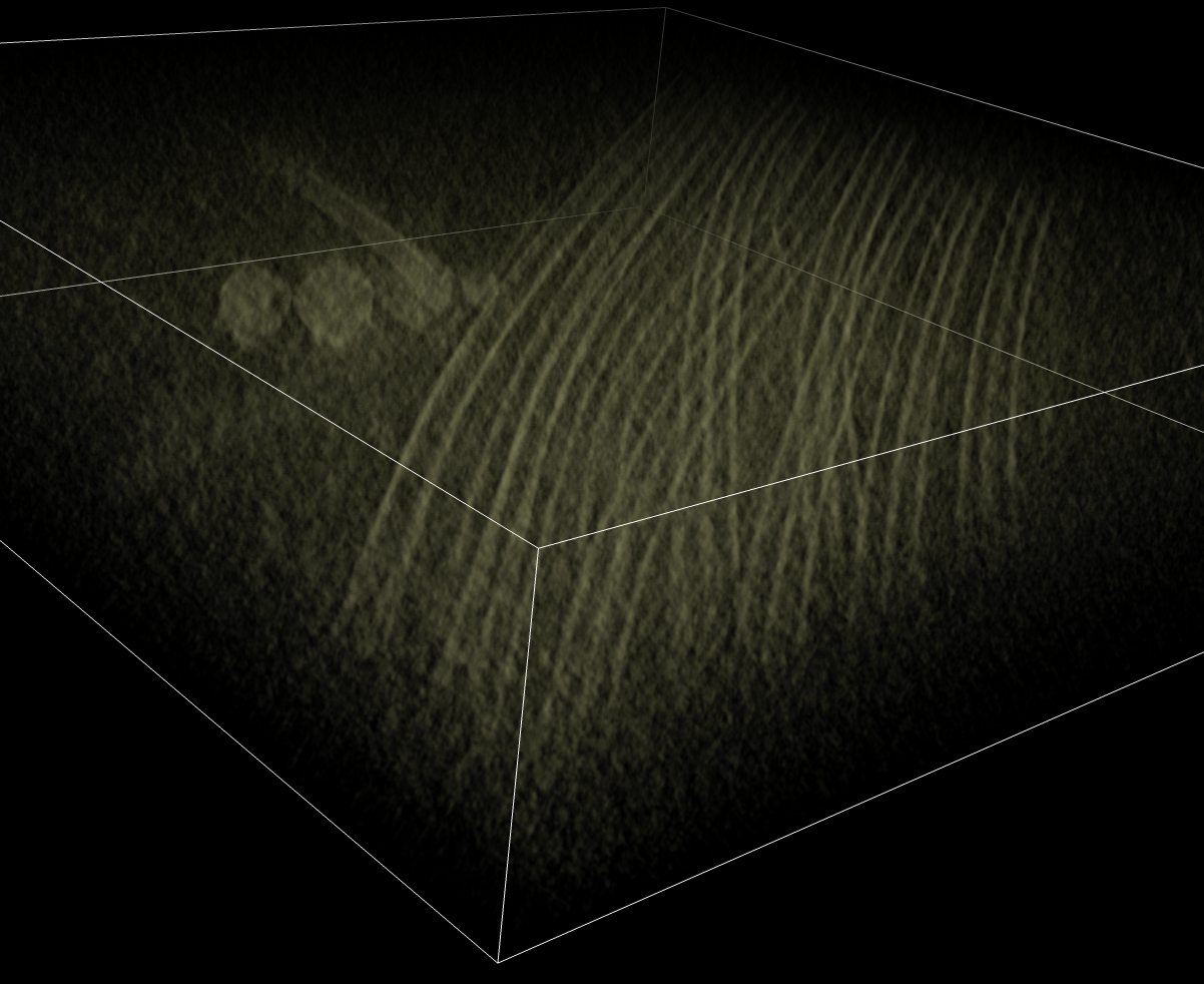
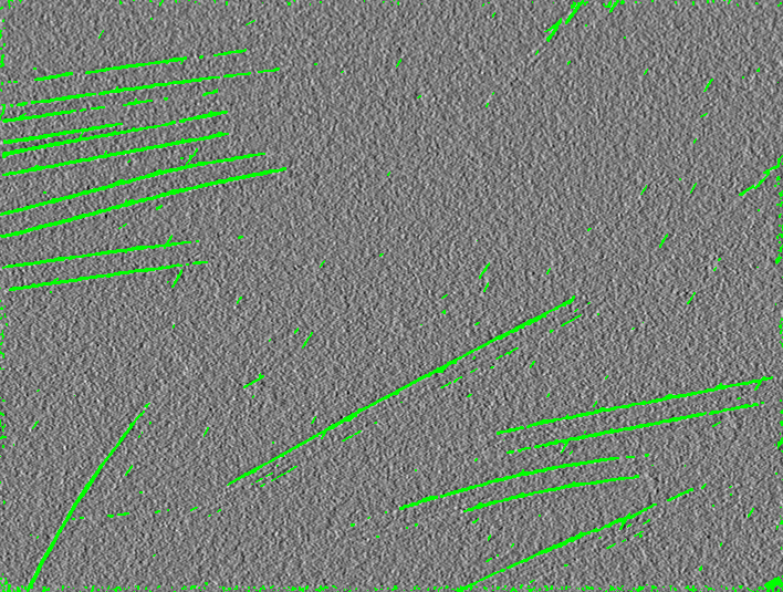
The Cryo-Seg software written in c ++ has been developed to detect microtubule structures and helical structures in 2D cryo-electron microscope images (see Figure 2 ). Cryo-electron tomography allows 3D observation of biological specimens in their hydrated state. Segmentation is formulated as Maximum A Posteriori estimation problem and exploits image patches to take into account spatial contexts (Markov Random Fields). Because of the contrast anisotropy in the specimen thickness direction, the whole tomogram is segmented section by section, with an automatic update of reference patches. This algorithm has been evaluated on synthetic data and on cryo-electron tomograms of in vitro microtubules. On real data, this segmentation method extracts the most contrasted regions of microtubules, and 3D visualization is improved.
Partners: S. Blestel and D. Chrétien (UMR 6026 CNRS University of Rennes 1)