

Section: New Results
Neuroprostheses
Distributed Measurement Unit for Closed-Loop Functional Electrical Stimulation: Prototype for Muscular Activity Detection
Participants : Guillaume Coppey, David Andreu, David Guiraud.
One way to face centralized Functional Electrical Stimulation (FES) architecture limitations is to distribute electronics close to electrodes. These Distributed Stimulation and Measurement Units (DSU and DMU) are interconnected by a network. Different DSU have been designed and prototyped. We started the design and prototyping of a DMU dedicated to ElectroMyoGramm (EMG) activity reading.
To validate both the digital architecture of the DMU and the digital processing it performs, we prototyped a DMU in charge of muscular activity detection. This DMU is able to detect a threshold crossing on an EMG input signal. The experimental setup is schematically represented on figure 11 , showing also the digital architecture of the prototyped DMU.
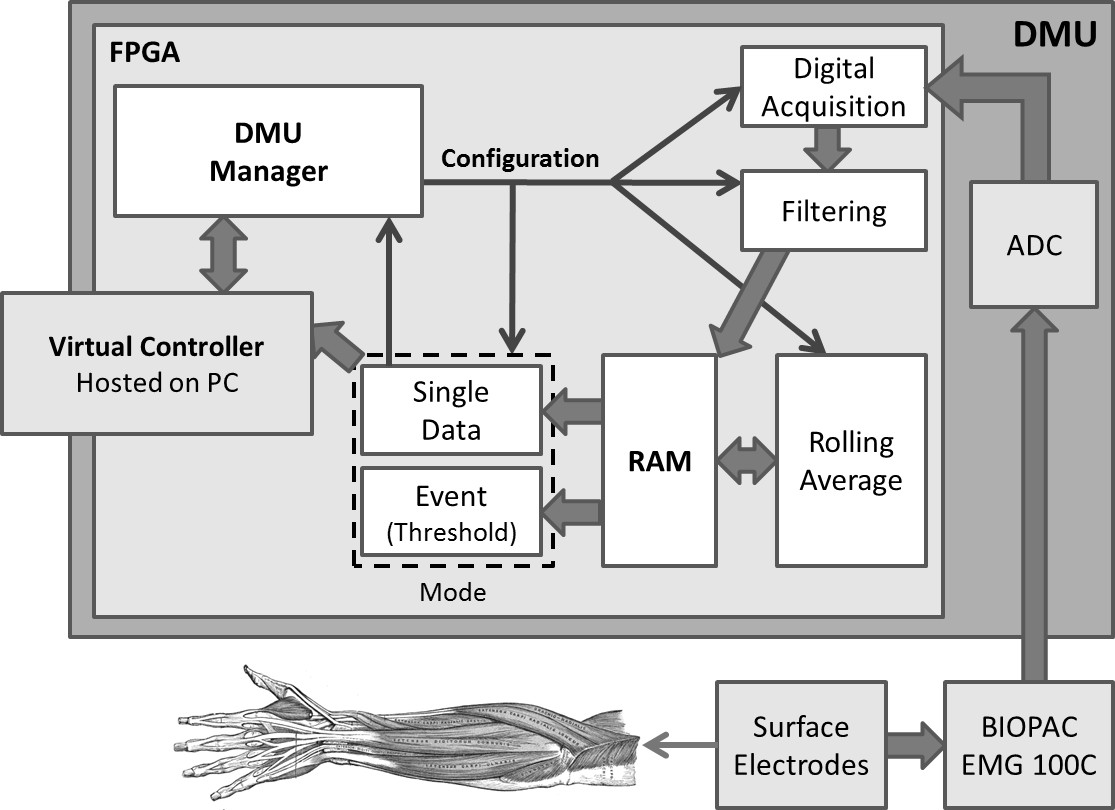
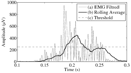
This DMU is able to accurately detect EMG activity after filtering and then processing a rolling average. Figure 12 shows intermediate signals: (a) is the absolute value of the filtered EMG signal, (b) is the rolling average, and (c) is the threshold used for activity detection.
This DMU prototype showed that digital processing chain dedicated to EMG activity detection can be embedded within a distributed measurement unit using a programmable logical device (FPGA), like we did for distributed stimulation unit. The embedded architecture of this unit is designed according to a Petri Net based methodology. This allows to exploit effective parallelism offered by FPGA devices, and to reach expected performances even at low frequency. The embedded processing chain is configurable and parameters can be adjusted, in order to optimize performance.
Future works will consist in adding the protocol stack to the digital architecture of the DMU, allowing integrating it within our distributed FES architecture. This will allow us to measure effective latencies and other performances from a closed-loop point of view. This work is necessary to ensure that such a distributed EMG activity detection is adequate with FES requirements. After that, we will investigate the trade-off between the global performances versus the implantable device constraints, like its size and power consumption.
Abstraction and composition for formal design of neuroprotheses
Participants : Hélène Leroux, David Andreu, Karen Godary.
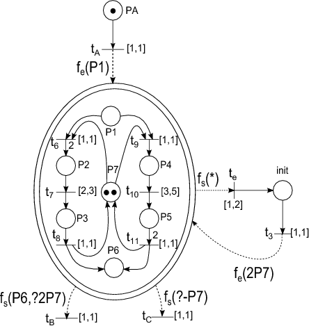
In the framework of specification and implementation of complex digital systems on FPGA, we have developped an approach based on components whose behavior and composition are specified by generalized interpreted T-time Petri nets. One of the inherent difficulties for designer is to take into account, on the behavioral part, exceptions. This leads often to a complex modeling and is a source of human errors. Indeed, it is intricate to express all the possible situations (i.e. current state of model). We have defined a way to model exception handling by integrating the well-know concept of macroplace into the formalism. The analysability of the model and the efficiency of the implementation on FPGA (reactivity and surface, ie number of logic blocks) have been preserved. An example of macroplace is given in figure 13 ; it contains a sub-net (set of places of its refinement) from which exception handling is simply described by a dedicated output transition (transition on fig. 13 ), whatever is the current state of the sub-net.
Increasing stimulation selectivity using the Transversal Intrafascicular Multichannel Electrode (TIME)
Participants : Pawel Maciejasz, David Andreu, David Guiraud, Xavier Navarro, Jordi Badia (Universitat Autònoma de Barcelona), Winnie Jensen, Kristian Rauhe Harreby, Aritra Kundu, Bo Geng (Aalborg University), Thomas Stieglitz, Tim Boretius (University of Freiburg), Ken Yoshida (Purdue University Indianapolis).
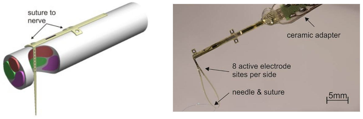
The electrical stimulation of nerve fibres may allow to restore or augment some body functions lost due to disease or injury. However, in typical peripheral nerves there are thousands of nerve fibres innervating various organs. Therefore, it is necessary to develop interfaces and methods allowing for selective activation of only desired population of nerve fibres. Various neural interfaces have been already proposed for that purpose, including multipolar cuff electrodes, longitudinal intrafascicular electrodes (LIFE) and the Utah Slanted Electrode Array (USEA), all with different selectivity and invasiveness ratios. Recently a new electrode concept of a transversal intrafascicular multichannel electrode (TIME) has been proposed (Fig. 14 ). This electrode has been developed in frame of the European Project TIME in which the DEMAR team is participating (grant CP-FP-INFSO 224012 from the European Union). It is intended to be implanted transversally in the nerve and address several fascicles or subgroups of nerve fibres with one device. It has longitudinal shape and has several independent stimulation sites equally spread on both sides of the electrode.
|
It has been already shown that TIME allows to achieve high selectivity of stimulation when using monopolar configuration, i.e. when current is delivered through one of the sites of the TIME against small needle electrode placed in the proximity of the nerve. We have performed investigation in the sciatic nerve of rat to verify if the use of bipolar configuration, i.e. when current is delivered through one of the TIME sites against an other site of the same electrode, could allow to further enhance selectivity of stimulation. The results of our studies suggest that using bipolar configuration do allow to increase selectivity of stimulation. However, higher charge of the stimulation may be necessary to achieve similar level of muscle activation, as compared to the monopolar configuration [24] .
When applied in the rat model the Transverse Intrafascicular Multi-channel Electrode (TIME) showed selective nerve fascicle recruitment. But results from the larger and poly-fasicular median nerves in pigs indicated that a single TIME could not reach the entire nerve and could only selectively recruit a subset of the nerve fasicles. The use of multiple TIME structures could offer a means to achieve highly selective fascicular stimulation while reaching a larger percentage of the fascicles in the nerve. Therefore we have investigated the use of pairs of TIMEs implanted in the median nerves of anesthesized pigs. TIME structures were implanted at different angles relative to each other or in parallel with one another. Electrical stimuli was passed through each contact of each TIME and the resulting electromyograms were recorded from seven muscles innervated by the median nerve. The ability to recruit these muscles was used to assess the stimulation selectivity of each contact using a selectivity index comparing the root-mean-square of the the evoked EMG of individul muscles. Results showed a signifcant increase in the selectivity index, when using two TIMEs compared to one. The optimal improvement was obeserved when TIMEs were placed in parallel to each other in such a way that they interfaced non-overlapping nerve regions [18] .
Nerve model for ENG recording
Participants : Olivier Rossel, Guy Cathébras, Fabien Soulier, Serge Bernard.
In the context of selective electroneurogram recording, we showed last year the efficiency of a small tripole filtering (the distance between contacts is ) thanks to simulated signals. This recording is locally sensitive and greatly increases the selectivity of the electrode. This year, we realized an experiment to verify the simulated results. Theoritical study of the small tripole sensitivity was realized for a single-fiber action potential (sfap ). We wanted to proceed in the same way for the experiment by trying to measure a sfap .
However, actual biological sfap would be hardly measurable by a small tripole in an in-vitro experiment. So, we decided to choose an approach based on an artificial axon. In this artificial model, every parameter is under control. The position of the fiber, the nodes of Ranvier, the position of the measuring electrode, as well as the involved currents are perfectly known. This allows us to implement and to estimate with accuracy the filtering realized by the small tripole with exactly the same configurations as in simulations. And by performing measures for several radial distances, we can verify the influence of this distance, in order to estimate the sensibility the small tripole. Moreover, we can increase the activity amplitude to be higher than in real fiber and then achieve a beneficial signal to noise ratio.
Method
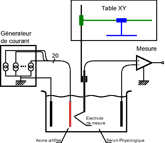
The experimental setup consists in an artificial model emulating an axon and in a system measuring the action potential across the surrounding medium, as sketched in Fig. 15 . Biological tissues are modeled by a saline solution having a conductivity close to the human body. The space and time behavior of the current generated by a natural axon is reproduced on the artificial axon. The latter is emulated by an cochlear electrode. It exhibits 20 contacts, being an accurate image of the nodes of Ranvier on an axon of diameter regarding the spatial periodicity.
In order to generate electric activity on several contacts that can be compared to the one of several nodes of Ranvier during the conduction of an action potential, we realized a custom multi-current generator with asynchronous outputs. The chosen amplitude was times that of an human axon.
The measure of the sfap in the space, is done using a punctual monopolar electrode. The position of the measuring electrode relative to the fiber is automatically set by a programmable micromanipulator. Then, the measure is repeated every along on the radial axis.
Monopolar and tripolar sensitivity
|
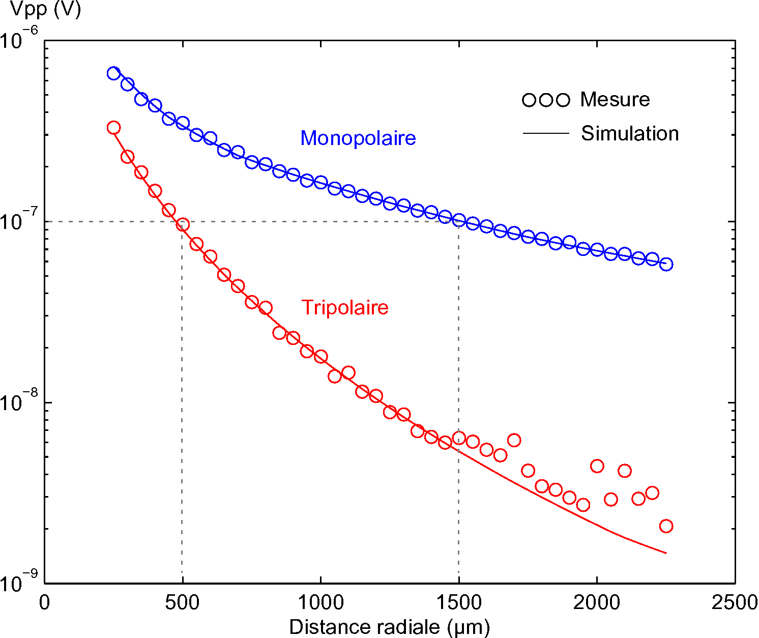
To verify quantitatively the sensibility results obtained by simulation for monopolar and tripolar electrodes, we have first estimated the amplitude of a sfap measured by a monopolar electrode. The measured sfap amplitude directly gives the monopolar recording that varies according to the radial distance between the artificial axon and the electrode. Then, the small-tripole recording results from an off-line process combining three points of measure spaced out of . The two kind of recordings are represented in Fig. 16 .
By comparing the recording sensitivities either estimated or measured, we can conclude that measures perfectly fit the theoretical results. We can also notice that the attenuation relative to the radial distance is more important for the small tripole than for the monopolar measure. This confirms that the measure performed by one small tripole electrode is exclusively sensitive to the closest fibers.