Section: New Results
Algorithms for dejittering and deconvolving large fluorescence and Tissue MicroArray (TMA) images
Participants : Hoai Nam Nguyen, Giovanni Petrazzuoli, Aminata Diouf, Charles Kervrann.
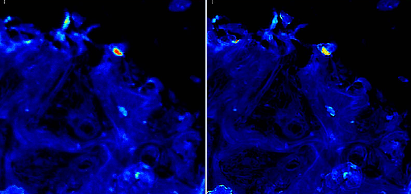
In fluorescence microscopy, the image quality is limited by out-of-focus blur and high noise. Traditionally, image deconvolution is needed to estimate a good quality version of the observed image. The result of deconvolution depends heavily on the choice of the regularization term and the noise dependent fidelity term. The regularization functional should be designed to remove noise while preserving image discontinuities. Accordingly, we investigated new regularization terms to preserve fine details of underlying structures and we studied appropriate proximal algorithms. The deconvolution method has been especially dedicated to large 2D images acquired with ISO scan imager (see Fig.3). The images are preliminary pre-processed to compensate non constant pixel displacement during acquisition/scanning (dejittering effect). The method has also been evaluated on 2D Vimentin filament images (UTSW, CytoDI Associated Team) to facilitate filament segmentation. The method is able to process a image in 250 ms with a non optimized implementation.
Collaborators: Vincent Paveau and Cyril Cauchois (Innopys company),
Philippe Roudot (UTSW, Dallas, USA).