Section: New Results
Denoising and compensation of the missing wedge in cryo electron tomography
Participants : Emmanuel Moebel, Charles Kervrann.
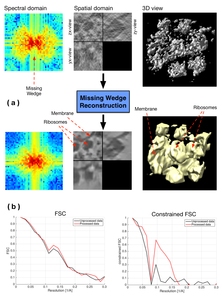
In this study, we address two important issues in cryo electron tomography (CET) images: reduction of noise and restoration of information in the missing wedge (MW). The MW is responsible for several type of imaging artifacts, and arises because of limited angle tomography: it is observable in the Fourier domain and is depicted by a region where Fourier coefficient values are unknown (see Fig. 9). The proposed stochastic method tackles the restoration problem by filling up the MW by iterating following steps : adding noise into the MW (step 1) and applying a denoising algorithm (step 2). The role of the first step is to propose candidates for the missing Fourier coefficients and the second step acts as a regularizer. A constraint is added in the spectral domain by imposing the known Fourier coefficients to be unchanged through iterations.
Several aspects of the method have been studied in order to gain a deeper understanding of this strategy: different kinds of noise as well as several denoising algorithms (BM3D, NL-Bayes, NL-means, Total Variation...) have been evaluated. Furthermore, different kinds of transforms have been tested in order to apply the constraint (Fourier transform, Cosine transform, pseudo-polar Fourier transform). Also, a process has been set up in order to evaluate the performance of the proposed method on experimental data. Thus, convincing results on experimental data have been achieved (see Fig. 9) using the Fourier Shell Correlation (FSC) as an evaluation metric. In order to measure the quality of the recovered MW only, we also compute the FSC over the MW support (“constrained FSC”).
Collaborators: Damien Larivière (Fondation Fourmentin-Guilbert),
Julio Ortiz (Max-Planck Institute, Martinsried, Germany).
|