Section: New Results
Detection of Brain Strokes Using Microwave Tomography
Participant : Laure Blanc-Féraud.
This work is done in collaboration with Vanna Lisa Coli and Juliette Leblond (EPI Factas, Inria Sophia), Pierre-Henri Tournier (Université Sorbonne, CNRS, LJLL, Inria, Paris) , Victorita Dolean (Université Côte d'Azur, CNRS, LJAD, Nice), Ibtissam El Kanfoud, Christian Pichot, Claire Migliaccio (Université Côte d'Azur, CNRS, LEAT, Sophia Antipolis).
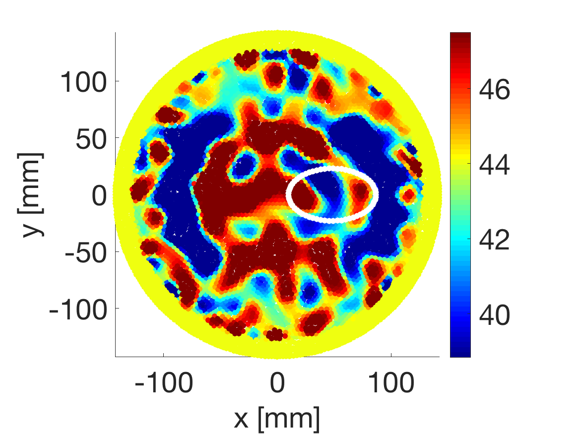
Brain strokes are one of the leading causes of disability and mortality in adults in developed countries. The ischemic stroke (85% of total cases) and hemorrhagic stroke (15%) must be treated with opposite therapies, so that the determination of the stroke nature must be made quickly to apply the appropriate treatment. Recent works in biomedical imaging showed that strokes produce variations on brain tissues complex electric permittivity that can be detected by means of microwave tomography.
We present here some synthetic results obtained with an experimental microwave tomography-based portable system for the early detection and monitoring of brain strokes (Figure 6). The determination of electric permittivity requires the solution of a coupled direct-inverse problem, where massive parallel computation from domain decomposition method and regularization techniques for optimization methods are employed. Synthetic data are obtained with electromagnetic simulations and a noise model developed for the specific problem, which has been derived from measurements errors with the experimental imaging system.