Section: New Results
3D flow estimation in 3D fluorescence microscopy image sequences
Participants : Sandeep Manandhar, Patrick Bouthemy, Charles Kervrann.
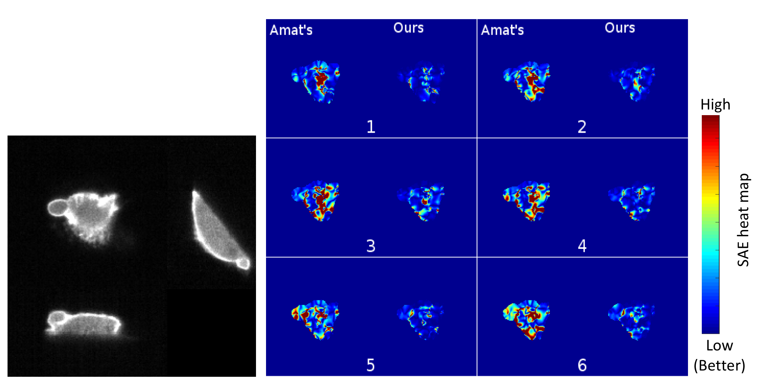
We have proposed a variational approach for 3D optical flow computation from a pair of fluorescence microscopy volumes. This computational method has been extensively evaluated on appropriate simulated data. To simulate a volume pair with a ground truth flow field, we extended the Horn-and-Schunck optical flow method to 3D (3DHS). The computed flow field by 3DHS between two input images is then used to generate a new pair volume. The new source and target volumes along with the corresponding 3DHS flow fields serve as the dataset with the ground truth. The latter was used for the parameterization of our method and for the comparative study with the state-of-the art method proposed by Amat et al., 2012 [44].
Meanwhile, we proposed a novel error measure named SAE (for Structural Angular Error) for 3D optical flow, in absence of any ground truth flow field (see Figure 7). SAE measures the angular difference between the principal orientations of the structures present in the backward warped target and the source volumes at each voxel. We found out that the average of SAE (ASAE) and average of the end-point-error (a standard optical flow error in presence of ground truth) behave similarly.
We also integrated regularization in our variational approach. In contrast to our previous regularization approach, this method preserves discontinuities of the flow field. However, both of our methods are time demanding and not parallelizable in implementation. Then, we integrated our Census Signature-based data term with total variation regularization that also produces discontinuous flow fields. Consequently, we took the splitting approach for optimization. A gain of four times was obtained in the calculation speed with 12-core implementation of the new method, compared to our previous two methods, for still similar ASAE score.
We have also proposed two new methods for the visualization of the 3D flow fields, named 3DHSV and MIP-flow respectively. The 3DHSV method color codes the 2D projections of the flow field in the Hue and the Saturation color spaces, while mapping the off-the-plane motion to the Value space. MIP-flow also encodes the 2D projections to the Hue and the Saturation spaces. However, it only considers encoding the 2D projections of the 3D vectors corresponding to the maximum intensity points in the direction perpendicular to the projection plane. This work is carried out in collaboration with UTSW Dallas in the frame of the Inria associated team CytoDI.
Software: FlowScope (see Section 6.7).
Collaborators: P. Roudot, E. Welf and G. Danuser (UTSW Medical Center, Dallas, USA).
|