

Section: New Results
Noise modeling and denoising for intensified camera in fluorescence imaging
Participants : Philippe Roudot, Charles Kervrann.
Two papers under review.
|
|
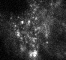
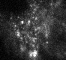

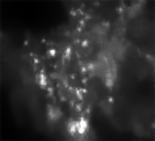
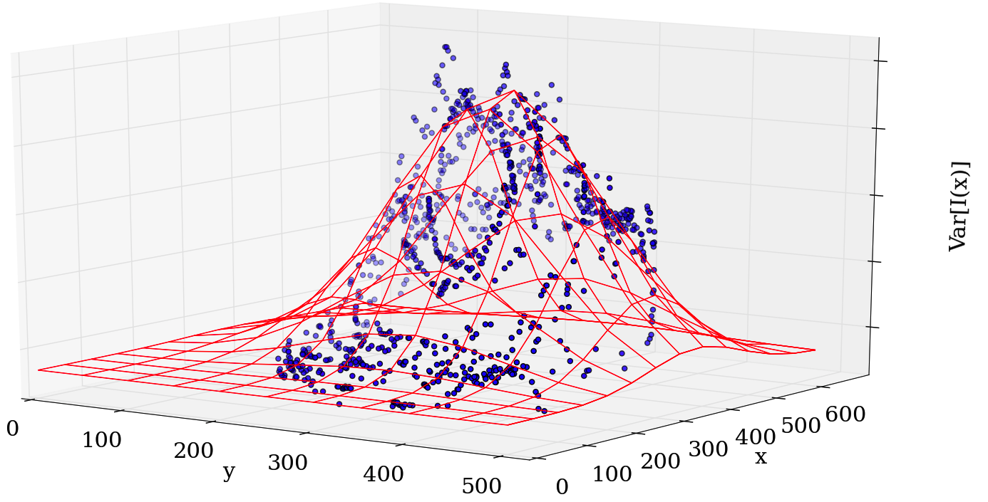
Image intensifiers are commonly used in low light level biological imaging, especially for fluorescence imaging. In this study, we have proposed a statistical framework for noise variance estimation dedicated to image sequences acquired with ICCD (Intensifier CCD). The model has been exploited for fluorescence lifetime estimation (Fluorescence lifetime imaging microscopy, FLIM) [13] , [12] and image denoising. We have investigated an alternative approach to [41] and we have shown that intensifier gain variation cannot be neglected in the variance estimation as opposed to a CCD sensor gain. Additionally, we have suggested to correct the noise model spatially to cope with microscopical aberration which are common in experimental setups (see Fig. 9 ). Finally, we have proposed a novel denoising algorithm based on the NL-means filter [23] which does rely on variance stabilization. The novel patch-based filter is able to adapt to local intensity-based noise statistics (see Fig. 10 ).
Partners: F. Waharte and J. Boulanger (UMR 144 CNRS PICT IBiSA Institut Curie)