Section: New Results
Vesicle segmentation method with automatic scale selection in TIRF microscopy
Participants : Antoine Basset, Charles Kervrann, Patrick Bouthemy.
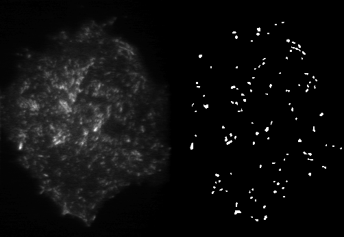
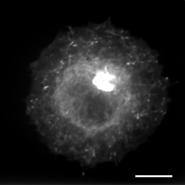
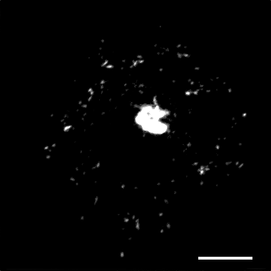
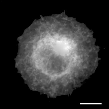
Accurately detecting cellular structures in fluorescence microscopy is of primary interest for further quantitative analysis such as counting, tracking or classification. We aimed at segmenting vesicles in Total Internal Reflection Fluorescence (TIRF) microscopy images.
In this study, we have proposed an original and efficient method – called SLT-LoG – for vesicle segmentation with fewer parameters than the state-of-the-art methods. It exploits the Laplacian of Gaussian (LoG) of the images at several scales. Since the vesicles size is almost constant in space and time, a prominent mode is expected in the empirical distribution of the scales at which the minima of LoG values are detected. It precisely corresponds to the optimal sought scale. The vesicle segmentation map is then derived by thresholding the LoG values obtained at this optimal scale. To set the threshold, we assume that the values of the LoG locally follow a normal distribution (see Fig. 7 ). For each point, we estimate the local mean and variance, and the threshold is deduced from a user-selected probability of false alarm.
We have evaluated our method on classical synthetic sequences for which the performances of many detection methods are available [52] , [56] . The comparative results on the dataset demonstrated that our method outperforms well-known unsupervised methods. We have also obtained very satisfactory results on real complex TIRF sequences.
Partners: Jean Salamero, J. Boulanger (UMR 144, PICT IBiSA, CNRS-Institut Curie)