Section: New Results
Classification of membrane dynamics in TIRF microscopy
Participants : Antoine Basset, Charles Kervrann, Patrick Bouthemy.
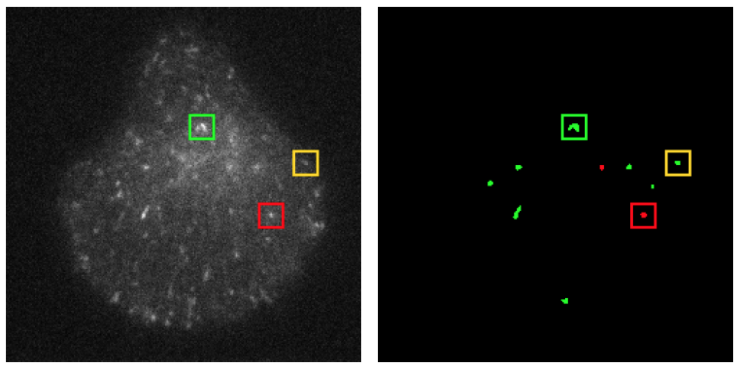
Recognizing dynamic protein behaviors in live cell fluorescence microscopy is of paramount importance to understand cell mechanisms. In the case of membrane traffic, cargo molecules are transferred from a donor to an acceptor compartments [49] . At each step, dedicated molecular platforms are acting to form, transport and address selected proteins. In microscopy imaging, this sequence of processes leads to a series of heterogeneous dynamics, which need to be untangled in order to understand the spatiotemporal coordination of the molecular actors. In this study, we aim at locating and recognizing temporal events in TIRF microscopy image sequences related to membrane dynamics. After segmenting the time-varying vesicles in the image, we exploit space-time information extracted from three successive images only to model, locate and recognize the two dynamic configurations of interest: translational motion or local fluorescence diffusion (see Fig. 11 ). A likelihood ratio test is defined to solve this issue. Results on synthetic sequences and real TIRF sequences demonstrated the accuracy and efficiency of the proposed method.
Partners: Jean Salamero, J. Boulanger (UMR 144, PICT IBiSA, CNRS-Institut Curie)
|