Section: New Results
Counting-based particle flux estimation for traffic analysis in live cell imaging
Participants : Thierry Pécot, Charles Kervrann.
In this study, we have proposed an original traffic analysis approach based on the counting of particles from frame to frame. Object tracking methods or optical flow methods are generally considered to analyze the dynamic contents of intracellular video-microscopy. The suggested method lies between these two well-known approaches. Instead of tracking each moving particle, we estimate fluxes of particles between predefined and adjacent regions. Our three-step counting-based approach is as follows:
-
The cell is uniformly partitioned into fixed-size and fixed-shape regions.
-
The moving particles are automatically detected using an appropriate algorithm.
-
The fluxes are estimated with sparse constraints from an image pair at each time step from temporal variations of the number of particles in each region of the uniform tessellation. Except for some trivial cases, the flux estimation is actually an ill-posed problem and additional constraints are necessary to find the optimal solution.
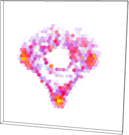
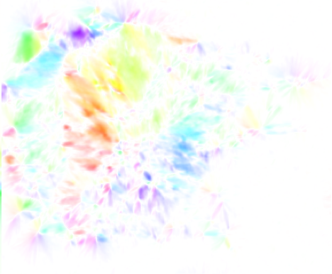
The problem is formulated as the minimization of a global cost function and the approach allows us to process image sequences with a high number of particles and a high rate of particle appearances and disappearances. We studied the influence of object density, image partition scale, motion amplitude and particle appearances/disappearances in a large variety of simulations. The potential of the method has been demonstrated on real image sequences showing GFP-tagged Rab6 trafficking in confocal microscopy.
Reference: [26]
Collaborators: Jean Salamero (UMR 144 CNRS-Institut Curie, PICT-IBiSA),
Jérôme Boulanger (UMR 144 CNRS-Institut Curie).