Section: New Results
Brain-Computer Interfaces
BCI Methods and Techniques
SimBCI: Novel Software Framework for Studying BCI Methods
Participants: Jussi Lindgren and Anatole Lécuyer
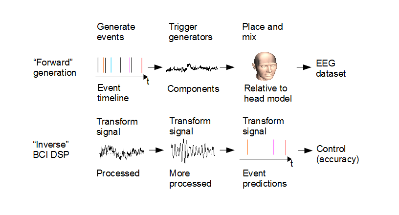
How to investigate the applicability of physiology-based source reconstruction for Brain-Computer Interfaces (BCIs)? The classic way is human experiments, but these unfortunately lack ground truth. The electrical activity inside the human brain is not fully described by external EEG measurements. In the CominLabs project SABRE, we have developed a BCI simulator framework called simBCI [22] to help in such studies. The framework allows modifying and changing generative models and their parameters inside a model brain, and studying what effects such changes have on the signal and subsequently the BCI signal processing. The modifiable parameters can include artifact properties, generative source locations, background activity characteristics and so on. We have released the framework as open source to the community (https://gitlab.inria.fr/sb/simbci/).
This work was done in collaboration with IMT Atlantique.
Novel Control Strategy for BCI Exploiting Visual Imagery and Attention
Participants: Jussi Lindgren and Anatole Lécuyer
Current paradigms for Brain-Computer Interfaces (BCIs) leave a lot to be desired in their accuracy and usability. We studied visual imagery as a potential new paradigm. In visual imagery, the user imagines objects or scenes visually, and the BCI is based on trying to classify the imagination type based on the EEG measurements. In [20], we studied to what extent can we distinguish the different mental processes of observing visual stimuli and imagining them based on the EEG. We found in a study of 26 users that we could somewhat differentiate (i) visual imagery vs. visual observation task (71% of classification accuracy), (ii) visual observation task towards different visual stimuli (classifying one observation cue versus another observation cue with an accuracy of 61%) and (iii) resting vs. observation/imagery (77% accuracy between imagery task versus resting state, and the accuracy of 75% between observation task versus resting state). All reported accuracies are averages over the users. Our results suggest that the presence of visual imagery and related alpha power changes may be useful to broaden the range of BCI control strategies.
|

BCI Applications
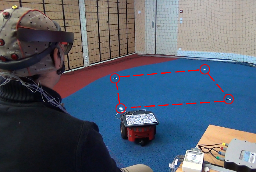
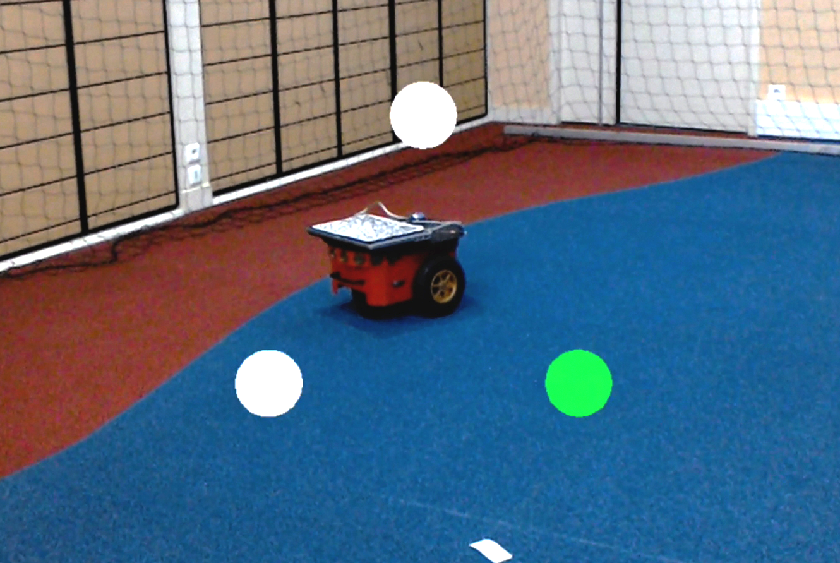
BCI-based Interfaces for Augmented Reality: Feasibility, Design and Evaluation
Participants: Hakim Si-Mohammed, Camille Jeunet, Ferran Argelaguet and Anatole Lécuyer
In [25], we have studied the combination of BCI and Augmented Reality (AR). We first tested the feasibility of using BCI in AR settings based on Optical See-Through Head-Mounted Displays (OST-HMDs). Experimental results showed that a BCI and an OST-HMD equipment (EEG headset and Hololens in our case) are well compatible and that small movements of the head can be tolerated when using the BCI. Second, we introduced a design space for command display strategies based on BCI in AR, when exploiting a famous brain pattern called Steady-State Visually Evoked Potential (SSVEP). Our design space relies on five dimensions concerning the visual layout of the BCI menu ; namely: orientation, frame-of-reference, anchorage, size and explicitness. We implemented various BCI-based display strategies and tested them within the context of mobile robot control in AR. Our findings were finally integrated within an operational prototype based on a real mobile robot that is controlled in AR using a BCI and a HoloLens headset. Taken together our results (from four user studies) and our methodology could pave the way to future interaction schemes in Augmented Reality exploiting 3D User Interfaces based on brain activity and BCIs.
This work was done in collaboration with Loki Inria team.
|
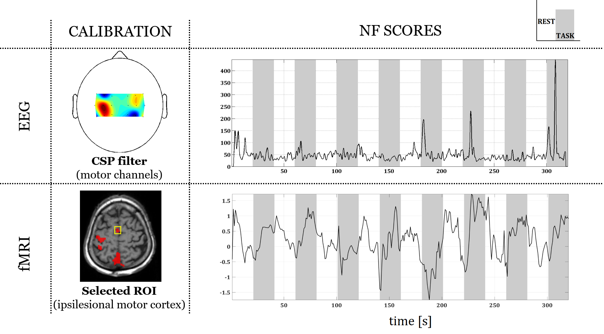
Neurofeedback for Stroke Rehabilitation: A Case Report
Participants: Giulia Lioi, Mathis Fleury and Anatole Lécuyer
Neurofeedback (NF) consists in training self-regulation of brain activity by providing real-time information about the participant brain function. Few works have shown the potential of NF for stroke rehabilitation however its effectiveness has not been investigated yet. NF approaches are usually based on real-time monitoring of brain activity using a single imaging technique. Recent studies have revealed the potential of combining EEG and fMRI to achieve a more efficient and specific self-regulation. In this case report [49], we tested the feasibility of applying bimodal EEG-MRI NF on two stroke patients affected by left hemiplegia participated. The protocol included a calibration step (motor imagery of hemiplegic hand) and two NF sessions (5 minutes each). The experiment was run using a NF platform performing real-time EEG-fMRI processing and NF presentation. Both patients were found able to self-regulate their brain activity during the NF sessions. The EEG activity was harder to modulate than the BOLD activity. The patients were highly motivated to engage and satisfied with the NF animation, as assessed with a qualitative questionnaire. These results showed the feasibility and the potential of applying EEG-fMRI NF for stroke rehabilitation.
This work was done in collaboration with Visages Inria team.
|
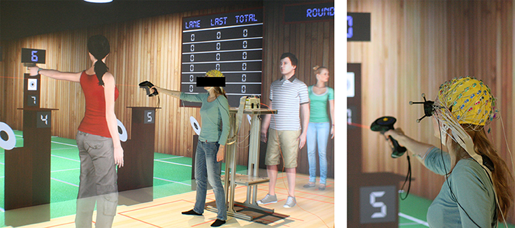
Using EEG in Sport Performance Analysis
Participants: Ferran Argelaguet and Anatole Lécuyer
Competition changes the environment for athletes. The difficulty of training for such stressful events can lead to the well-known effect of “choking” under pressure, which prevents athletes from performing at their best level. To study the effect of competition on the human brain we recorded [24] pilot electroencephalography (EEG) data while novice shooters were immersed in a realistic virtual environment representing a shooting range. We found a differential between-subject effect of competition on mu (8–12 Hz) oscillatory activity during aiming; compared to training, the more the subject was able to desynchronize his mu rhythm during competition, the better was his shooting performance. Because this differential effect could not be explained by differences in simple measures of the kinematics and muscular activity, nor by the effect of competition or shooting performance per se, we interpret our results as evidence that mu desynchronization has a positive effect on performance during competition. It remains to show whether this effect can be generalized to expert shooters. Our findings could be relevant in sports training to help athletes avoid choking under pressure during competition. Confirmation through further experimental validation is however needed.
This work was done in collaboration with EPFL.