Section: New Results
Detection of transitions between diffusion models along biomolecule trajectories
Participants : Antoine Salomon, Charles Kervrann.
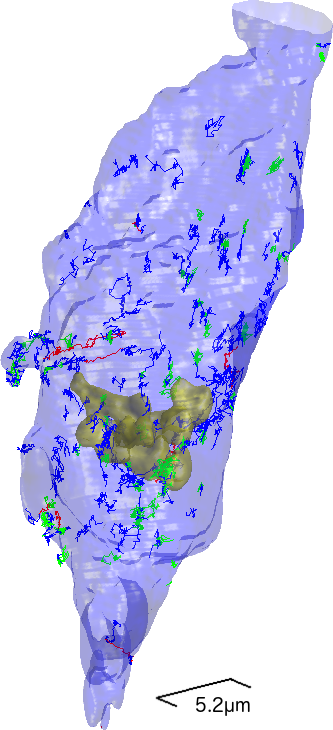
Recent advances in molecular biology and fluorescence microscopy imaging have made possible the inference of the dynamics of single molecules in living cells. When we observe a long trajectory (more than 100 points), it is possible that the particle switches mode of motion over time. Then, a goal is to estimate the temporal change-points, that is the distances at which a change of dynamics occurs. To address this issue, we proposed a non-parametric procedure based on test statistics [16], computed on local windows along the trajectory, to detect the change-points. This algorithm controls the number of false change-point detections in the case where the trajectory is fully Brownian. Our algorithm is user-friendly as there is only one parameter to tune, namely the sliding window size. A Monte Carlo study is proposed to demonstrate the performances of the method and also to compare the procedure to two competitive algorithms. Our method is much faster than previous methods which is an advantage when dealing with a large numbers of trajectories. With this computational approach, we analyzed real data depicting neuronal mRNPs (mRNAs in complex with mRNA-binding), and another very complex biological example, Gal-3 trafficking from the plasma membrane to different cellular compartments (acquired with Lattice Light Sheet microscopy). The analysis of multiple Gal-3 trajectories demonstrates nicely that there is not one typical signature. Biological trafficking events are very multifaceted. The algorithm was capable of identifying and characterizing the multistep biological movement, switching several times between subdiffusive, superdiffusive and Brownian motion.
Collaborators: Vincent Briane (UNSW Sydney, School of Medical Sciences, Australia),
Myriam Vimond (CREST ENSAI Rennes),
C.A. Valades Cruz and C. Wunder, (Institut Curie, PSL Research University, Cellular and
Chemical Biology, U1143 INSERM / UMR 3666 CNRS).
|