Section: New Results
Computational Physiology
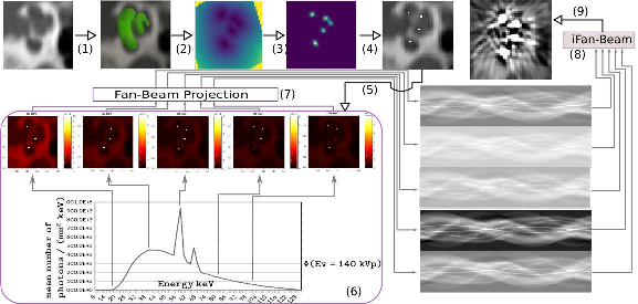
Deep Learning based Metal Artifacts Reduction in post-operative Cochlear Implant CT Imaging
Participants : Zihao Wang [Correspondant] , Clair Vandersteen, Thomas Demarcy, Dan Gnansia, Charles Raffaelli, Nicolas Guevara, Hervé Delingette.
This work is funded by the Provence-Alpes-Côte-d'Azur region, the Université Côte d’Azur and Oticon Medical through CIMPLE https://team.inria.fr/epione/en/research/cimple/ research project.
Generative Adversarial Network, Metal Artifacts Reduction, Cochlea Implantation
We propose a 3D metal artifact reduction method using convolutional neural networks for post-operative cochlear implant imaging.[44]
-
Learn metal artifacts reduction by using pre-operative images and metal artifacts simulation to create image pairs for training GANs.
-
Metal artifacts simulation starts from a cochlea implantation fusion image and ends with the simulated post-operative image.(Fig. 15)
-
A 3D generative adversarial network (MARGANs) to create an image with a reduction of metal artifacts.
-
Evaluations on ten patients show the effectiveness of artefact reduction compared to two classical methods.
|
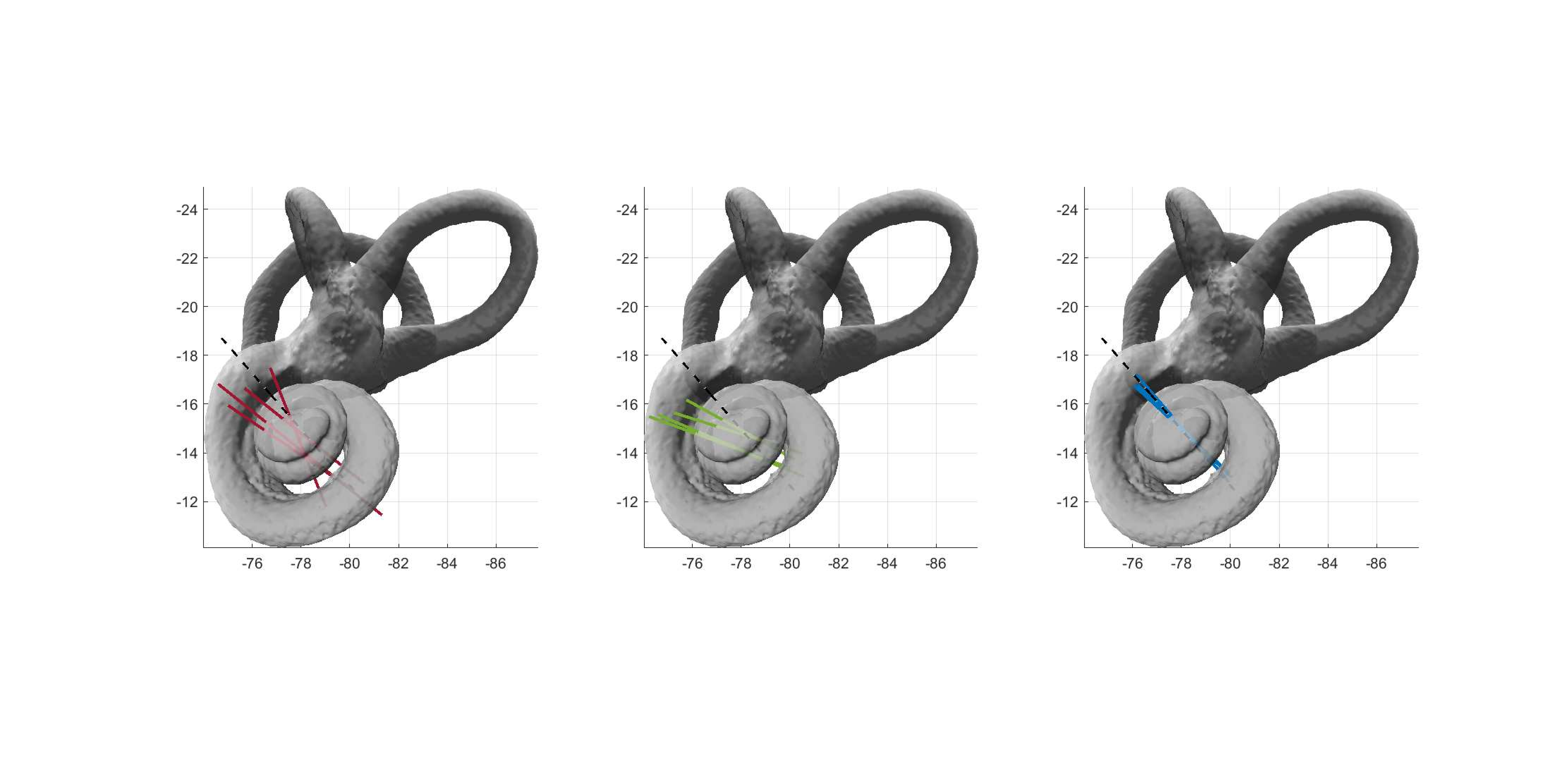
Kinematic Spiral Shape Recognition in the Human Cochlea
Participants : Wilhelm Wimmer [Correspondant] , Clair Vandersteen, Nicolas Guevara, Marco Caversaccio, Hervé Delingette.
Supported by the Swiss National Science Foundation (no. P400P2_180822) and the French government (UCA JEDI - ANR-15-IDEX-01).
Approximate maximum likelihood, kinematic surface recognition, natural growth
To improve therapies for hearing loss and deafness, e.g., with auditory neuroprostheses, we developed a reliable detection algorithm for the cochlear modiolar axis in CT images (Fig. 16). The algorithm was tested in an experimental study with 4 experts in 23 human cochlea CT data sets [45] [27]. Our experiments showed that the algorithm reduces the alignment error providing more reliable modiolar axis detection for clinical and research applications.